

Hashimoto's thyroiditis: celebrating the centennial through the lens of the Johns Hopkins hospital surgical pathology records
- PMID: 23151083
- PMCID: PMC3569966
- DOI: 10.1089/thy.2012.0554
Hashimoto's thyroiditis: celebrating the centennial through the lens of the Johns Hopkins hospital surgical pathology records
Abstract
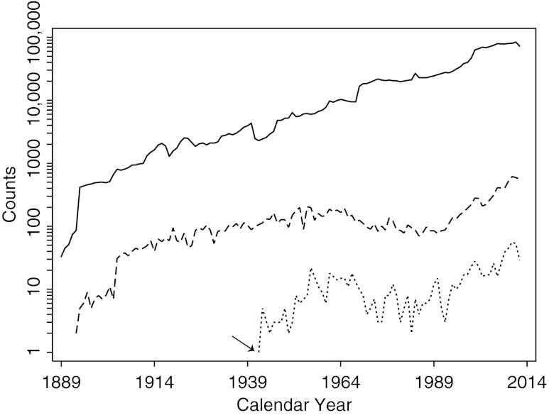
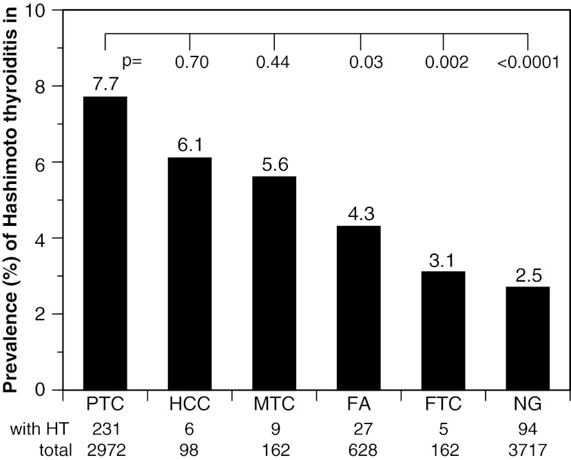
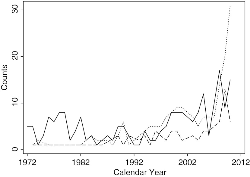
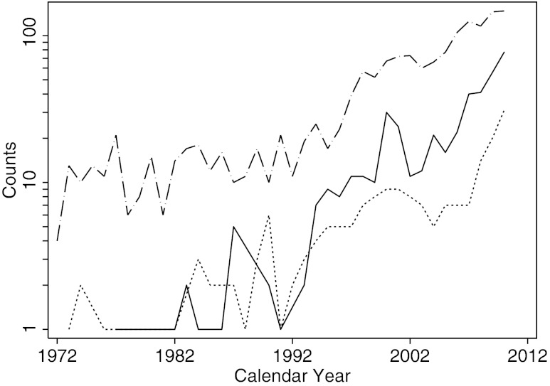
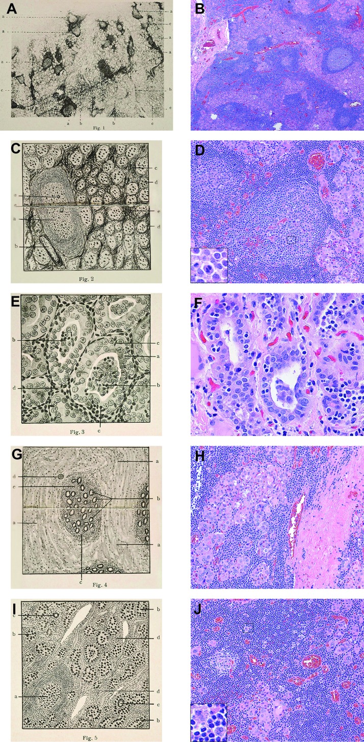
Hashimoto's thyroiditis is now considered the most prevalent autoimmune disease, as well as the most common endocrine disorder. It was initially described in 1912, but only rarely reported until the early 1950s. To celebrate this centennial, we reviewed the surgical pathology archives of the Johns Hopkins hospital for cases of Hashimoto's thyroiditis, spanning the period from May 1889 to October 2012. Approximately 15,000 thyroidectomies were performed at this hospital over 124 years. The first surgical case was reported in 1942, 30 years after the original description. Then, 867 cases of Hashimoto's thyroiditis were seen from 1942 to 2012, representing 6% of all thyroidectomies. Hashimoto's thyroiditis was the sole pathological finding in 462 cases; it accompanied other thyroid pathologies in the remaining 405 cases. The most commonly associated pathology was papillary thyroid cancer, an association that increased significantly during the last two decades. The most common indication for thyroidectomy was a thyroid nodule that was cytologically suspicious for malignancy. Hashimoto's thyroiditis remains a widespread, intriguing, and multifaceted disease of unknown etiology one century after its description. Advances in the understanding of its pathogenesis and preoperative diagnosis will improve recognition and treatment of this disorder, and may one day lead to its prevention.
Figures
Comment in
-
A hundred years of Hashimoto's thyroiditis.Thyroid. 2013 Feb;23(2):135-6. doi: 10.1089/thy.2013.2302.ed1. Thyroid. 2013. PMID: 23398159 No abstract available.
-
On the association between Hashimoto's thyroiditis and papillary thyroid carcinoma: looking 100 years back and, hopefully, fewer years ahead to sort out this association.Thyroid. 2013 Sep;23(9):1180-1. doi: 10.1089/thy.2013.0126. Thyroid. 2013. PMID: 23607293 No abstract available.
References
-
- LiVolsi VA. The pathology of autoimmune thyroid disease: a review. Thyroid. 1994;4:333–339. - PubMed
-
- Jansson R. Totterman TH. Sallstrom J. Dahlberg PA. Intrathyroidal and circulating lymphocyte subsets in different stages of autoimmune postpartum thyroiditis. J Clin Endocrinol Metab. 1984;58:942–946. - PubMed
-
- Kakudo K. Li Y. Taniguchi E. Mori I. Ozaki T. Nishihara E. Matsuzuka F. Miyauchi A. IgG4-related disease of the thyroid gland. Endocr J. 2011;59:273–281. - PubMed
-
- Hahsimoto H. Zur Kenntniss der lymphomatösen Veränderung der Schilddrüse (Struma lymphomatosa) Arch Klin Chir. 1912;97:219–248.
-
- Sawin CT. Hakaru Hashimoto (1881–1934) and his disease. Endocrinologist. 2001;11:73–76.
Publication types
MeSH terms
Grants and funding
LinkOut - more resources
Full Text Sources
Other Literature Sources
