. 2012 Nov 1;8(11):1607-19.
doi: 10.4161/hv.22574. Epub 2012 Nov 1.

Generation of antigen-specific immunity following systemic immunization with DNA vaccine encoding CCL25 chemokine immunoadjuvant

Affiliations

Generation of antigen-specific immunity following systemic immunization with DNA vaccine encoding CCL25 chemokine immunoadjuvant

Noshin Kathuria et al. Hum Vaccin Immunother. .

Abstract

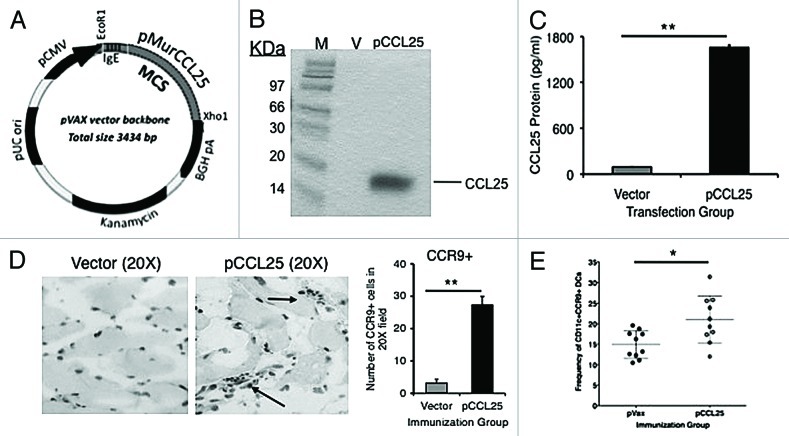
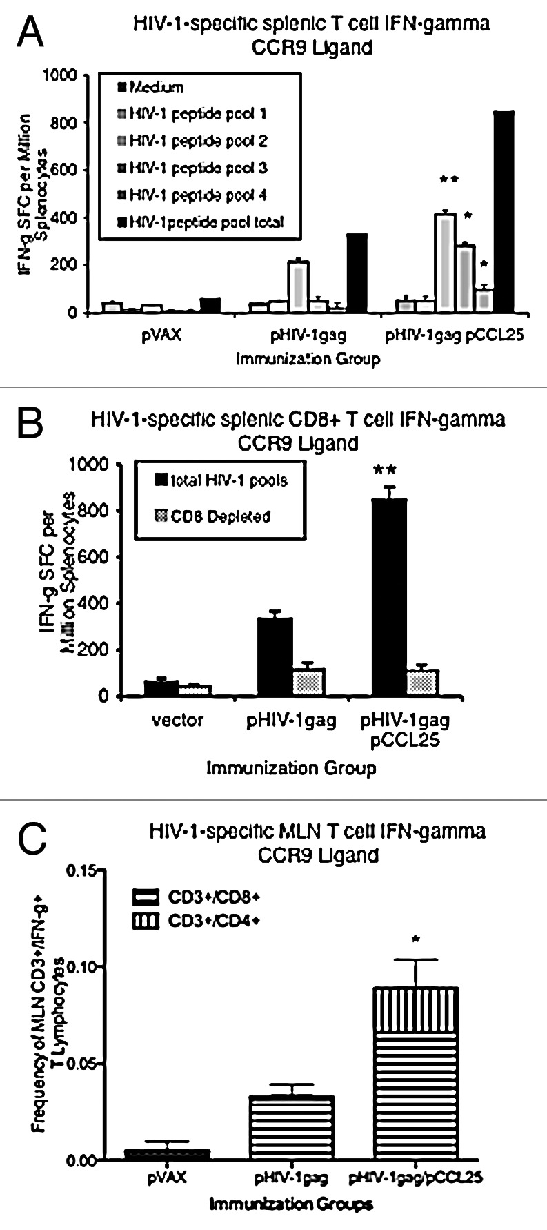
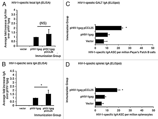
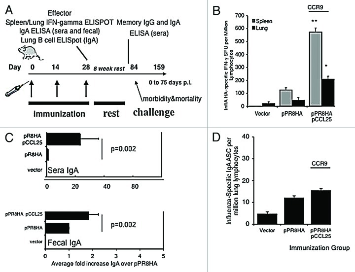
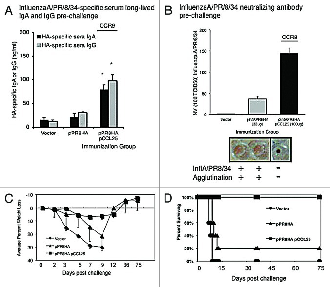
A significant hurdle in vaccine development for many infectious pathogens is the ability to generate appropriate immune responses at the portal of entry, namely mucosal sites. The development of vaccine approaches resulting in secretory IgA and mucosal cellular immune responses against target pathogens is of great interest and in general, requires live viral infection at mucosal sites. Using HIV-1 and influenza A antigens as models, we report here that a novel systemically administered DNA vaccination strategy utilizing co-delivery of the specific chemokine molecular adjuvant CCL25 (TECK) can produce antigen-specific immune responses at distal sites including the lung and mesenteric lymph nodes in mice. The targeted vaccines induced infiltration of cognate chemokine receptor, CCR9+/CD11c+ immune cells to the site of immunization. Furthermore, data shows enhanced IFN-λ secretion by antigen-specific CD3+/CD8+ and CD3+/CD4+ T cells, as well as elevated HIV-1-specific IgG and IgA responses in secondary lymphoid organs, peripheral blood, and importantly, at mucosal sites. These studies have significance for the development of vaccines and therapeutic strategies requiring mucosal immune responses and represent the first report of the use of plasmid co-delivery of CCL25 as part of the DNA vaccine strategy to boost systemic and mucosal immune responses following intramuscular injection.

Keywords: DNA vaccine; IgA; TECK/CCL25; chemokine; molecular adjuvant; mucosal.


Figures

None
Figure 1. Expression of bioactive chemokines from plasmid encoded mucosal chemokine and the induction of infiltration, after intramuscular injection, by cognate receptor positive cells (A). All genes were cloned into the multiple cloning region of the expression vector pVAX (kanamycin resistance) using the restriction enzyme sites, EcoR1 and Xho1. (B) Expression of pCCL25 was confirmed by a T7 coupled transcription/translation reticulocyte lysate system. The blotting gel shows size markers (designated M, lane 1), pVAX background control protein (V, lane 2), 14.2 kDa CCL25 protein (lane 3). (C) Expression of bioactive chemokine protein translated from the plasmid forms of CCL25. ELISA was performed using supernatants from pCCL25-transfected RD cells (pg/ml). Vector background control is included (gray bar) vs. pCCL25 (black bar). Data are shown as pg/ml of chemokine protein ± SD of triplicate wells. (D) Infiltration of CCR9 positive cells induced following intramuscular injection of 100 μg of pCCL25. Immunohistochemical staining of quadriceps sections 7 d post immunization with 100 μg of vector backbone control (left panel) or pCCL25. Infiltrating cells were enumerated following a visual count by microscope of brown positive cells over 6 (20×) fields, and the total number of CCR9+ cells was averaged and shown as ± SD for vector (gray bar) and pCCL25 injection (black bar). (E) Frequency of CD11c+ cells that express CCR9 in the popliteal and inguinal DLNs of individual immunized mice in vector backbone immunized (solid black symbols) vs. pCCL25 immunized mice (open circles). (*p < 0.05, **p < 0.01, for comparisons between vector backbone pVAX or pCCL25 immunized groups.) Data are representative of three independent experiments, and in panel (E), ± SEM is shown because individual mouse DLNs were tested.
None
Figure 2. Induction of mucosal antigen-specific IFN-λ in secondary lymphoid organs and MLNs after mucosal chemokine systemic co-immunization. (A) Mice (n = 4 per group) were immunized intramuscularly three times, with each injection separated by 2 weeks. Splenocytes were harvested from vector control, pHIV-1gag, pHIV-1gag/pCCL25, 1 week following the last injection. Splenocytes from vaccinated animals from each experimental group were pooled and were cultured overnight in the presence of medium (gray bar, negative control) or 10 μg/ml HIV-1 peptide pools 1 through 4 (hatched bars). The solid black bar represents the total number of HIV-1-specific IFN-λ SFCs per million splenocytes for all 4 HIV-gag pools. (B) CD8+ T cell depletion partially abrogates HIV-1gag-specific IFN-λ secreted by the lymphocytes from immunized mice. Total HIV-1gag-specific IFN-λ SFCs are shown as black bars and following CD8+ T cell depletion (gray bars). Values represent SFCs per million splenocytes and error represents the mean ± SD of the triplicate cultures and are representative of 6 independent experiments. (C) pCCL25 co-immunization enhances HIV-1-specific IFN-λ secretion by CD3+/CD4 and CD3+/CD8 MLN T lymphocytes. Mice were immunized intramuscularly three times, with each injection separated by 2 weeks. MLNs were harvested from vector control, pHIV-1gag, pHIV-1gag/pCCL25, 1 week following the last injection. Mucosal lymphocytes were harvested and stimulated with either medium background control, HIV-1gag peptides or PMA and ionomycin. Cells were stained as described in “Methods” and 50,000 CD3+ live lymphocytes (as determined by the LIVE/DEAD stain) were collected. Total IFN-λ secreting CD3+/CD4 (hatched upper stack) or CD3+/CD8 (gray lower stack) are graphed as the mean ± SEM of individual mice for vaccination groups. Responses from the negative control wells were subtracted from the antigenic stimulations prior to graphing (*p ≤ 0.05, ** p ≤ 0.01, for statistical comparisons between pCCL25 co-immunized groups vs. pHIV-1gag alone for similar treatment conditions). Data are representative of three independent experiments.
None
Figure 3. Augmented secretory IgA in the periphery and in the GALT following systemic co-immunization with mucosal chemokine. (A–B) The average fold increase of anti-HIVgag-specific IgA elicited by the chemokine immune adjuvants over pHIV-1gag antigenic plasmid alone is shown in panel A (fecal IgA) and panel B (sera IgA). Data generated from 6 independent experiments of 4 mice per group were averaged and fold increases were determined. Levels of HIV-1 specific IgA in fecal extract (A) or sera (B) harvested 10 d following the last immunization were determined by an ELISA assay against HIV-1gag p24 protein. Quantitation of fecal and serum IgA (μg/ml) was determined using a recombinant IgA standard of known concentration as the standard curve, and fold increase values were determined. (C–D) Mucosal chemokine enhanced IgA antibody secretion cell frequency in Peyer’s patches (C) and in the spleen (D). Whole Peyer’s patch (C) or splenocyte (D) immune cells purified from vector, pHIV-1gag or pCCL25 co-immunized mice were placed in an HIV-1gag p24 protein coated, 96 well ELISpot blocked plate and incubated for 5 h. Secreted anti-HIV-1gag IgA by antibody secreting plasmablasts was captured on plate bound P24, counted using an ELISpot reader and graphed as HIV-1-specific IgA ASCs per million Peyer’s patch B cells or splenocytes. Error represents the mean ± SD of triplicate wells (*p ≤ 0.05, ** p ≤ 0.01, for statistical comparisons between pCCL25 co-immunized groups vs. pHIV-1gag alone for similar treatment conditions). Data are representative of three independent experiments.
None
Figure 4. pCCL25 immune adjuvant-induced CTL and IgA immunity specific against influenza A/PR/8/34 in a mouse model of mucosal lung infection. (A) Schedule for influenza A (PR8) immunization, immune analysis and lethal mucosal challenge. Mice (n = 10 per group) were immunized as previously described with 33 μg of HA or in combination with our pCCL25. Effector immune responses were measured 1 week following the third immunization, and long-lived responses were examined 8 weeks after the final boost (pre-challenge). (B) Mucosal chemokines enhanced the secretion of IFN-λ influenza specific A/PR/8/34 by lung and splenic T cells. Lung lymphocytes (black bar) or splenocytes (gray bar) were harvested from immunized mice and utilized in an IFN-λ ELISpot assay stimulated with either medium control or the CD8+ T cell epitope peptide encoding influenza A/PR/8/34 (H1N1) hemagglutinin (IYSTVASSL amino acid 518–526). Values depicted in panel (B) are resulting values after medium is subtracted and error represents the mean ± SD of triplicate wells (*p < 0.05, **p < 0.01 for statistical comparisons of chemokine/pPR8HA groups with pPR8HA antigenic vector alone). (C) pCCL25 co-immunization augments mucosal-specific IgA in sera and fecal extracts. Pooled sera and fecal pellets from 4 mice per group were analyzed, by ELISA, for the presence of influenza A/PR/8/34 hemagglutinin specific IgA and data generated from six independent experiments were averaged and shown in panel (C). The fold increase observed from vaccinated sera (upper panel) and feces (lower panel) IgA by ELISA using a recombinant IgA standard of known concentration and the fold increase in IgA levels are shown, and statistical analysis was performed between the chemokine adjuvanted groups in relation to pPR8HA antigenic plasmid alone across 6 independent experiments. (D) pCCL25 co-immunization does not increase the frequency of IgA secreting plasmablasts in the lung. Lung lymphocytes isolated from vector, pPR8HA or pCCL25 co-immunized (perfused) mice were added to the wells of UV-inactivated influenza A/PR/8/34-coated, 96 well ELISpot blocked plates and incubated for 5 h. Secreted anti-HA IgA plasmablasts were captured on plate bound influenza A, counted using an ELISpot reader and graphed as influenza-specific IgA ASCs per million lung lymphocytes. Error represents the mean ± SD between triplicate wells. (The data are a representation of three independent experiments.)
None
Figure 5. pCCL25 co-immunization induces long-lived IgG, IgA, neutralizing antibody and protects mice against influenza A/PR/8/34 lethal mucosal challenge. (A) Influenza specific long-lived IgG and IgA titers were determined prior to challenge. Individual sera samples were harvested from mice from each immunization group (n = 10 mice per group) and (ng/ml) levels of pPR8HA-specific IgG (gray bar) and IgA (black bar) were determined by ELISA using a recombinant standard of known concentration. Error represents ± SEM since individual mouse sera samples were analyzed (*p < 0.05 for statistical comparisons between chemokine plasmid adjuvanted groups, n = 10 mice per experimental group, vs. n = 10 pPR8HA alone). (B) Co-immunization with pCCL25 elicits anti-influenza A/PR/8/34 neutralizing antibodies. Serial 4-fold dilutions of individual serum samples from immunized mouse groups are incubated with influenza A/PR/8/34 (100 TCID50 virus per well), and results from the chicken erythrocyte sedimentation assay are shown as the serum dilution at which 2000TCID50/ml of influenza A/PR/8/34 can be neutralized. Data represent individual mouse sera (n = 10) in 6 replicates per dilution for each immunized mouse group. Representative wells of non-neutralized virus (agglutination) are shown for control and pR8HA immunized groups, and a single well depicting neutralized virus (no agglutination or a pool of red blood cells at bottom of well) representing pCCL25 co-immunized group. (C–D) Systemic co-immunization with pCCL25 protects mice from influenza A/PR/8/34 intranasal challenge. Average percent weight loss (morbidity) and death (survival) in groups of mice immunized and challenged were as depicted in the immunization schedule shown in Figure 4A. Vector immunized mice (diamonds) lost weight rapidly (C) and 100% (n = 10) died by day 9 post infection (D). Mice that were immunized with pPR8HA (triangles) alone lost an average 18% body weight (C) by day 10 and 80% died (D) by day 10 post challenge. The two surviving mice in the pPR8HA (triangles) immunized group regained weight and survived. Mice immunized with the CCR9 ligand, pCCL25, had a slight reduction in weight (4.7% weight loss) (squares, (C), but all ultimately survived challenge [squares, in panel (D)]. Using log rank analysis, pVAX vs. pR8HA (p = 0.0005), pVAX vs. CCL25 (p < 0.0001), pR8HA vs. pR8HA/CCL25 (p = 0.0003).

Similar articles

Cited by

References

    1. Schoenly KA, Weiner DB. Human immunodeficiency virus type 1 vaccine development: recent advances in the cytotoxic T-lymphocyte platform “spotty business”. J Virol. 2008;82:3166–80. - PMC - PubMed
    1. Kodihalli S, Kobasa DL, Webster RG. Strategies for inducing protection against avian influenza A virus subtypes with DNA vaccines. Vaccine. 2000;18:2592–9. doi: 10.1016/S0264-410X(99)00485-5. - DOI - PubMed
    1. Kutzler MA, Robinson TM, Chattergoon MA, Choo DK, Choo AY, Choe PY, et al. Coimmunization with an optimized IL-15 plasmid results in enhanced function and longevity of CD8 T cells that are partially independent of CD4 T cell help. J Immunol. 2005;175:112–23. - PubMed
    1. Chattergoon MA, Saulino V, Shames JP, Stein J, Montaner LJ, Weiner DB. Co-immunization with plasmid IL-12 generates a strong T-cell memory response in mice. Vaccine. 2004;22:1744–50. doi: 10.1016/j.vaccine.2004.01.036. - DOI - PubMed
    1. Chattergoon MA, Kim JJ, Yang JS, Robinson TM, Lee DJ, Dentchev T, et al. Targeted antigen delivery to antigen-presenting cells including dendritic cells by engineered Fas-mediated apoptosis. Nat Biotechnol. 2000;18:974–9. doi: 10.1038/79470. - DOI - PubMed

Publication types

MeSH terms