. 2012 Dec 11;109(50):E3463-72.
doi: 10.1073/pnas.1210373109. Epub 2012 Nov 14.

Cholesterol increases kinetic, energetic, and mechanical stability of the human β2-adrenergic receptor

Affiliations

Cholesterol increases kinetic, energetic, and mechanical stability of the human β2-adrenergic receptor

Michael Zocher et al. Proc Natl Acad Sci U S A. .

Abstract

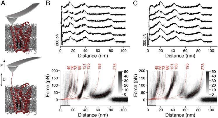
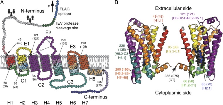
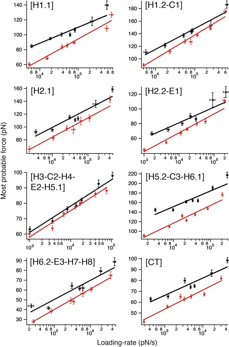
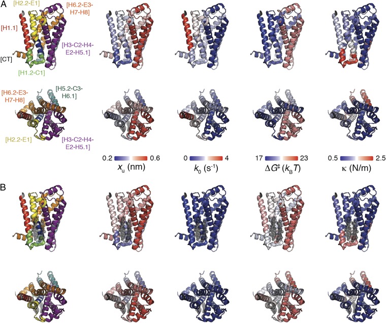
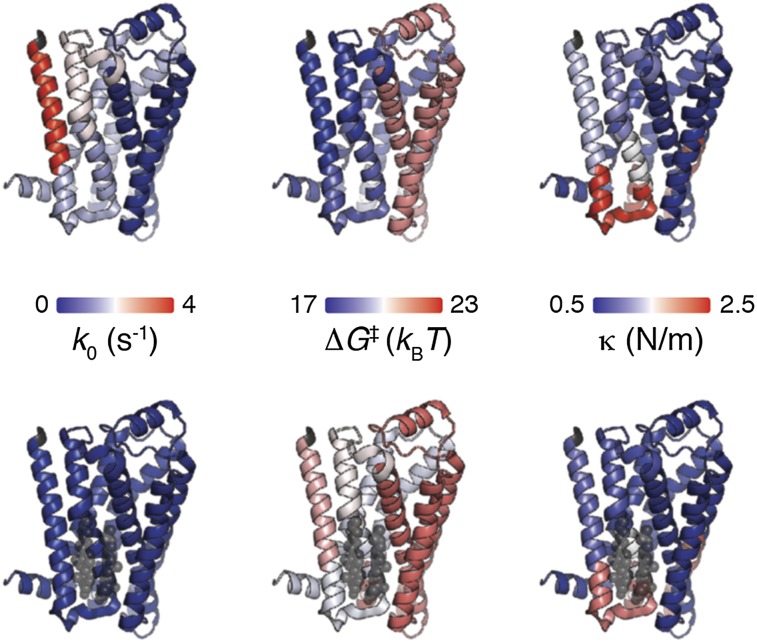
The steroid cholesterol is an essential component of eukaryotic membranes, and it functionally modulates membrane proteins, including G protein-coupled receptors. To reveal insight into how cholesterol modulates G protein-coupled receptors, we have used dynamic single-molecule force spectroscopy to quantify the mechanical strength and flexibility, conformational variability, and kinetic and energetic stability of structural segments stabilizing the human β(2)-adrenergic receptor (β(2)AR) in the absence and presence of the cholesterol analog cholesteryl hemisuccinate (CHS). CHS considerably increased the kinetic, energetic, and mechanical stability of almost every structural segment at sufficient magnitude to alter the structure and functional relationship of β(2)AR. One exception was the structural core segment of β(2)AR, which establishes multiple ligand binding sites, and its properties were not significantly influenced by CHS.


Conflict of interest statement

The authors declare no conflict of interest.

Figures

Fig. 1.
Fig. 1.
SMFS of β2AR reconstituted into liposomes composed of either phospholipids (DOPC) or phospholipids and cholesterol (DOPC/CHS). (A) Pressing the AFM stylus onto the proteoliposomes promotes the unspecific attachment of a single β2AR polypeptide to the stylus. Withdrawal of the AFM cantilever stretches the polypeptide and induces the sequential unfolding of β2AR. (B and C) Selection of F-D curves recorded on N-terminal unfolding of β2AR reconstituted into DOPC (B, Upper) and DOPC/CHS (C, Upper) liposomes. Density plots of superimposed F-D curves (B, Lower and C, Lower) highlight their common features. Number of superimposed F-D curves: n = 100 (B) and n = 100 (C). Red numbers on top of each WLC curve (red dashed lines) indicate the average contour lengths (in amino acids) revealed from fitting each force peak of each superimposed F-D curve. Gray scale bars allow evaluation of how frequently individual force peaks were populated.
Fig. 2.
Fig. 2.
Structural segments stabilizing human β2AR. Secondary (A) and tertiary (B) structure models of β2AR. Each color represents a structural segment that is stabilized by inter- and intramolecular interactions. (A) Black amino acids highlight the end of the previous and the beginning of the next stable structural segment. This position corresponds to the mean contour length (given in parentheses) revealed from WLC curves fitting the force peaks common in every F-D curve. Amino acids colored at less intensity give the SD of locating the average force peak (Table S1). Membrane compensation (Materials and Methods) was applied for the boundaries of stable structural segments that had to be assumed to be within the membrane or at the membrane surface opposite to the puling AFM stylus. All seven transmembrane α-helices of β2AR are labeled with bold numerals (H1–H7). Cytoplasmic and extracellular loops are indicated C1, C2, and C3 and E1, E2, and E3, respectively. H8 denotes the short C-terminal α-helix 8 at the cytoplasmic side. The secondary structure model (A) of C-terminally truncated β2AR carrying an N-terminal FLAG epitope (blue) followed by a tobacco etch virus (TEV) protease cleavage site (green) was taken from ref. . The tertiary structure model (B) was taken from Protein Data Bank ID code 3D4S.
Fig. 3.
Fig. 3.
Loading rate-dependent interactions stabilizing structural segments of β2AR depend on cholesterol. DFS plots of each stable structural segment of β2AR reconstituted into DOPC (red) and DOPC/CHS (black) liposomes. Shown is the most probable unfolding force (Figs. S8 and S9) against the most probable loading rate (Figs. S10 and S11). Solid lines show the DFS fits from which xu and k0 were obtained (Table 1). Error bars indicate SE of the most probable unfolding force and loading rate.
Fig. 4.
Fig. 4.
Mapping the kinetic, energetic, and mechanical properties of β2AR in the absence (A) and presence (B) of cholesterol. Structural segments stabilizing β2AR (Protein Data Bank ID code 3D4S) are mapped on the left. Transition state distance xu, transition rate k0, free-energy barrier height ΔG, and spring constant κ of stable structural segments in the absence of cholesterol (A) and the presence of cholesterol (B). The color of the β2AR backbone roughly indicates the value for each parameter as indicated by the scale bars. A, Upper and B, Upper show β2AR from the side view; A, Lower and B, Lower show β2AR from the extracellular view. Values were taken from Table 1.
Fig. P1.
Fig. P1.
Mapping the kinetic, energetic, and mechanical properties of human β2AR in the absence (Upper) and presence (Lower) of cholesterol. Properties shown are kinetic stability (characterized by the reciprocal of transition rate k0), unfolding free energy (characterized by ΔG), and mechanical rigidity (characterized by the spring constant κ). The color of the β2AR backbone (Protein Data Bank ID code 3D4S) roughly indicates the value for each parameter, which is indicated by the color scale bars in the center. Cholesterol, as revealed by X-ray crystallography, is shown as semitransparent space-filling models (black). The properties shown were quantified from DFS experiments.

References

    1. Milligan G, Svoboda P, Brown CM. Why are there so many adrenoceptor subtypes? Biochem Pharmacol. 1994;48(6):1059–1071. - PubMed
    1. Takeda S, Kadowaki S, Haga T, Takaesu H, Mitaku S. Identification of G protein-coupled receptor genes from the human genome sequence. FEBS Lett. 2002;520(1-3):97–101. - PubMed
    1. Bai TR. Beta 2 adrenergic receptors in asthma: A current perspective. Lung. 1992;170(3):125–141. - PubMed
    1. Barnes PJ. Beta-adrenoceptors on smooth muscle, nerves and inflammatory cells. Life Sci. 1993;52(26):2101–2109. - PubMed
    1. Smiley RM, Finster M. Do receptors get pregnant too? Adrenergic receptor alterations in human pregnancy. J Matern Fetal Med. 1996;5(3):106–114. - PubMed

Publication types

MeSH terms