. 2012 Nov 15;491(7424):406-12.
doi: 10.1038/nature11544. Epub 2012 Sep 18.

Broad and potent neutralization of HIV-1 by a gp41-specific human antibody

Affiliations

Broad and potent neutralization of HIV-1 by a gp41-specific human antibody

Jinghe Huang et al. Nature. .

Abstract

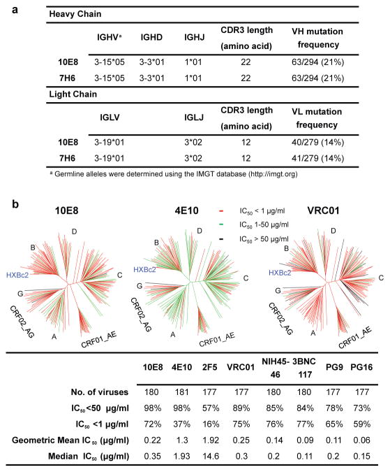
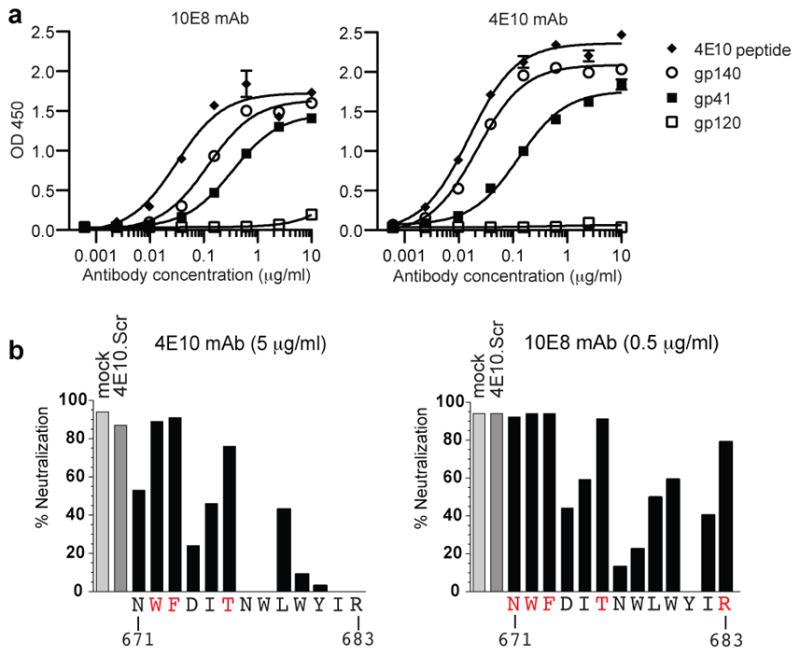
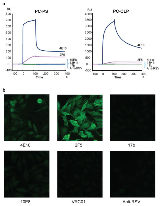
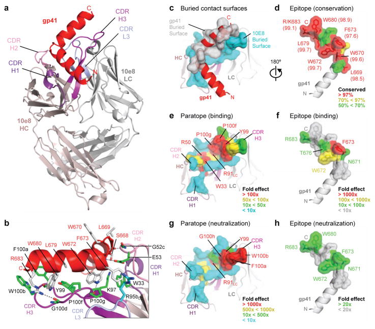
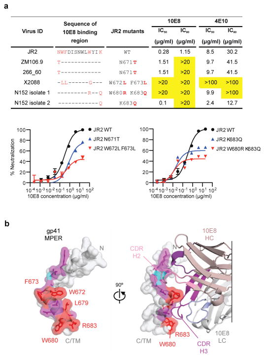
Characterization of human monoclonal antibodies is providing considerable insight into mechanisms of broad HIV-1 neutralization. Here we report an HIV-1 gp41 membrane-proximal external region (MPER)-specific antibody, named 10E8, which neutralizes ∼98% of tested viruses. An analysis of sera from 78 healthy HIV-1-infected donors demonstrated that 27% contained MPER-specific antibodies and 8% contained 10E8-like specificities. In contrast to other neutralizing MPER antibodies, 10E8 did not bind phospholipids, was not autoreactive, and bound cell-surface envelope. The structure of 10E8 in complex with the complete MPER revealed a site of vulnerability comprising a narrow stretch of highly conserved gp41-hydrophobic residues and a critical arginine or lysine just before the transmembrane region. Analysis of resistant HIV-1 variants confirmed the importance of these residues for neutralization. The highly conserved MPER is a target of potent, non-self-reactive neutralizing antibodies, suggesting that HIV-1 vaccines should aim to induce antibodies to this region of HIV-1 envelope glycoprotein.


Conflict of interest statement

Competing financial interests The authors declare no competing financial interests.

Figures

Figure 1
Figure 1. Analyses of 10E8 sequence and neutralization
a, Inferred germline genes encoding the variable regions of 10E8 and 7H6. b, Neutralizing activity of antibodies against a 181-isolate Env-pseudovirus panel. Dendrograms indicate the gp160 protein distance of HIV-1 primary isolate Envs. Data below the dendrogram show the number of tested viruses, the percentage of viruses neutralized, and the geometric mean IC50 for viruses neutralized with an IC50<50 μg/ml. Median titers are based on all tested viruses, including those with IC50 >50ug/ml, which were assigned a value of 100.
Figure 2
Figure 2. Binding specificity of 10E8
a, ELISA binding of mAb 10E8 or 4E10 to gp140, gp120, gp41, or 4E10 peptide. Error bars denote one standard error of the mean (SEM). b, Inhibition of mAb 10E8 or 4E10 neutralization of C1 HIV-2/HIV-1 MPER virus by 4E10 alanine scanning peptides. Residues shown in red indicate positions for which the alanine mutant peptide did not block neutralization.
Figure 3
Figure 3. Analysis of 10E8 autoreactivity
a, SPR analysis of 10E8 binding to anionic phospholipids. 10E8 was injected over PC-CLP liposomes or PC-PS liposome immobilized on the BIAcore L1 sensor chip. 4E10 and 2F5 were used as positive controls and 13H1, 17b, and anti-RSV F protein as negative controls. b, Reactivity of 10E8 with HEP-2 epithelial cells. Controls are as above with VRC01 added as an additional negative control. Antibody concentration was 25 μg/ml. All pictures are shown at 400x magnification.
Figure 4
Figure 4. Crystal structure of 10E8 Fab in complex with its gp41 MPER epitope
a. 10E8 recognizes a highly conserved gp41 helix to neutralize HIV-1. Fab 10E8 is shown in ribbon representation (shades of violet for heavy chain and of gray for light chain) in complex with a gp41 peptide (red) that encompasses the MPER (Asn656-Arg683). b, Interface between 10E8 and gp41 with select 10E8-side chains (green, heavy chain; cyan, light chain) and gp41-side chains (gray) in stick representation. In analogy to a hand, the hinge can be viewed as being gripped by a thumb (represented by the CDR H2), the C-terminal helix as being suspended along a corresponding extended forefinger (represented by the CDR H3), and residues that commence the C-terminal helix as being caught in the cleft between the thumb and forefinger (represented by the juncture of the CDR loops). c-d, Buried contact surfaces and epitope conservation. An examination of the buried contact surface on gp41 (gray; c) reveals that epitope residues (labeled, d) that are directly contacted by 10E8 are highly conserved across 2870 examined strains (Supplementary Tables 10–12; conservation percentages provided in parentheses). e-h, Alanine mutagenesis of paratope and epitope. Residues at the tip of the 10E8 CDR H3 loop and within the hydrophobic cleft are crucial for recognition of gp41 and for virus neutralization (Supplementary Tables 15–16), as mapped onto the buried 10E8-contact surface (e,g). These results mirror the effects of alanine scan mutations of the 10E8 epitope (Supplementary Tables 2–3), as mapped onto the buried gp41-contact surface (f,h). A comparison of the effects of the alanine mutagenesis of the paratope and epitope reveal that residues of the epitope that are most crucial for 10E8 recognition and neutralization, are also the most highly conserved (d).
Figure 5
Figure 5. A site of gp41 vulnerability
a, Impact of sequence variation on 10E8 neutralization. Predicted amino acid sequences within the binding epitope of 10E8 for 3 10E8 resistant viruses and the patient virus are shown. The 10E8 epitope and differences in sequence compared to the JR2 virus are labeled in red. IC50 and IC80 values that are >20-fold than JR2 wild-type pseudovirus are highlighted in yellow. Error bars denote one SEM. b, Structural definition of a highly conserved region of gp41 recognized by neutralizing antibodies. Atoms of highly conserved residues that make direct contacts with 10E8 are colored red and shown in stick representation, atoms buried by 10E8 are colored purple, and main chain-contacting atoms are colored cyan. Semi-transparent surfaces of the gp41 MPER are colored according to the underlying atoms. 90° views are shown, with bound antibody 10E8 in the right panel. The 10E8 CDR H3 interacts with highly conserved hydrophobic residues, whereas the CDR H2 contacts main chain atoms at the juncture between the N- and C-terminal helices. Many of the unbound residues of the MPER (gray) are hydrophobic, especially those within the C-terminal helix. In the structure of a late fusion intermediate (Supplementary Fig. 8) these residues face towards the outside of a helical coiled-coil; in the pre-fusion conformation of the viral spike, these may interact with the viral membrane or with other hydrophobic regions of Env.

References

    1. Doria-Rose NA, et al. Breadth of human immunodeficiency virus-specific neutralizing activity in sera: clustering analysis and association with clinical variables. J Virol. 2010;84:1631–1636. - PMC - PubMed
    1. Stamatatos L, Morris L, Burton DR, Mascola JR. Neutralizing antibodies generated during natural HIV-1 infection: good news for an HIV-1 vaccine? Nature medicine. 2009 - PubMed
    1. Sather DN, et al. Factors associated with the development of cross-reactive neutralizing antibodies during human immunodeficiency virus type 1 infection. J Virol. 2009;83:757–769. - PMC - PubMed
    1. Walker LM, et al. A limited number of antibody specificities mediate broad and potent serum neutralization in selected HIV-1 infected individuals. PLoS Pathog. 2010;6:e1001028. - PMC - PubMed
    1. Simek MD, et al. Human immunodeficiency virus type 1 elite neutralizers: individuals with broad and potent neutralizing activity identified by using a high-throughput neutralization assay together with an analytical selection algorithm. J Virol. 2009;83:7337–7348. - PMC - PubMed

Publication types