

Review
doi: 10.1136/heartjnl-2012-301962.
Left ventricular thrombus formation after acute myocardial infarction
Affiliations
- PMID: 23151669
- PMCID: PMC3505867
- DOI: 10.1136/heartjnl-2012-301962
Item in Clipboard
Review
Left ventricular thrombus formation after acute myocardial infarction
Heart.
2012 Dec.
Free PMC article
No abstract available
Conflict of interest statement
Figures
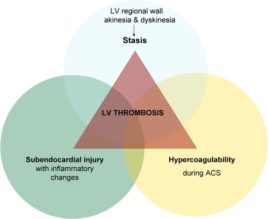
The three components of the Virchow's triad in left ventricular thrombus formation. ACS, acute coronary syndrome; LV, left ventricular.
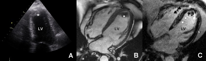
Left ventricular (LV) thrombus formation on delayed gadolinium contrast cardiac MRI and transthoracic echocardiography. Transthoracic echocardiographic appearance of a thrombus (asterisk) in the apex of the left ventricle (A); cine cardiovascular magnetic resonance of the same patient also delineates the apical thrombus (B); late gadolinium enhancement imaging clearly confirms the avascular non-enhancing thrombus (asterisk, dark) close to the transmural infarcted myocardium (bright hyperenhanced, black arrowheads) with areas of microvascular obstruction (black, white arrowheads) (C). Courtesy of Dr A C van Rossum, Dr R Nijveldt, Department of Cardiology, VU University Medical Center, Amsterdam, the Netherlands, and Dr B J Bouma, Department of Cardiology, Academic Medical Center, Amsterdam, the Netherlands.
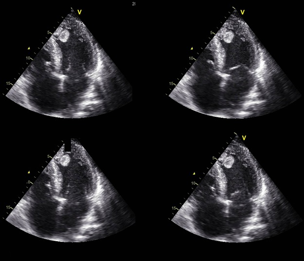
Transthoracic echocardiographic appearance of a mobile, protruding left ventricular thrombus. Courtesy of J Vleugels and Rianne H A de Bruin, Department of Cardiology, Department of Cardiology, Academic Medical Center, Amsterdam, the Netherlands.
References
-
- Keren A, Goldberg S, Gottlieb S, et al. Natural history of left ventricular thrombi: their appearance and resolution in the posthospitalization period of acute myocardial infarction. J Am Coll Cardiol 1990;15:790–800 - PubMed
-
Clinical study assessing appearance and resolution of LV thrombi on echocardiographic follow-up.
-
- Asinger RW, Mikell FL, Elsperger J, et al. Incidence of left-ventricular thrombosis after acute transmural myocardial infarction. Serial evaluation by two-dimensional echocardiography. N Engl J Med 1981;305:297–302 - PubMed
-
Eminent clinical study on the incidence of LV thrombus after AMI.
-
- Visser CA, Kan G, Lie KI, et al. Left ventricular thrombus following acute myocardial infarction: a prospective serial echocardiographic study of 96 patients. Eur Heart J 1983;4:333–7 - PubMed
-
Clinical study evaluating the incidence of LV thrombus formation after AMI.
-
- Jugdutt BI, Sivaram CA. Prospective two-dimensional echocardiographic evaluation of left ventricular thrombus and embolism after acute myocardial infarction. J Am Coll Cardiol 1989;13:554–64 - PubMed
-
Clinical study evaluating LV thrombi and systemic embolism after AMI.
-
- Chiarella F, Santoro E, Domenicucci S, et al. Predischarge two-dimensional echocardiographic evaluation of left ventricular thrombosis after acute myocardial infarction in the GISSI-3 study. Am J Cardiol 1998;81:822–7 - PubMed
-
Substudy of the GISSI-3 study providing one of the largest datasets to date on the incidence and risk factors of LV thrombus formation.
Publication types
MeSH terms
LinkOut - more resources
Full Text Sources
Medical