Building vascular networks
- PMID: 23152325
- PMCID: PMC3569029
- DOI: 10.1126/scitranslmed.3003688
Building vascular networks
Abstract
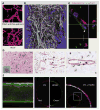
Only a few engineered tissues-skin, cartilage, bladder-have achieved clinical success, and biomaterials designed to replace more complex organs are still far from commercial availability. This gap exists in part because biomaterials lack a vascular network to transfer the oxygen and nutrients necessary for survival and integration after transplantation. Thus, generation of a functional vasculature is essential to the clinical success of engineered tissue constructs and remains a key challenge for regenerative medicine. In this Perspective, we discuss recent advances in vascularization of biomaterials through the use of biochemical modification, exogenous cells, or microengineering technology.
Conflict of interest statement
Figures


References
-
- Khademhosseini A, Vacanti JP, Langer R. Progress in tissue engineering. Sci Am. 2009;300:64–71. - PubMed
-
- Stenman JM, Rajagopal J, Carroll TJ, Ishibashi M, Mc-Mahon J, McMahon AP. Canonical Wnt signaling regulates organ-specific assembly and differentiation of CNS vasculature. Science. 2008;322:1247–1250. - PubMed
-
- Ehrbar M, Djonov VG, Schnell C, Tschanz SA, Martiny-Baron G, Schenk U, Wood J, Burri PH, Hubbell JA, Zisch AH. Cell-demanded liberation of VEGF121 from fibrin implants induces local and controlled blood vessel growth. Circ Res. 2004;94:1124–1132. - PubMed
-
- Peters MC, Polverini PJ, Mooney DJ. Engineering vascular networks in porous polymer matrices. J Biomed Mater Res. 2002;60:668–678. - PubMed
-
- Black AF, Berthod F, L’heureux N, Germain L, Auger FA. In vitro reconstruction of a human capillary-like network in a tissue-engineered skin equivalent. FASEB J. 1998;12:1331–1340. - PubMed
Publication types
MeSH terms
Substances
Grants and funding
- EB024618/EB/NIBIB NIH HHS/United States
- R01 EB012597/EB/NIBIB NIH HHS/United States
- R01 HL099073/HL/NHLBI NIH HHS/United States
- EB012597/EB/NIBIB NIH HHS/United States
- U54 CA143837/CA/NCI NIH HHS/United States
- R01 HL092836/HL/NHLBI NIH HHS/United States
- U54-CA-143837/CA/NCI NIH HHS/United States
- R01 EB000246/EB/NIBIB NIH HHS/United States
- DE021468/DE/NIDCR NIH HHS/United States
- EB012726/EB/NIBIB NIH HHS/United States
- R01 GM043337/GM/NIGMS NIH HHS/United States
- R01 DE021468/DE/NIDCR NIH HHS/United States
- HL099073/HL/NHLBI NIH HHS/United States
- R01 AR057837/AR/NIAMS NIH HHS/United States
- HL092836/HL/NHLBI NIH HHS/United States
- R21 EB012726/EB/NIBIB NIH HHS/United States
LinkOut - more resources
Full Text Sources
Other Literature Sources
Miscellaneous

