. 2012 Dec 1;26(23):2567-79.
doi: 10.1101/gad.200758.112. Epub 2012 Nov 14.

Whole-genome microRNA screening identifies let-7 and mir-18 as regulators of germ layer formation during early embryogenesis

Affiliations

Whole-genome microRNA screening identifies let-7 and mir-18 as regulators of germ layer formation during early embryogenesis

Alexandre R Colas et al. Genes Dev. .

Abstract

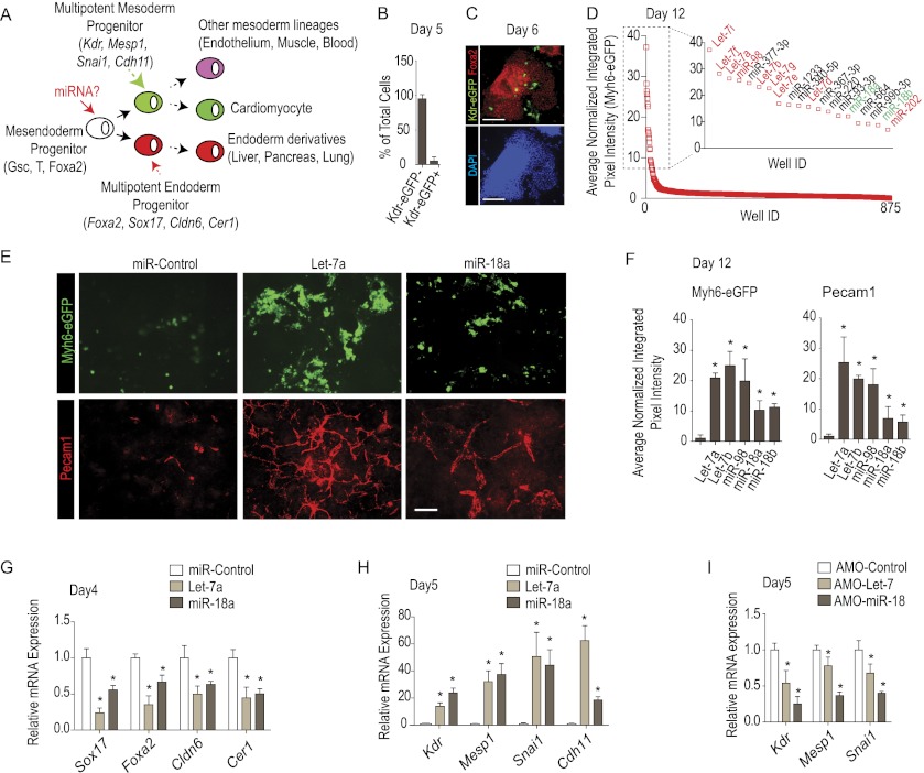
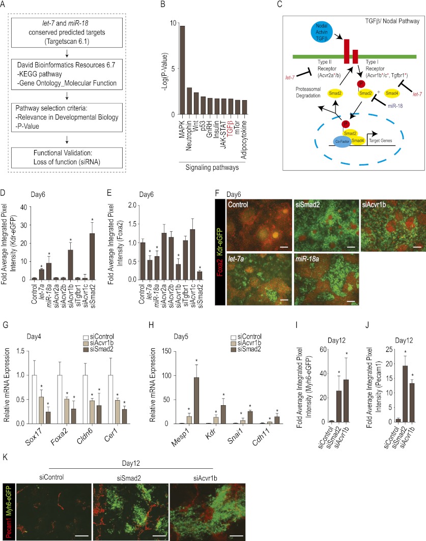
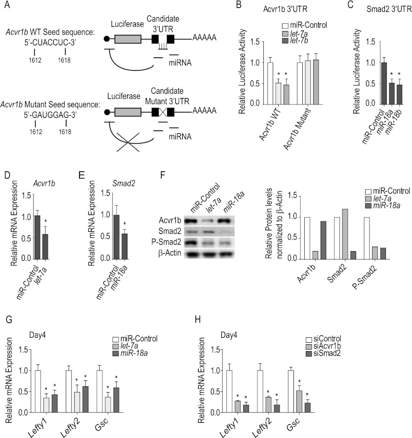
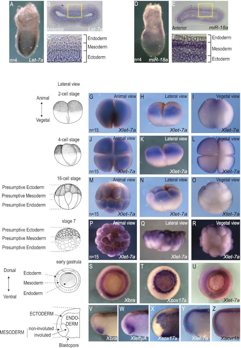
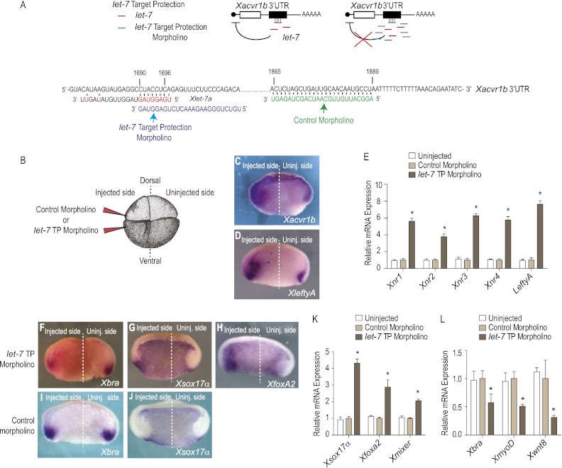
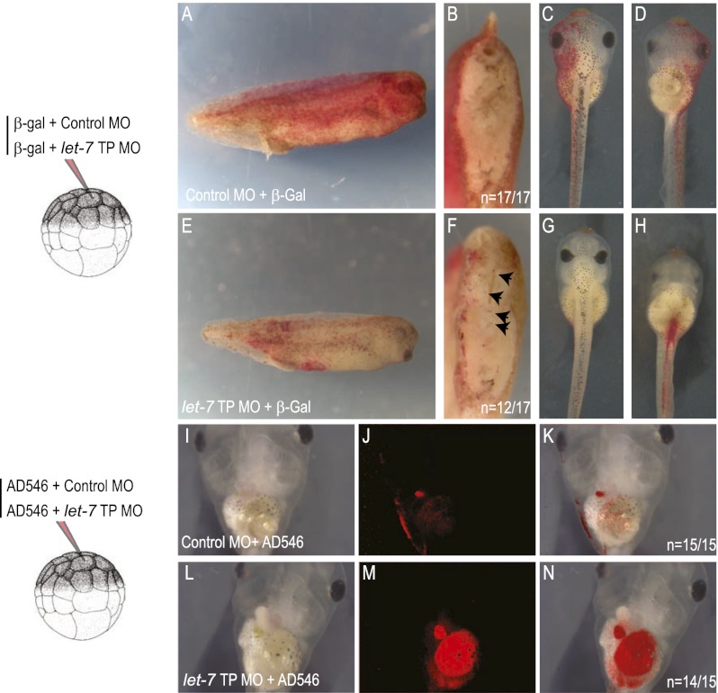
Tight control over the segregation of endoderm, mesoderm, and ectoderm is essential for normal embryonic development of all species, yet how neighboring embryonic blastomeres can contribute to different germ layers has never been fully explained. We postulated that microRNAs, which fine-tune many biological processes, might modulate the response of embryonic blastomeres to growth factors and other signals that govern germ layer fate. A systematic screen of a whole-genome microRNA library revealed that the let-7 and miR-18 families increase mesoderm at the expense of endoderm in mouse embryonic stem cells. Both families are expressed in ectoderm and mesoderm, but not endoderm, as these tissues become distinct during mouse and frog embryogenesis. Blocking let-7 function in vivo dramatically affected cell fate, diverting presumptive mesoderm and ectoderm into endoderm. siRNA knockdown of computationally predicted targets followed by mutational analyses revealed that let-7 and miR-18 down-regulate Acvr1b and Smad2, respectively, to attenuate Nodal responsiveness and bias blastomeres to ectoderm and mesoderm fates. These findings suggest a crucial role for the let-7 and miR-18 families in germ layer specification and reveal a remarkable conservation of function from amphibians to mammals.


Figures

Figure 1.
Figure 1.
Screen for miRs that control endoderm and mesoderm fate. (A) Schematic representation of the screening strategy. (B,C) FACS analysis of differentiating mESCs with eGFP under the control of Kdr promoter (n = 3) revealed that most cells are Kdr-eGFP (shown in B). (C) Immunostaining confirmed that most cells express the endoderm marker Foxa2 at day 6. See Supplemental Figure 1 for additional marker profiling showing endodermal and mesodermal marker expression in the differentiating cells. (D) Ranking of 875 miRs screened for induction of the Myh6-eGFP reporter indicative of cardiomyogenic cells. The inset shows the top 19 hits. (E,F) Confirmation that let-7 and miR-18 bias Myh6-eGFP and Alexa Fluor568-Pecam1 fluorescence levels (integrated pixel intensity) (see the Materials and Methods) in mESCs transfected at day 3 and assayed at day 12. Fluorescence values of responses to let-7 (let-7a, let-7b, and miR-98) and miR-18 (miR-18a and miR-18b) plotted relative to that for the control (scrambled sequence) miR. (F) Representative images of Myh6-eGFP and Alexa Fluor568-Pecam1. (GI) qRT–PCR of early endoderm (G,I) and mesoderm (H) marker genes in response to transfection with let-7, miR-18 (G,H), or specific anti-miRs (I). Note that miRs repress endoderm and stimulate mesoderm marker gene expression. The anti-miRs repress mesoderm markers; endodermal markers were not examined, since the assay baseline has maximal endodermal marker expression and is insensitive to any increase. All qRT–PCR data were normalized to β-actin mRNA levels. All data are presented as mean ± SD. (*) P < 0.05.
Figure 2.
Figure 2.
let-7 and miR-18 modulate cell fate through inhibition of Nodal signaling. (A) miR target identification strategy (see the Materials and Methods). (B) Top 10 computationally predicted signaling pathways targeted by let-7 and miR-18 ranked by −log(P-value). (C) Predicted let-7 and miR-18 targets in the Nodal/TGFβ signaling pathway (red asterisk). (DF) Kdr-eGFP (D) and Alexa Fluor568-Foxa2 (E) fluorescence measurements at day 6 in mESCs transfected with siRNAs against Nodal/TGFβ signaling pathway components, quantified by automated microscopy, and plotted relative to values obtained with control (scrambled sequence) siRNA. (F) Representative images of Kdr-eGFP and Alexa Fluor568-Foxa2 fluorescence as in D and E. Note that siRNAs to Acvr1b and Smad2 specifically enhanced Kdr-eGFP while reducing Foxa2 levels. (G,H) Effect of Smad2 and Acvr1b knockdown on early endoderm (G) and mesoderm (H) marker genes by qRT–PCR. Note repression of endoderm and stimulation of mesoderm marker genes. (IK) Myh6-eGFP (I) and Alexa Fluor568-Pecam1 (J) at day 12 of differentiation after transfection of siRNAs against Smad2 or Acvr1b at day 3, relative to control (scrambled) siRNA. Representative image of Myh6-eGFP and Alexa Fluor568-Pecam1, as in I and J. Quantitative data are presented as means ± SD. (*) P < 0.05.
Figure 3.
Figure 3.
let-7 and miR-18 directly target Acvr1b and Smad2. (A) Schematic showing that mutation of the mRE is predicted to abolish the effect of the miR against the luc reporter-3′ UTR fusion construct, validating dependence on a specific miR:mRE interaction. (B,C) Mutation of the putative recognition elements in Acvr1b (B) and Smad2 (C) 3′ UTRs. Note the reduction of luciferase activity in HEK293T cells 24 h after transfection with specific miR relative to control miR. (D,E) Decrease in endogenous Acvr1b (D) or Smad2 (E) transcript levels after transfection of mESCs at day 3 with let-7a or miR-18a, respectively, relative to control miR. (F) Western blot analysis of Acvr1b, Smad2, P-Smad2, and β-Actin protein levels in mESCs transfected at day 3 with let-7a, miR-18a, or control miRs. (G,H) qRT–PCR of transcriptional targets (Lefty1, Lefty2, and Gsc) in mESCs transfected at day 3 with either with let-7a or miR-18a relative to control miR (G) and siRNAs against Acvr1b or Smad2 relative to control siRNA (H).
Figure 4.
Figure 4.
Endogenous localization of let-7a and miR-18a in early vertebrate embryos. (AF) In situ hybridization showing endogenous let-7a and miR-18a in E7 mouse embryos viewed in whole-mount (A,D) or transverse histological section (B,E). (C,F) High-magnification view distinguishing epiblast mesoderm and endoderm layers. Note the abundant expression of let-7a (A–C) and miR-18a (D–F) in ectoderm and mesoderm but not in endoderm. (GR) Endogenous Xlet-7a in cleavage stage Xenopus embryos showing expression in the animal hemisphere (presumptive ectoderm and mesoderm). (SZ) Endogenous Xlet-7a expression in gastrula stage (stage 10.5) Xenopus embryos. View of the mesoderm in the equatorial region marked by Xbra (S) and involuting endoderm marked by Xsox17α (T) reveals overlap with Xlet-7a prominently in mesoderm (U). (V–Z) Transverse sections show expression domains of Xbra (V), XleftyA (W), and Xsox17α (X) relative to Xlet-7a (Y), revealing that the miR is present in both ectoderm and mesoderm but not in endoderm and is nonoverlapping with XleftyA but coincident with Xacvr1b (Z) mRNAs.
Figure 5.
Figure 5.
Endogenous Xlet-7 discriminates mesoderm and endoderm by repressing Xacvr1b. (A) Diagram of the TP loss-of-function strategy to block interaction of the miR with the mRE in vivo. (B) Either Xlet-7 TP or control morpholinos were injected equatorially into two blastomeres on one side of four-cell stage embryos. (C,D) Unilaterally injected embryos (as in B) cultured to gastrula stage (stage 10.5), bisected transversely, and probed for Xacrv1b (C) and XleftyA (D) expression. Injection of Xlet-7 TP morpholino up-regulated and expanded the domains of Xacrv1b and XleftyA expression in the marginal zone mesoderm and underlying deep endoderm. (E) Xlet-7 TP injection up-regulated Xnr-1, Xnr-2, Xnr-3, and Xnr-4 as well as XleftyA by qRT–PCR on dissected lateral marginal zone explants relative to the control morpholino, which has no effect on development or gene expression. (FH) Unilateral Xlet-7 TP morpholino injection repressed a marker of mesoderm, Xbra (F), while concomitantly expanding the domain of endodermal marker genes Xsox17α (G) and Xfoxa2 (H) into the mesoderm domain. (I,J) Unilateral control morpholino injection had no effect on both mesodermal (I) and endodermal (J) markers. (K,L) qRT–PCR analyses on dissected lateral marginal zone explants show that the Xlet-7 TP morpholino up-regulated expression of endodermal genes (XSox17α, Xfoxa2, and Xmixer) (K) and repressed mesodermal genes (Xbra, XmyoD, and Xwnt8) (L) as compared with the control morpholino.
Figure 6.
Figure 6.
Blocking endogenous let-7 converts ectoderm to endoderm in embryos. (AH) Embryos at the 16-cell stage were injected in one animal blastomere (presumptive ectoderm) together with Xlet-7 TP or control morpholino and LacZ mRNA dextran as a lineage label. Whereas the progeny of control morpholino-injected blastomeres contributed exclusively to epidermis (A–D), Xlet-7 TP morpholino-injected blastomeres contributed to endoderm, examined at the tailbud stage (stage 35) in lateral view (A,D), transverse bisection (B,F), dorsal view (C,G), and ventral view (D,H). (IN) The same as in AH, but examined at the tadpole stage (stage 45) and injected with Alexa Fluor546 dextran as a lineage label. Note the pronounced diversion of ectoderm to gut cell fate conversion, viewed ventrally.
Figure 7.
Figure 7.
miR control of germ layer segregation in vertebrate embryos. let-7 and miR-18 family members repress Acvr1b and Smad2 protein production, rendering cells less sensitive to Nodal signaling and sharpening a border between endoderm versus ectoderm and mesoderm. By acting cell-autonomously to titrate Nodal responsiveness, the miRs function nonredundantly with protein inhibitors (e.g., Lefty and Cerberus proteins) and the miR-302/427/430 family, previously shown to influence mesendoderm specification by repressing secreted modulators of Nodal signaling (see the text).

References

    1. Agius E, Oelgeschlager M, Wessely O, Kemp C, De Robertis EM 2000. Endodermal Nodal-related signals and mesoderm induction in Xenopus. Development 127: 1173–1183 - PMC - PubMed
    1. Armes NA, Smith JC 1997. The ALK-2 and ALK-4 activin receptors transduce distinct mesoderm-inducing signals during early Xenopus development but do not co-operate to establish thresholds. Development 124: 3797–3804 - PubMed
    1. Bartel DP 2009. MicroRNAs: Target recognition and regulatory functions. Cell 136: 215–233 - PMC - PubMed
    1. Ben-Ami O, Pencovich N, Lotem J, Levanon D, Groner Y 2009. A regulatory interplay between miR-27a and Runx1 during megakaryopoiesis. Proc Natl Acad Sci 106: 238–243 - PMC - PubMed
    1. Branford WW, Yost HJ 2002. Lefty-dependent inhibition of Nodal- and Wnt-responsive organizer gene expression is essential for normal gastrulation. Curr Biol 12: 2136–2141 - PubMed

Publication types