. 2013 Feb;87(3):1301-11.
doi: 10.1128/JVI.02519-12. Epub 2012 Nov 14.

Epstein-Barr virus LMP1 modulates lipid raft microdomains and the vimentin cytoskeleton for signal transduction and transformation

Affiliations

Epstein-Barr virus LMP1 modulates lipid raft microdomains and the vimentin cytoskeleton for signal transduction and transformation

David G Meckes Jr et al. J Virol. 2013 Feb.

Abstract

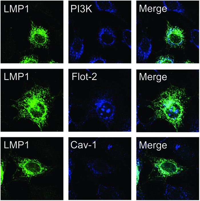
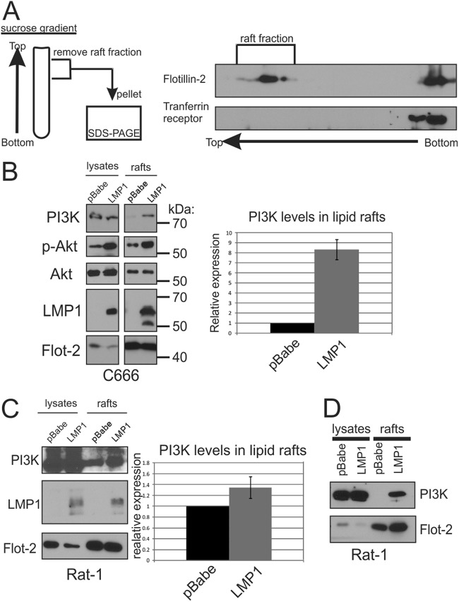
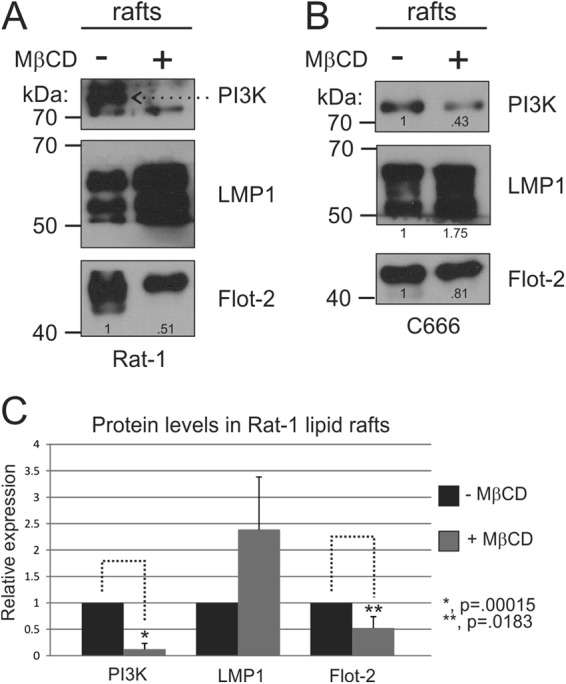
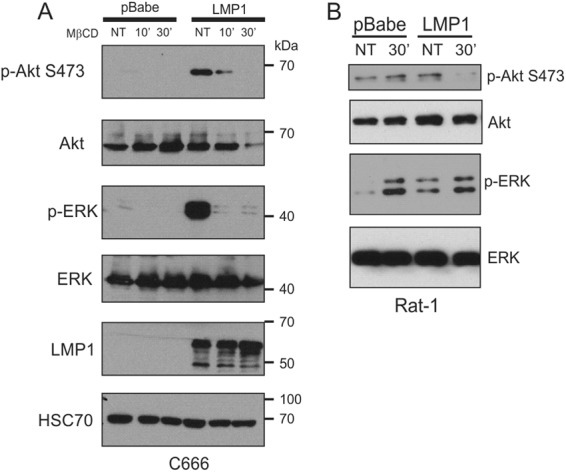
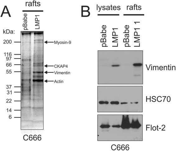
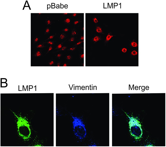
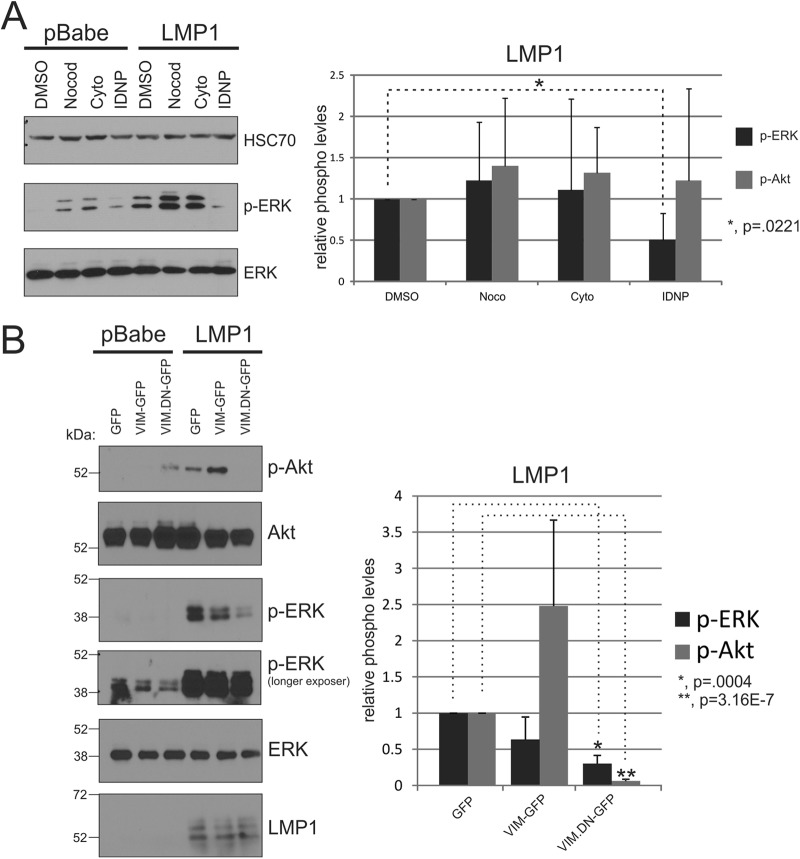
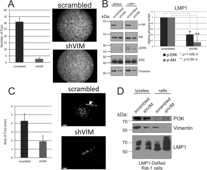
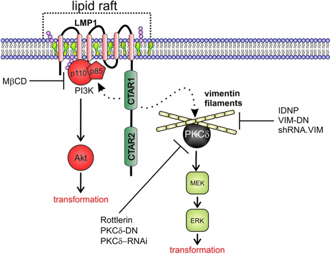
The Epstein-Barr virus (EBV) is an important human pathogen that is associated with multiple cancers. The major oncoprotein of the virus, latent membrane protein 1 (LMP1), is essential for EBV B-cell immortalization and is sufficient to transform rodent fibroblasts. This viral transmembrane protein activates multiple cellular signaling pathways by engaging critical effector molecules and thus acts as a ligand-independent growth factor receptor. LMP1 is thought to signal from internal lipid raft containing membranes; however, the mechanisms through which these events occur remain largely unknown. Lipid rafts are microdomains within membranes that are rich in cholesterol and sphingolipids. Lipid rafts act as organization centers for biological processes, including signal transduction, protein trafficking, and pathogen entry and egress. In this study, the recruitment of key signaling components to lipid raft microdomains by LMP1 was analyzed. LMP1 increased the localization of phosphatidylinositol 3-kinase (PI3K) and its activated downstream target, Akt, to lipid rafts. In addition, mass spectrometry analyses identified elevated vimentin in rafts isolated from LMP1 expressing NPC cells. Disruption of lipid rafts through cholesterol depletion inhibited PI3K localization to membranes and decreased both Akt and ERK activation. Reduction of vimentin levels or disruption of its organization also decreased LMP1-mediated Akt and ERK activation and inhibited transformation of rodent fibroblasts. These findings indicate that LMP1 reorganizes membrane and cytoskeleton microdomains to modulate signal transduction.


Figures

Fig 1
Fig 1
LMP1 colocalizes with PI3K in internal membranes containing lipid raft resident proteins. Rat-1 cells stably expressing LMP1 were fixed and stained with primary antibodies specific for LMP1 (CS1-4), p85 (PI3K), flotillin-2 (Flot-2), or caveolin-1 (Cav-1). Cells were visualized by confocal microscopy with a ×60 oil objective lens.
Fig 2
Fig 2
LMP1 induces the lipid raft localization of PI3K. (A) Schematic diagram of lipid raft isolation protocol. Detergent-resistant membranes (DRMs) or lipid rafts were isolated from cell lysates separated on a discontinuous sucrose gradient by ultracentrifugation. Due to their buoyant density, lipid rafts and associated proteins migrate to the top of the gradient. Fractions were collected from the gradient, separated by SDS-PAGE, and analyzed by immunoblot for flotillin-2 and transferrin receptor as controls. The lipid raft fraction or whole-cell lysates of C666 cells (B) or Rat-1 cells (C) expressing LMP1 or vector control (pBabe) were separated by SDS-PAGE and analyzed by immunoblot analysis for the indicated proteins. The band intensities of PI3K were determined from four independent experiments using ImageJ software, normalized to flotillin-2 levels, and represented relative to the pBabe control level. (D) Rat-1 cells were grown in the absence of serum for 24 h prior to the isolation of lipid rafts and analysis of PI3K levels.
Fig 3
Fig 3
Lipid raft disruption through cholesterol depletion inhibits PI3K DRM localization. Rat-1 (A) or C666 (B) cells expressing LMP1 were serum starved for 1 h and then treated with MβCD for 30 min at 37°C. DRMs were then isolated, and equivalent amounts by volume were analyzed by immunoblotting for PI3K, LMP1, and flotillin-2 (Flot-2) levels. The band intensities of PI3K, LMP1, and flotillin-2 were determined using ImageJ software and are represented relative to the untreated control level. (C) The results of three independent experiments were graphed as mean averages with standard errors of the mean. Statistical significance was determined by using the paired two-tailed Student t test.
Fig 4
Fig 4
Lipid raft disruption inhibits LMP1-mediated Akt and ERK activation. C666 (A) or Rat-1 (B) cells expressing pBabe or LMP1 were serum starved for 1 h and then treated with vehicle (NT) or MβCD for the indicated times. After treatment, cell lysates were prepared and separated in SDS–10% polyacrylamide gels, transferred to nitrocellulose, and subjected to immunoblot analysis with the indicated primary antibodies.
Fig 5
Fig 5
LMP1 increases vimentin levels within DRMs isolated from NPC cells. DRMs were isolated from C666 cells expressing pBabe or LMP1 and separated in an SDS–4 to 20% gradient polyacrylamide gel. The gel was fixed and stained with Coomassie blue to visualize proteins. The predominant bands in the LMP1 rafts not detected in the corresponding region within pBabe rafts were excised, and proteins within that band were digested with trypsin and analyzed by MALDI MS. The major protein components of these bands based on significant Mascot ion sores were determined to be myosin-9, CKAP4, vimentin, and actin. (B) The increased levels of vimentin within lipid rafts were confirmed by immunoblot analysis of pBabe and LMP1 lipid rafts, with HSC70 and Flot-2 serving as loading controls.
Fig 6
Fig 6
LMP1 colocalizes with and reorganizes vimentin within cells. Rat-1 cells expressing pBabe or LMP1 were fixed and subjected to immunofluorescence using primary antibodies against vimentin (A) or LMP1 (CS1-4) and vimentin (B). Cells were visualized by confocal microscopy after incubation with fluorescently labeled secondary antibodies.
Fig 7
Fig 7
Vimentin organization is essential for LMP1-mediated ERK activation. (A) pBabe or LMP1 stable Rat-1 cells were serum starved for 1 h, treated for 1 h with the indicated compounds, and then analyzed by immunoblot for the activation of ERK and Akt with phospho-specific antibodies. HSC70 and total ERK and Akt antibodies were used as loading controls. (B) pBabe or LMP1 Rat-1 cells were transfected with GFP, VIM-GFP, or VIM.DN-GFP using FugeneHD. At 24 h posttransfection, the cells were harvested, and protein lysates were analyzed for p-Akt, p-ERK, and total protein levels by immunoblotting. The band intensities of p-Akt and p-ERK from were determined using ImageJ software, normalized to their corresponding total protein levels, and represented relative to DMSO-treated cells. The results of three independent experiments were graphed as mean averages with the standard errors of the mean. Statistical significance was determined using the paired two-tailed Student t test.
Fig 8
Fig 8
Vimentin is important for the ability of LMP1 to transform cells and activate ERK and Akt. Rat-1 cells stably expressing an shRNA against vimentin (shVIM) or scrambled control (pGIVZ) were transduced with retrovirus particles expressing HA-LMP1 (A) or LMP1-DsRed (C) and incubated 10 to 14 days with medium being replaced every 2 days. After incubation, the cells were visualized for focus formation by crystal violet staining (A) or fluorescence microscopy (C). (A) The average numbers of foci per field were counted and are represented as average means of 10 fields with the standard errors of the mean. (B) Cell lysates were prepared from pBabe or LMP1 Rat-1 cells expressing pGIVZ or shVIM constructs and analyzed by immunoblot for vimentin, p-Akt, p-ERK, ERK, and Akt. The band intensities of p-ERK and p-Akt were determined using ImageJ software, normalized to total Akt and ERK protein levels, and represented relative to the shRNA scrambled cells. The results of three independent experiments were graphed as mean averages with standard errors of the mean. The statistical significance was determined using the paired two-tailed Student t test. (C) The foci formed using the fluorescent DsRed LMP1 construct were measured to determine the relative sizes of each focus by measuring the diameter of the fluorescent focus. The graph represents the mean focus diameters of 25 foci with standard errors of the mean. An individual fluorescent focus formed in the presence of the scrambled or vimentin-specific shRNA is shown to indicate the greater number of LMP1 expressing cells in the larger focus formed in the presence of the scrambled shRNA. (D) The levels of expression of LMP1, PI3K, and vimentin in total lysates or purified rafts in the presence of the scrambled or vimentin-specific shRNAs were determined by immunoblotting.
Fig 9
Fig 9
LMP1 modulation of membrane rafts and the vimentin cytoskeleton to activate Akt and ERK signal transduction pathways. LMP1 self-aggregates in lipid raft microdomains rich in cholesterol and sphingolipids. LMP1 expression leads to the recruitment of PI3K to lipid rafts and the activation of downstream target Akt that is essential for transformation. These effects can be disrupted through cholesterol depletion from cellular membranes with MβCD. LMP1 also induces vimentin expression and raft localization. Disruption of vimentin organization or depletion of vimentin expression blocks LMP-mediated ERK and Akt activation and transformation. PKCδ binds vimentin filaments and is also required for LMP1-mediated ERK activation. These results suggest a two component model that requires the localization of LMP1 and associated molecules into lipid rafts and that the interaction of LMP1 with vimentin may facilitate signaling complexes containing PKCδ and downstream kinases important for ERK signaling.

References

    1. Raab-Traub N. 2012. Novel mechanisms of EBV-induced oncogenesis. Curr. Opin. Virol. 2:453–458 - PMC - PubMed
    1. Raab-Traub N. 2007. EBV-induced oncogenesis, p 986–1006 In Arvin A, Campadelli-Fiume G, Mocarski E, Moore PS, Roizman B, Whitley R, Yamanishi K. (ed), Human herpesviruses: biology, therapy, and immunoprophylaxis. Cambridge University Press, Cambridge, United Kingdom - PubMed
    1. Kaye KM, Izumi KM, Kieff E. 1993. Epstein-Barr virus latent membrane protein 1 is essential for B-lymphocyte growth transformation. Proc. Natl. Acad. Sci. U. S. A. 90:9150–9154 - PMC - PubMed
    1. Wang D, Liebowitz D, Kieff E. 1985. An EBV membrane protein expressed in immortalized lymphocytes transforms established rodent cells. Cell 43:831–840 - PubMed
    1. Mei Y-P, Zhou J-M, Wang Y, Huang H, Deng R, Feng G-K, Zeng Y-X, Zhu X-F. 2007. Silencing of LMP1 induces cell cycle arrest and enhances chemosensitivity through inhibition of AKT signaling pathway in EBV-positive nasopharyngeal carcinoma cells. Cell Cycle 6:1379–1385 - PubMed

Publication types

MeSH terms

Substances