. 2012 Nov 14;32(46):16181-92.
doi: 10.1523/JNEUROSCI.0228-12.2012.

β-secretase cleavage of the fly amyloid precursor protein is required for glial survival

Affiliations

β-secretase cleavage of the fly amyloid precursor protein is required for glial survival

Bonnie J Bolkan et al. J Neurosci. .

Abstract

β-secretase (or BACE1) is the key enzyme in the production of β-amyloid (Aβ), which accumulates in the senile plaques characteristic for Alzheimer's disease. Consequently, the lack of BACE1 prevents β-processing of the amyloid precursor protein and Aβ production, which made it a promising target for drug development. However, the loss of BACE1 is also detrimental, leading to myelination defects and altered neuronal activity, functions that have been associated with the cleavage of Neuregulin and a voltage-gated sodium channel subunit. Here we show that the Drosophila ortholog of BACE, dBACE, is required for glial survival. Cell-specific knockdown experiments reveal that this is a non-cell autonomous function, as a knockdown of dBACE in photoreceptor neurons leads to progressive degeneration of glia in their target zone, the lamina. Interestingly, this phenotype is suppressed by the loss of the fly amyloid precursor protein (APPL), whereas a secretion-deficient form of APPL enhances the degeneration. This shows that full-length APPL in neurons promotes the death of neighboring glial cells and that β-processing of APPL is needed to prevent glial death. These results therefore not only demonstrate a novel function for an APP protein in glia, but they also show this function specifically requires regulation by β-cleavage.


Figures

Figure 1.
Figure 1.
dBACE mRNA and protein levels. A, Western blot using an anti-dBACE serum shows expression of the ∼38 kDa dBACE protein in head extracts from wild-type (WT) flies (lanes 1 and 3). Neither dBACE2045 (lane 2) nor dBACE2525 (lane 4) show significant reductions in the levels of dBACE, whereas dBACE is reduced in heterozygous dBACE5243 flies (lane 5) when compared with WT. B, No mRNA-derived dBACE PCR product is detectable in flies expressing the UAS-dBACERNAi construct via actin-GAL4 (lane 1). In contrast, it is present in flies carrying only the UAS-dBACERNAi or the actin-GAL4 construct (lanes 2 and 3). A control reaction using primers against the swiss-cheese transcript is shown below. C, Flies expressing UAS-dBACE with GMR-GAL4 show a strong expression of dBACE (lane 1, arrow) compared with WT (lane 2). GMR-GAL4; UAS-dBACERNAi knockdown flies show lower levels of dBACE (lane 3). Bottom, Loading controls for the Western blots using anti-tubulin.
Figure 2.
Figure 2.
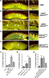
dBACE mutant and knockdown flies show degeneration in the lamina. A, GMR-GAL4 control flies show an intact lamina neuropil (la n), which mostly consists of neuronal fibers, and lamina cortex (la c), which houses the cell bodies of the neuronal monopolar cells and various glial cells. The basement membrane, which separates the retina (re) from the lamina, is indicated by the white lines in the magnifications on the right side. B, C, Vacuoles have formed in the lamina cortex (arrowheads) after induction of the UAS-dBACERNAi construct via Appl-GAL (B, arrowheads) or GMR-GAL4 (C, arrowheads). A similar phenotype is detectable in flies carrying the dBACE2045 allele over the Df(2L)Exel7038 deficiency (D) or over the dBACE5243 allele (E). In addition, we can detect some vacuoles in the lamina neuropil of these flies (D, arrow). F, Quantification of the degeneration in the dBACE knockdown flies versus controls. G, The degeneration in the retinal dBACE knockdown increases significantly with aging (3-d-old knockdown flies are not significantly different from controls). H, Quantification of the degeneration in the dBACE point mutations compared with wild type Canton S (CS). *p < 0.05, **p < 0.01, ***p < 0.001. A–E, All flies were 28 d old. Scale bar, 10 μm.
Figure 3.
Figure 3.
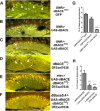
The degeneration is rescued by expressing dBACE. A, A GMR-GAL4; UAS-dBACERNAi fly shows the characteristic row of vacuoles in the lamina cortex (arrowheads). B, In flies overexpressing dBACE in photoreceptors we find vacuoles in the lamina neuropil (la n), which can extend into the lamina cortex, but not the characteristic row of vacuoles seen in the retinal (re) knockdown flies (A). C, Coexpressing UAS-dBACERNAi with UAS-dBACE reduced the vacuoles in the lamina neuropil and cortex to the level in controls (Table 1). D, dBACE2045 over the Df(2L)Exel7038 showing vacuoles in the lamina cortex (arrowheads) and neuropil (arrow). E, Expression of UAS-dBACE in dBACE2045/Df(2L)Exel7038 via elav-GAL4 reduces the degenerative phenotype although some vacuoles can still be found (arrow). F, Expressing dBACE with the dBACE5.4-GAL4 promoter construct rescued the phenotype in dBACE2045/Df(2L)Exel7038 flies. G, H, Quantification of the vacuoles in rescue flies versus dBACE knockdown flies (G) and point mutations (H). re, Retina. **p < 0.01, ***p < 0.001. All flies were 4 weeks old. Scale bar, 10 μm.
Figure 4.
Figure 4.
Electron microscopy reveals a separation of the lamina cortex from the basement membrane. A, Schematic of the lamina cortex (la c) region shown in B and where it is localized in the fly head (box in E). The retina (re) is separated from the lamina cortex (light gray) by the basement membrane (arrowheads). The layer directly beneath the basement membrane (from the basement membrane to the black line) is filled with pigment vesicles and contains trachea (t) and the fenestrated glia (not seen in this picture), but no neuronal cell bodies. The cell bodies of the neuronal monopolar cells (n) and the satellite glia (g) are found in the layer adjacent to the lamina neuropil (la n; darker gray). The lamina neuropil houses the cell bodies of the epithelial glia (g) as well as axons from photoreceptors (a) and neurites from monopolar cells. B, The comparable region in the lamina cortex of a 14-d-old GMR-GAL4 fly. The magnification (F) shows the basement membrane (arrowheads) and pigment vesicles (asterisks). C, The lamina cortex of a 24- to 36-h-old GMR-GAL4; UAS-dBACERNAi fly still appears quite normal at the lower magnification (C), with glia (g), photoreceptor axons (a), and monopolar cells (n) present and only small lesions (arrows) detectable close to the basement membrane (arrowhead). G, A higher magnification of this area reveals that these lesions seem to be due to a loosening of the lamina tissue from the basement membrane (arrowhead). In addition, finger-like extensions from the basement membrane (arrow) are detectable. D, H, In a 14-d-old GMR-GAL4; UAS-dBACERNAi fly the gaps at the basement membrane have widened (black arrows in D) and vacuoles have formed in more proximal layers (white arrows in D). H, A magnification shows that the finger-like structures persist but are more stunted (arrow; the arrowhead points to the basement membrane). Scale bar in B applies to B–D; scale bar in F applies to F–H. me, medulla.
Figure 5.
Figure 5.
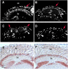
Loss of glial staining in the dBACE knockdown and mutant. A–D, Fifty micrometer vibratome head sections stained with anti-REPO. The image shows a stack of 10 confocal sections taken at 0.5 μm steps. A, In a 3-week-old GMR-GAL4 control fly, anti-REPO labels distal rows of glial cells (red arrow), consisting of the fenestrated glia, pseudocartridge glia, and outer satellite glia. Proximal to this region, additional glial rows are detectable, with the inner satellite glia localized in the lamina cortex (arrowhead) and the epithelial glia in the lamina neuropil (white arrow). B, In a 3-d-old GMR-GAL4; UAS-dBACERNAi fly, gaps appear in the rows of glial cells in the subretinal layer (red arrow). The row of satellite (arrowhead) and epithelial glia (white arrow) still seems intact. C, The phenotype is more prominent after 3 weeks with only a few cells still stained in the subretinal layer (red arrows). Most of the remaining glia appears to be satellite glia (arrowheads). D, A 3-week-old dBACE2045/Df(2L)Exel7038 fly also shows loss of glial staining, with gaps in the distal layer (red arrow) and a loss of most of the epithelial glia (white arrow), whereas the satellite glia still seemed to be present (arrowheads). E, F, Ten micrometer cryostat head sections stained with anti-ELAV show no significant difference in the staining pattern between the 3-week-old GMR-GAL4 control fly and an age-matched GMR-GAL4; UAS-dBACERNAi fly. re, retina; la, lamina. Scale bar: (in A, E) 10 μm.
Figure 6.
Figure 6.
Loss of dBACE causes apoptotic cell death of glia. A, B, Vibratome sections stained with anti-cleaved caspase-3. A, Whereas a 3- to 5-d-old wild-type control does not show any anti-caspase staining (the weak green autofluorescence is caused by the eye pigment), several immunopositive cells are detectable in the lamina cortex of an age-matched GMR-GAL4; UAS-dBACERNAi fly (B, C), including cells in the subretinal region (arrows). The arrowhead points to a more proximally localized cell. D–F, Three-day-old GMR-GAL4; UAS-dBACERNAi fly double stained with anti-cleaved caspase-3 and anti-REPO. As seen in D (arrows) all the caspase-positive cells (red) are also labeled by anti-REPO (green) and most of them are found in the layer adjacent to the retina (arrows). Cells only stained by anti-REPO are mostly localized in more proximal layers (arrowheads). A, B, and C are 2 μm confocal sections and D–F is a 1 μm section. re, retina; la, lamina. Scale bars: (in A) 10 μm; (in C) 5 μm; (in D) 4 μm.
Figure 7.
Figure 7.
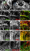
dBACE is expressed in neurons. A, B, Vibratome sections stained with anti-dBACE reveal expression in the brain cortex (arrow in A; stack of 25 0.5 μm confocal sections) and in the neuropils of the optic system (A, arrowheads) and central brain (B, shown is a stack of 11 0.5 μm confocal sections). C–F, Single 0.5 μm confocal sections. Shown are dBACE staining (left), staining against the indicated marker, and the overlay of both stainings (right). C, A double staining with the neuronal marker 22C10 (red) reveals significant colocalization with dBACE (green), including staining in axons (arrowheads). Shown is the region indicated by the red box in A. D, E, dBACE (green) also colocalizes with APPL (red) in the retina (E) and in the brain, including staining in axons (D, arrows) and cell bodies (D, arrowheads). The region shown in D is indicated by the green box in A. F, A glial marker (mCD8-GFP expressed by the glial repo-GAL4, in red) shows limited colocalization with dBACE (green) in a few areas (arrows). The region shown is indicated by the blue box in B. la, lamina; me, medulla; lo, lobula; lp, lobula plate; cc, central complex. Scale bar, 20 μm.
Figure 8.
Figure 8.
APPL levels modify the degenerative phenotype. A, Expressing UAS-GFP in GMR-GAL4; UAS-dBACERNAi flies does not alter the degenerative phenotype (left) nor does GFP expression alone cause a phenotype (right). B, Coexpression of full-length APPL does not affect the vacuolization caused by the dBACE knockdown (left), although we now detect a few vacuoles in the lamina neuropil (arrow) in addition to the vacuoles in the lamina cortex (arrowheads). Expression of UAS-APPL alone results in a few vacuoles in the lamina cortex (arrowheads, right). C, Inducing UAS-dBACERNAi in flies lacking APPL (Appld) suppresses the degenerative phenotype and the occasional remaining vacuoles are very small (arrowhead, left) and comparable to those observed in the Appld control flies shown (arrowhead, right). D, In contrast, coexpression of the secretion-deficient form of APPL causes a strong enhancement of the lamina cortex phenotype (arrowheads, left). Expression of this construct alone results in vacuole formation in the lamina cortex (arrowheads, right), similar to expression of the dBACERNAi construct alone (A, left). E, Quantification of the vacuoles in dBACE knockdown flies with changes in APPL levels (left) and vacuolization caused by changes in APPL levels without knocking down dBACE (right). **p < 0.01, ***p < 0.001. All flies were 4 weeks old. Scale bar, 10 μm.
Figure 9.
Figure 9.
dBACE knockdown in neurons suppresses APPL-induced phenotypes. A, A Western blot using an anti-APPL serum against the intracellular domain reveals increased levels of the endogenous full-length APPL protein in dBACE knockdown flies compared with controls. B, Using the same C-terminal antibody reveals the two alternative C-terminal cleavage products of APPL in flies expressing APPL via GMR-GAL4: the α-cleaved CTF of 14.5 kDa and the 14 kDa β-CTF (lane 1). Inducing the dBACERNAi construct in GMR-GAL4; UAS-APPL flies reduces the levels of the β-CTF (lane 2), as does heterozygosity for dBACE5243 (lane 3). As expected, flies lacking APPL (Appld) show neither cleavage product (lane 4). A loading control using anti-β-tubulin is shown below each blot. C, Fast phototaxis assays reveal a dramatic decline in the PI of flies expressing APPL pan-neuronally using elav-GAL4, whereas their performance is substantially improved by expressing APPL in flies heterozygous for the dBACE5243 allele. D, Heterozygosity for dBACE5243also significantly enhances the viability of these flies. SEMs are indicated.

References

    1. Bennett BD, Babu-Khan S, Loeloff R, Louis JC, Curran E, Citron M, Vassar R. Expression analysis of BACE2 in brain and peripheral tissues. J Biol Chem. 2000;275:20647–20651. - PubMed
    1. Benzer S. Behavioral mutants of Drosophila isolated by countercurrent distripution. Proc Natl Acad Sci U S A. 1967;58:1112–1119. - PMC - PubMed
    1. Bettencourt da Cruz A, Schwärzel M, Schulze S, Niyyati M, Heisenberg M, Kretzschmar D. Disruption of the MAP1B-related protein FUTSCH leads to changes in the neuronal cytoskeleton, axonal transport defects, and progressive neurodegeneration in Drosophila. Mol Biol Cell. 2005;16:2433–2442. - PMC - PubMed
    1. Bogart K, Andrews J. CGB Technical Report 10. Bloomington, IN: The Center for Genomics and Bioinformatics; 2006. Extraction of Total RNA from Drosophila.
    1. Cai H, Wang Y, McCarthy D, Wen H, Borchelt DR, Price DL, Wong PC. BACE1 is the major beta-secretase for generation of Abeta peptides by neurons. Nat Neurosci. 2001;4:233–234. - PubMed

Publication types

MeSH terms

LinkOut - more resources