. 2012:7:5671-82.
doi: 10.2147/IJN.S34635. Epub 2012 Nov 8.

Enhancing microparticle internalization by nonphagocytic cells through the use of noncovalently conjugated polyethyleneimine

Affiliations

Enhancing microparticle internalization by nonphagocytic cells through the use of noncovalently conjugated polyethyleneimine

Tania Patiño et al. Int J Nanomedicine. 2012.

Abstract

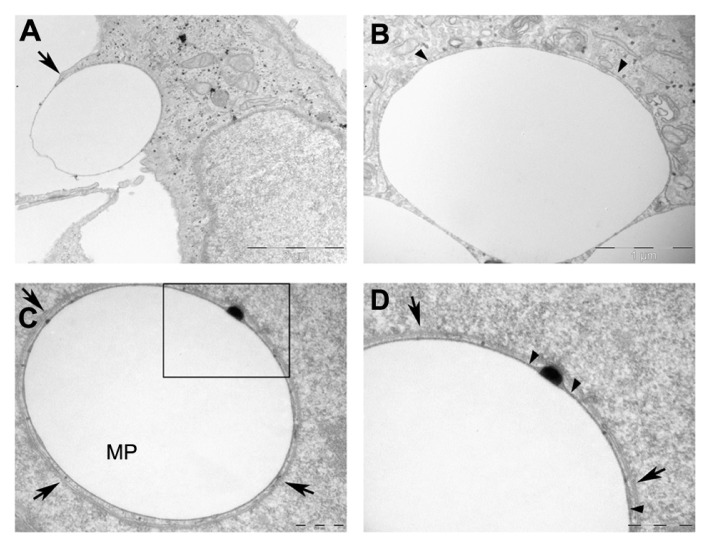
Development of micro- and nanotechnology for the study of living cells, especially in the field of drug delivery, has gained interest in recent years. Although several studies have reported successful results in the internalization of micro- and nanoparticles in phagocytic cells, when nonphagocytic cells are used, the low internalization efficiency represents a limitation that needs to be overcome. It has been reported that covalent surface modification of micro- and nanoparticles increases their internalization rate. However, this surface modification represents an obstacle for their use as drug-delivery carriers. For this reason, the aim of the present study was to increase the capability for microparticle internalization of HeLa cells through the use of noncovalently bound transfection reagents: polyethyleneimine (PEI) Lipofectamine™ 2000 and FuGENE 6(®). Both confocal microscopy and flow cytometry techniques allowed us to precisely quantify the efficiency of microparticle internalization by HeLa cells, yielding similar results. In addition, intracellular location of microparticles was analyzed through transmission electron microscopy and confocal microscopy procedures. Our results showed that free PEI at a concentration of 0.05 mM significantly increased microparticle uptake by cells, with a low cytotoxic effect. As determined by transmission electron and confocal microscopy analyses, microparticles were engulfed by plasma-membrane projections during internalization, and 24 hours later they were trapped in a lysosomal compartment. These results show the potential use of noncovalently conjugated PEI in microparticle internalization assays.

Keywords: HeLa cells; drug delivery; endocytosis; internalization efficiency.


Figures

Figure 1
Figure 1
Cytotoxicity of transfection treatments. (A) Percentage of cells that remained attached to the dish after treatments, normalized to the control. (B) Percentage of viable attached cells after treatments. (C) Percentage of viable cells. Note: a, b, c denote significant differences among groups. Abbreviations: PEI, polyethyleneimine; LF, Lipofectamine™.
Figure 2
Figure 2
Z potential of control microparticles and of microparticles treated with PEI, LF2000 and FuGENE 6®. Note: The Zeta potential of the functionalized microparticle is also shown. Abbreviations: FMP, functionalized microparticle; PEI, polyethyleneimine; LF, Lipofectamine™.
Figure 3
Figure 3
Flow cytometry analysis. Percentage of cells in contact with at least one microparticle. Notes: a, b, c, d denote significant differences among groups. Abbreviations: PEI, Polyethyleneimine; LF, Lipofectamine™.
Figure 4
Figure 4
Flow cytometry analysis. Transfection agents’ effect on the internalization of fluorescent microparticles by HeLa cells. (A), (C), (E), (G), (I) and (K) Dot plots showing two populations of cells: associated (cells + MP) or not associated (cells) with green fluorescent microparticles. (B), (D), (F), (H), (J) and (L) Histograms showing the fluorescence intensity of the different populations of cells. Notes: Cells = cell population not associated with microparticles. Cells + MP = cell population associated with at least one microparticle. P2, P3, P4, P5 = cell population associated with one, two, three, or more than three microparticles, respectively. Abbreviations: MP, microparticle; PEI, polyethyleneimine; LF, Lipofectamine™.
Figure 5
Figure 5
CSLM analyses of microparticle location. (A and B) CSLM-obtained images (middle) and their orthogonal projections of the z-stack reconstructions (right and bottom) of consecutive focal planes (0,5 μm each). (C) Percentage of cells in contact with microparticles, either attached or internalized, analyzed by CSLM. Notes: Green arrows indicate a microparticle located inside the cell, clearly surrounded by the plasma membrane. The red arrow points to a microparticle attached to the plasma membrane but outside the cell. Plasma membrane appears in red (WGA-Texas Red® Staining) and chromatin in blue (Hoescht 33258). Abbreviations: CSLM, confocal scanning laser microscopy; PEI, polyethyleneimine; LF, Lipofectamine™.
Figure 6
Figure 6
Microparticle internalization efficiency. Percentage of viable cells with one or more internalized microparticles after the different transfection treatments. Notes: Results were obtained from internalization analyses by both CSLM and FC, taking into account the number of cells that remained attached to the dish after treatments and their viability. Abbreviations: CSLM, confocal scanning laser microscopy; FC, flow cytomtery; PEI, polyethyleneimine; LF, Lipofectamine™.
Figure 7
Figure 7
Microparticle localization by transmission electron microscopy. Micrographs of HeLa cells with internalized microparticles. (A) Illustration of the internalization of microparticles; arrow indicates a cell membrane evagination, typical of macropinocytosis. (B) A single membrane surrounding an internalized microparticle. (C) Microparticle surrounded by a double membrane. (D) Enlarged view of the microparticle shown in C. Notes: Arrow heads point to a single membrane tightly associated to microparticle. Arrows indicate the two membrane complex. Scale bars: (A) 2 μm (B) 1 μm (C) and (D) 500 nm. Abbreviation: MP, microparticle.
Figure 8
Figure 8
LAMP-1 immunogold detection. Cryotransmission electron micrographs of LAMP-1 immunogold performed in HeLa cells. (AC) Arrow heads indicate positive LAMP-1 marks. (D) Arrow heads indicate positive LAMP-1 marks whereas arrows point to the lysosome membrane. Abbreviation: LAMP-1, lysosome associated membrane protein 1; L, lysosome.
Figure 9
Figure 9
Intracellular location analysis of functionalized microparticles by CSLM. (A) Endosomal labeling with EEA-1. (B) Lysosomal labeling with LAMP-1. (C and D) Microparticles functionalized with an Alexa Fluor®-594 conjugated antibody. (E and F) Merged images of compartment and microparticles. Abbreviations: CSLM, confocal scanning laser microscopy; EEA-1, early endosome antigen 1 protein; LAMP-1, lysosome associated membrane protein 1.

References

    1. Grayson ACR, Shawgo RS, Johnson AM, et al. A BioMEMS review: MEMS technology for physiologically integrated devices. Proc IEEE. 2004;92(1):6–21.
    1. Fernandez-Rosas E, Gómez R, Ibanez E, et al. Intracellular polysilicon barcodes for cell tracking. Small. 2009;5(21):2433–2439. - PubMed
    1. Novo S, Barrios L, Santaló J, et al. A novel embryo identification system by direct tagging of mouse embryos using silicon-based barcodes. Hum Reprod. 2011;26(1):96–105. - PubMed
    1. Tao Z, Toms B, Goodisman J, Asefa T. Mesoporous silica microparticles enhance the cytotoxicity of anticancer platinum drugs. ACS Nano. 2010;4(2):789–794. - PubMed
    1. Tasciotti E, Liu X, Bhavane R, et al. Mesoporous silicon particles as a multistage delivery system for imaging and therapeutic applications. Nat Nanotechnol. 2008;3(3):151–157. - PubMed

Publication types