. 2012;7(11):e48567.
doi: 10.1371/journal.pone.0048567. Epub 2012 Nov 12.

Knockdown of Ki-67 by dicer-substrate small interfering RNA sensitizes bladder cancer cells to curcumin-induced tumor inhibition

Affiliations

Knockdown of Ki-67 by dicer-substrate small interfering RNA sensitizes bladder cancer cells to curcumin-induced tumor inhibition

Sivakamasundari Pichu et al. PLoS One. 2012.

Abstract

Transitional cell carcinoma (TCC) of the urinary bladder is the most common cancer of the urinary tract. Most of the TCC cases are of the superficial type and are treated with transurethral resection (TUR). However, the recurrence rate is high and the current treatments have the drawback of inducing strong systemic toxicity or cause painful cystitis. Therefore, it would be of therapeutic value to develop novel concepts and identify novel drugs for the treatment of bladder cancer. Ki-67 is a large nucleolar phosphoprotein whose expression is tightly linked to cell proliferation, and curcumin, a phytochemical derived from the rhizome Curcuma longa, has been shown to possess powerful anticancer properties. In this study, we evaluated the combined efficacy of curcumin and a siRNA against Ki-67 mRNA (Ki-67-7) in rat (AY-27) and human (T-24) bladder cancer cells. The anticancer effects were assessed by the determination of cell viability, apoptosis and cell cycle analysis. Ki-67-7 (10 nM) and curcumin (10 µM), when treated independently, were moderately effective. However, in their combined presence, proliferation of bladder cancer cells was profoundly (>85%) inhibited; the rate of apoptosis in the combined presence of curcumin and Ki-67-7 (36%) was greater than that due to Ki-67-7 (14%) or curcumin (13%) alone. A similar synergy between curcumin and Ki-67-7 in inducing cell cycle arrest was also observed. Western blot analysis suggested that pretreatment with Ki-67-7 sensitized bladder cancer cells to curcumin-mediated apoptosis and cell cycle arrest by p53- and p21-independent mechanisms. These data suggest that a combination of anti-Ki-67 siRNA and curcumin could be a viable treatment against the proliferation of bladder cancer cells.


Conflict of interest statement

Competing Interests: The authors have declared that no competing interests exist.

Figures

Figure 1
Figure 1. Effect of various DsiRNA constructs on a) Ki-67 mRNA expression and b) cell proliferation in bladder cancer cells; c) Dose-response studies with Ki-67-7. a) Rat TCC cells (AY-27) were cultured in 12-well plates (∼0.8
×105 cells/well) as described in Materials and Methods. After 24 h, they were transfected with each of the siRNA constructs (Ki-67-2, Ki-67-7 and Ki-67-9) targeted against Ki-67 mRNA. After 48 h, RNA was extracted and the levels of mRNA were determined by quantitative PCR as described in Methods. Cells treated with the transfection reagent alone were used as the control. The rate of expression of Ki-67 mRNA was normalized to the expression of β-actin. Values represent data from one (of two) experiment using the average of triplicate determinations. b) AY-27 cells were cultured in 12-well (0.8×10⁵ cells/well) plates for 24 h and transfected with various siRNA constructs as described in Methods. Cell viability was determined 48 h after transfection by MTT assay. Cells treated with transfection reagent alone served as the control ( = 100%). Bars indicate values which are the means ± S.E of three determinations. (*P<0.05). c) AY-27 cells were cultured in 12-well plates (0.8×10⁵ cells/well) as described in Methods. Forty eight hours after siRNA transfection, the effect of various concentrations of Ki-67-7 on cell viability was determined by MTT assay. Cells treated with transfection reagent alone served as the control. Cell viability values are expressed as percent control and are the means ± S.E of 3-8 determinations (*P<0.05, ***P<0.005).
Figure 2
Figure 2. Dose-dependent effects of curcumin on bladder cancer cell proliferation.
T-24 (2a) and AY-27 (2b) cells were plated in 12-well plates (0.8×10⁵ cells/well) and cultured for 48 hr (40–50% confluence) as described in Methods. Following exposure to various concentrations of curcumin for 24 h, cells were washed with PBS and incubated for another 24 h in curcumin-free medium. Cells incubated with DMSO (0.1%) alone were treated as controls. DMSO alone had little impact on tumor cell growth (data not shown). Cell proliferation was assessed on the basis of cell viability, measured by MTT assay. Cell viability was expressed as percent of viability observed in DMSO-treated control cells. Values are from a representative (of two) (2a) or 3–8 (mean ± S.E) determinations (2b). *P<0.05, **P<0.01, ***P<0.005.
Figure 3
Figure 3. Effect of the combined treatment of anti-Ki-67 DsiRNA and curcumin on bladder cancer cells.
Equal number (0.8×10⁵ cells/well) of bladder cancer cells T-24 (3a) and AY-27 (3b) cultured as described in Methods were either transfected with Ki-67–7 or control DsiRNA (10 nM) or treated with the transfection reagent alone for 24 h. Subsequently, where indicated, curcumin (10 µM) was added to the cells and incubated for 24 h followed by an additional 24 h incubation in the absence of curcumin. Cell viability was determined by MTT assay at the end of 72 h from transfection. Cells treated with DMSO plus transfection reagent served as the control. Data presented are the mean ± S.E of 3–5 determinations (*P<0.05, **P<0.01). 3c) AY-27 cells (0.8×10⁵ cells/well) were treated exactly as described above in legend to Figures 3A and 3B and at the end of 24, 48 and 72 h after curcumin treatment (which is same as 48, 72 and 96 h after siRNA transfection), cell growth was assessed based on cell numbers as described in Methods. Data are mean ± S.E of three determinations.
Figure 4
Figure 4. Effect of Ki-67-7 and curcumin on Ki-67 a) gene and b) protein expression in bladder cancer cells.
a) AY-27 cells were treated with Ki-67 siRNA in the presence or absence of curcumin exactly as described in legend to Figures 3A and 3B. At the end of the treatment, RNA was extracted and Ki-67 mRNA expression was determined by quantitative PCR using β-actin as the internal standard as described in Methods. Ki-67 mRNA levels are expressed as percent of the rate of expression in control cells which were treated with transfection reagent and DMSO. Bars indicate values which are the mean ± S.E of three determinations. b) AY-27 cells were grown on glass coverslips placed inside 12-well plates and treated with siRNA (Ki-67-7), curcumin or as a combination, as described in the legend to Figures 3A and 3B. At the end of the treatment period, cells were exposed to Ki67 primary antibody (M19) and FITC-labeled secondary antibody followed by PI (Propidium iodide), and observed under confocal microscopy as described in Methods. Scale bar inserts  = 5 µm. Increased down-regulation of Ki-67 protein due to Ki-67-7 was observed.
Figure 5
Figure 5. Determination of Ki-67-7 and curcumin-induced apoptosis by flow cytometry.
AY-27 cells were treated with anti-Ki-67 siRNA, curcumin or in the combined presence of both as described in legend to Figures 3A and 3B. At the end of the treatment period, the cells were harvested, stained with fluorescence-conjugated Annexin V and 7AAD as described in Methods. Following flow cytometric analysis, the percentage (number inserts in the figure) of normal cells or those undergoing apoptosis and necrosis was calculated using appropriate software (Flow Jo v.9). The rate of apoptosis was measured by adding the percentages in the right two quadrants in each of the figures. The data are from a representative (of two identical) experiment.
Figure 6
Figure 6. Effect of Ki-67-7 and curcumin on cell cycle phases.
AY-27 cells were treated with Ki-67-7, curcumin or with both, as described earlier in legend to Figures 3A and 3B. At the end of the incubation period, the cells were treated with PI and the distribution of cells in various cell cycle phases were determined by flow cytometry as described in Methods. Cells were scored as percentage distribution of cells in each of the cell cycle phases (Go/G1, S and G2/M). Bars indicate values which are the means ± S.E of three determinations.
Figure 7
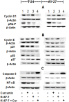
Figure 7. Effect of Ki-67-7 and curcumin on the regulatory proteins of cell cycle phases and apoptosis: Western blot analysis.
Bladder cancer cells (T-24 and AY-27) were cultured and exposed to Ki-67-7 and curcumin as described above in legend to Figures 3A and 3B. At the end of the treatment period, total protein was extracted and subjected to Western blotting using appropriate antibodies as described in Methods. For purposes of associating proteins with specific roles, they were grouped in separate categories, such as gene transcription (A), cell-cycle progression (B) and apoptosis (C). β-actin was used as the internal control. Since different proteins (e.g. Cyclin D1 and NF-κB) from the same membrane were identified in separate categories, the same β-actin blot is displayed on more than one occasion. Data presented are western blots from a single experiment, which is representative of 2–3 identical experiments.
Figure 8
Figure 8. Molecular targets of CusiRNA.
The scheme displays the possible molecular targets of Ki-67-7 and curcumin when used in combination (CusiRNA). Arrowheads indicate the activation of indicated proteins; hammer heads indicate inhibition and the broken lines represent the possible role for putative intermediates. Further details are provided in the text.

References

    1. Parker SL, Tong T, Bolden S, Wingo PA (1996) Cancer Statistics. CA Cancer J Clin: 5–27. - PubMed
    1. Lamm DL, Riggs DR, Traynelis CL, Nseyo UO (1995) Apparent failure of current intravesical chemotherapy prophylaxis to influence long-term course of superficial transitional cell carcinoma of the bladder. J Urol 153: 1444–1450. - PubMed
    1. Parkin DM, Bray F, Ferlay J, Pisani P (2005) Global Cancer Statistics, 2002. CA Cancer J Clin 55: 74–108. - PubMed
    1. Herr HW, Donat SM, Reuter VE (2007) Management of low grade papillary bladder tumors. J Urol 178: 1201–1205. - PubMed
    1. Guhasarkar S, Banerjee R (2010) Intravesical drug delivery: Challenges, current status, opportunities and novel strategies. J Control Release 48: 147–159. - PubMed

Publication types

MeSH terms