. 2012;7(11):e48636.
doi: 10.1371/journal.pone.0048636. Epub 2012 Nov 9.

Targeting the acute promyelocytic leukemia-associated fusion proteins PML/RARα and PLZF/RARα with interfering peptides

Affiliations

Targeting the acute promyelocytic leukemia-associated fusion proteins PML/RARα and PLZF/RARα with interfering peptides

Sabine Beez et al. PLoS One. 2012.

Abstract

In acute promyelocytic leukemia (APL), hematopoietic differentiation is blocked and immature blasts accumulate in the bone marrow and blood. APL is associated with chromosomal aberrations, including t(15;17) and t(11;17). For these two translocations, the retinoic acid receptor alpha (RARα) is fused to the promyelocytic leukemia (PML) gene or the promyelocytic zinc finger (PLZF) gene, respectively. Both fusion proteins lead to the formation of a high-molecular-weight complex. High-molecular-weight complexes are caused by the "coiled-coil" domain of PML or the BTB/POZ domain of PLZF. PML/RARα without the "coiled-coil" fails to block differentiation and mediates an all-trans retinoic acid-response. Similarly, mutations in the BTB/POZ domain disrupt the high-molecular-weight complex, abolishing the leukemic potential of PLZF/RARα. Specific interfering polypeptides were used to target the oligomerization domain of PML/RARα or PLZF/RARα. PML/RARα and PLZF/RARα were analyzed for the ability to form high-molecular-weight complexes, the protein stability and the potential to induce a leukemic phenotype in the presence of the interfering peptides. Expression of these interfering peptides resulted in a reduced replating efficiency and overcame the differentiation block induced by PML/RARα and PLZF/RARα in murine hematopoietic stem cells. This expression also destabilized the PLZF/RARα-induced high-molecular-weight complex formation and caused the degradation of the fusion protein. Targeting fusion proteins through interfering peptides is a promising approach to further elucidate the biology of leukemia.


Conflict of interest statement

Competing Interests: The authors have declared that no competing interests exist.

Figures

Figure 1
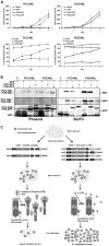
Figure 1. Co-expression of PCC/POZ-GFP reverses IL3-independent growth in PCC/ABL- or POZ/ABL-positive BA/F3 cells.
A, Infected BA/F3 cells were cultivated for 5 days without IL3. The total cell count and FACS measurement of the GFP signal were ascertained daily, n = 3. The upper panels represent the GFP positive population accumulation, and the lower panels represent the IL3-independent growth of the Ba/F3 cells expressing the ABL fusions in the presence or absence of the related peptides or GFP alone, as indicated. Mock: PIDE vector without BCC-ABL or POZ-ABL, expressing only GFP. B, Western blot of whole cell lysates of Phoenix and BA/F3 cells probed for ABL (α-ABL), phospho-ABL (α-ABL*) and GFP (α-GFP). Control: empty vector. C, Schematic diagram of IL3 growth dependency, as shown for PCC. By fusing ABL to PCC, BA/F3 cells can grow in the absence of IL3. The introduction of PCC-GFP inhibits the PCC/ABL complex formation and restores IL3-dependent growth.
Figure 2
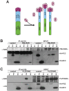
Figure 2. PCC and POZ bind to PML/RARα and PLZF/RARα, respectively.
A, Theory for the peptide binding to X-RARα, as exemplified by POZ and PLZF/RARα. By binding the oligomerization domain of PLZF/RARα, POZ disables the self-oligomerization and the high-molecular-weight complex formation. B, C, HA-tagged peptides were co-expressed with PML/RARα or PLZF/RARα in the 293 cells through the transfection of the corresponding combination of pCDNA3 vectors with calcium phosphate. An HA-empty plasmid and an HA-tagged BCC (coiled-coil domain of BCR) were used as a specificity control. In B, PCC was transfected at two different concentrations, 5 and 10 µG DNA (first and second BCC lane, respectively). Immunoprecipitation (IP) was performed with an anti-HA matrix. Western blots were probed with α-HA and α-RARα antibodies.
Figure 3
Figure 3. PCC and POZ influences on the HMW complex formation of PML/RARα and PLZF/RARα, respectively.
A and B, Size-exclusion chromatography fractions of in vitro translated PML/RARα (A) or PLZF/RARα (B) and HA-tagged peptide. Fractions were analyzed by western blot (on the right) and probed against RARα. The densitometric analysis of the western blot is shown on the left side as a percentage of the entire signal. C and D, Size-exclusion chromatography of the Phoenix whole cell lysates overexpressing PML/RARα (C) or PLZF/RARα (D) and GFP-tagged peptide. Fractions were analyzed by western blot (WB) and probed against RARα (α-RARα) (lower part). The densitometric analysis of the western blot is shown in the upper part as a percentage of the entire signal. HA-mock: control empty vector. Input (I), eluted fractions 14 to 29. The numbers above the arrows represent the molecular weight (KD) of the proteins used for the MW calibration: 136 KD for the BSA dimer, 440 KD for Ferritin, and 670 KD for Thyroglobulin.
Figure 4
Figure 4. PM/RARα and PLZF/RARα, degrade in the presence of PCC and POZ, respectively.
A and B, Western blot of the whole cell lysates of PML/RARα- (A) or PLZF/RARα (B)-positive Phoenix and BA/F3 cells probed against RARα (α-RARα) and GFP (α-GFP). C and D, BA/F3 cells were treated with 10 µM MG132 or 20 µM Calpain I for 16 h. Control: empty vector; tubulin: loading control. E. BA/F3 cells were treated with 500 nM Lactacystin for 24 h. Control: empty vector; GAPDH: loading control. F, The infection efficiency, measured as the percentage of GFP-positive cells, of the Ba/F3 cells after infection with PINCO (control) or PIDE carrying PLZF/RARα alone or in combination with GFP, GFP-POZ or GFP-PCC or PML/RARα alone or in combination with GFP, GFP-POZ or GFP-PCC peptides, as indicated. G, Western blot of the Phoenix whole cell lysate expressing PML/RARα or a sumoylation-deficient mutant (PML3-160/RARα) PCC-GFP or GFP, probed with α-GFP, α-RARα and α-GPADH as a loading control. The image shown is a representative of three separate experiments.
Figure 5
Figure 5. PCC and POZ reverse the differentiation block of PML/RARα- and PLZF/RARα-positive murine hematopoietic stem cells.
A, Schematic diagram of the experiment conditions. Sca1+/lin- murine bone marrow cells were isolated and infected with retroviral vectors containing PML/RARα or PLZF/RARα and GFP or PCC/POZ-GFP and then analyzed for differentiation. B, The infection efficiency, measured as the percentage of the GFP-positive cells to the retrovirally infected cells. C, Reverse transcriptase-PCR of the retrovirus-infected Sca+/lin bone marrow cells for PML/RARα and PLZF/RARα. Control: β-Actin. D, GIEMSA staining of Sca+/lin bone marrow cells seven days after infection with retroviral vectors. E, FACS analysis of the Sca1+/lin bone marrow cells infected with PML/RARα and GFP or PCC/POZ-GFP. Mock: empty vector. F, FACS analysis of the Sca1+/lin bone marrow cells infected with PLZF/RARα and GFP or PCC/POZ-GFP. Mock: empty vector.
Figure 6
Figure 6. PCC and POZ reverse the proliferation capacity of PML/RARα- and PLZF/RARα-positive murine hematopoietic stem cells.
A, Schematic diagram of the experimental conditions. Sca1+/lin bone marrow cells were isolated and infected with retroviral vectors containing PML/RARα or PLZF/RARα and GFP or PCC/POZ-GFP. Infected cells were plated in methylcellulose on day 3. Colony counts and replating were measured every 10 days. B and C, Colony count of Sca1+/lin bone marrow cells infected with PML/RARα (B) or PLZF/RARα (C) and GFP or PCC/POZ-GFP cultured in methylcellulose. Mock: empty vector.

References

    1. Puccetti E, Ruthardt M (2004) Acute promyelocytic leukemia: PML/RARalpha and the leukemic stem cell. Leukemia 18: 1169–1175. - PubMed
    1. Brown D, Kogan S, Lagasse E, Weissman I, Alcalay M, et al. (1997) A PMLRARalpha transgene initiates murine acute promyelocytic leukemia. Proc Natl Acad Sci U S A 94: 2551–2556. - PMC - PubMed
    1. He LZ, Merghoub T, Pandolfi PP (1999) In vivo analysis of the molecular pathogenesis of acute promyelocytic leukemia in the mouse and its therapeutic implications. Oncogene 18: 5278–5292. - PubMed
    1. Puccetti E, Obradovic D, Beissert T, Bianchini A, Washburn B, et al. (2002) AML-associated translocation products block vitamin D(3)-induced differentiation by sequestering the vitamin D(3) receptor. Cancer Res 62: 7050–7058. - PubMed
    1. Seshire A, Rossiger T, Frech M, Beez S, Hagemeyer H, et al. (2012) Direct interaction of PU.1 with oncogenic transcription factors reduces its serine phosphorylation and promoter binding. Leukemia doi:10.1038/leu.2011.331. - DOI - PubMed

Publication types

MeSH terms

Substances