. 2012;7(11):e48801.
doi: 10.1371/journal.pone.0048801. Epub 2012 Nov 9.

Counteracting roles of AMP deaminase and AMP kinase in the development of fatty liver

Affiliations

Counteracting roles of AMP deaminase and AMP kinase in the development of fatty liver

Miguel A Lanaspa et al. PLoS One. 2012.

Abstract

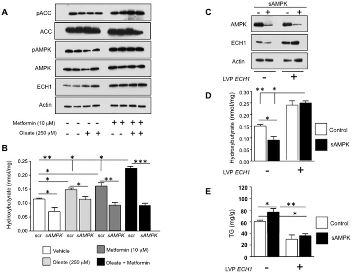
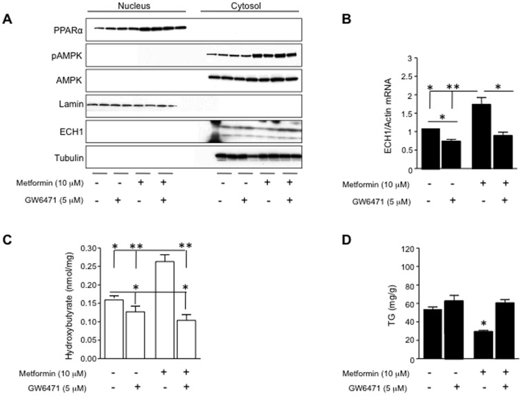
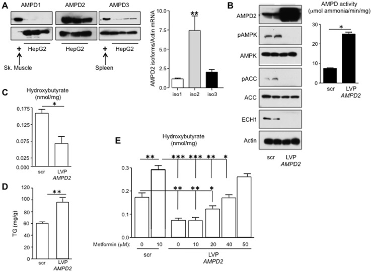
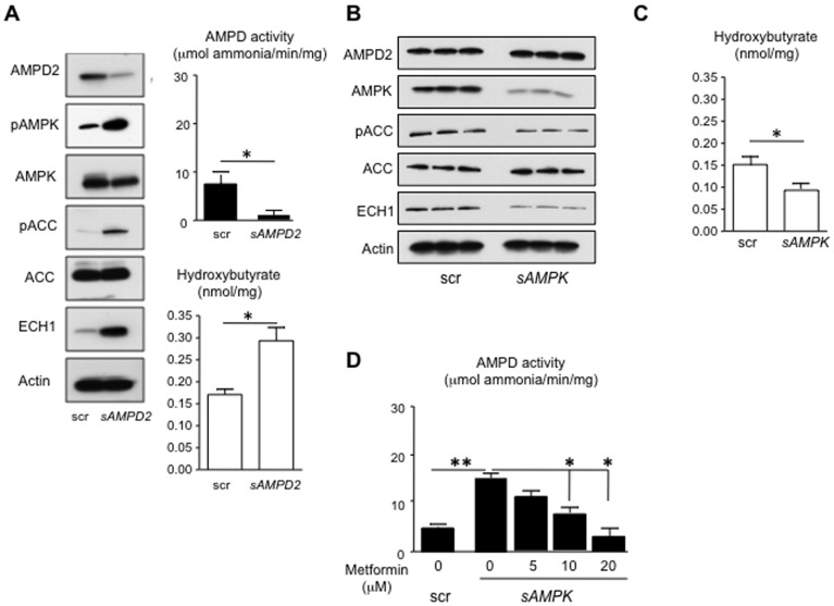
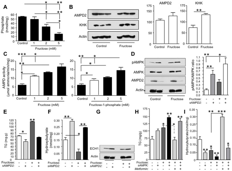
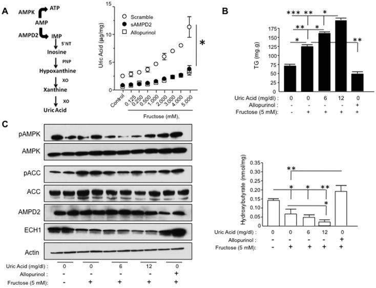
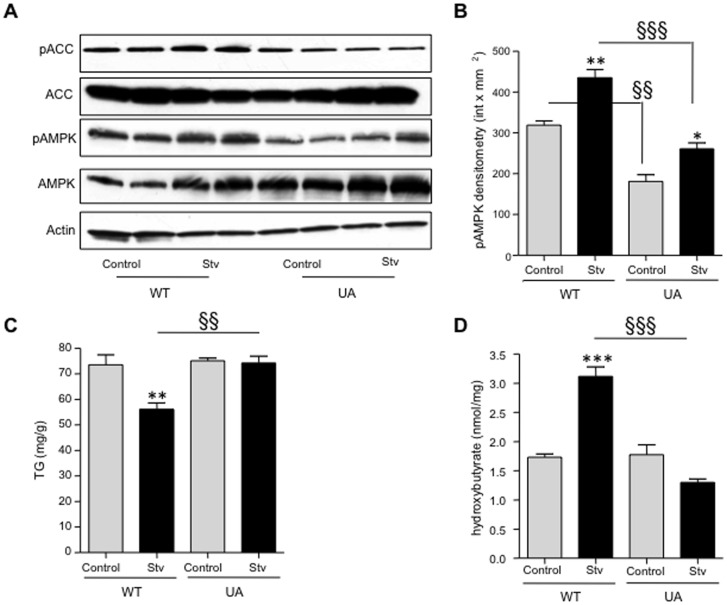
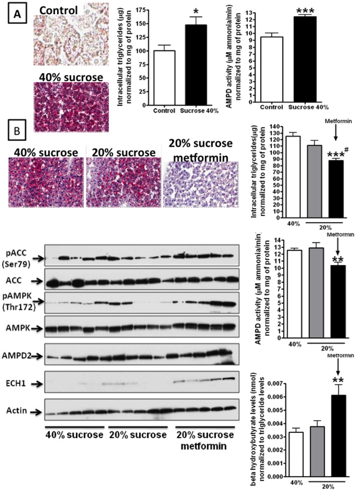
Fatty liver (hepatic steatosis) is associated with nucleotide turnover, loss of ATP and generation of adenosine monophosphate (AMP). It is well known that in fatty liver, activity of the AMP-activated kinase (AMPK) is reduced and that its stimulation can prevent hepatic steatosis by both enhancing fat oxidation and reducing lipogenesis. Here we show that another AMP dependent enzyme, AMPD2, has opposing effects on fatty acid oxidation when compared to AMPK. In human hepatocytres, AMPD2 activation -either by overexpression or by lowering intracellular phosphate levels with fructose- is associated with a significant reduction in AMPK activity. Likewise, silencing of AMPK spontaneously increases AMPD activity, demonstrating that these enzymes counter-regulate each other. Furthermore, we show that a downstream product of AMP metabolism through AMPD2, uric acid, can inhibit AMPK activity in human hepatocytes. Finally, we show that fructose-induced fat accumulation in hepatocytes is due to a dominant stimulation of AMPD2 despite stimulating AMPK. In this regard, AMPD2-deficient hepatocytes demonstrate a further activation of AMPK after fructose exposure in association with increased fatty acid oxidation, and conversely silencing AMPK enhances AMPD-dependent fat accumulation. In vivo, we show that sucrose fed rats also develop fatty liver that is blocked by metformin in association with both a reduction in AMPD activity and an increase in AMPK activity. In summary, AMPD and AMPK are both important in hepatic fat accumulation and counter-regulate each other. We present the novel finding that uric acid inhibits AMPK kinase activity in fructose-fed hepatocytes thus providing new insights into the pathogenesis of fatty liver.


Conflict of interest statement

Competing Interests: The authors have declared that no competing interests exist.

Figures

Figure 1
Figure 1. Activation of AMPK stimulates fat oxidation in HepG2 cells.
A) Stimulation of AMPK activity by metformin (10 µM) significantly increases the phosphorylation of AMPK at Thr172, the phosphorylation of ACC at ser79 as well as the expression of the fat oxidation-related protein ECH1 (Enoyl CoA-hydratase) B) Metformin up-regulates the accumulation of β-hydroxybutyrate -a marker of fat oxidation activity- in cells exposed to oleic acid (250 µM, 72 hour). Hydroxybutyrate accumulation, ACC phosphorylation and ECH1 up-regulation by metformin does not occur in AMPK-deficient cells. *p<0.05, **p<0.01, ***p<0.001 C) Representative western blot showing over-expression of ECH1 in HepG2 cells (lanes 3 and 4) in control cells (lanes 1 and 3) and AMPK deficient cells (lanes 2 and 4). ECH1 overexpression restores the loss of fat oxidation observed in AMPK deficient cells and reduces fat accumulation as determined by a significant up-regulation of β-hydroxybuyrate levels (D) with paralleled decrease in intracellular triglyceride levels (E) *p<0.05, **p<0.01.
Figure 2
Figure 2. Metformin-induced up-regulation of ECH1 is mediated by PPARα.
A) Representative western blot of nuclear and cytosolic extracts from hepatocytes exposed to metformin (10 µM) and the PPARα inhibitor, GW6471 (5 µM). As shown, GW6471 reduces metformin-induced ECH1 up-regulation in the cytoplasmic fraction but do not affet either PPARα nuclear expression or AMPK phosphorylation. B) ECH1 mRNA levels in hepatocytes exposed to metformin (10 µM) and the PPARα inhibitor, GW6471 (5 µM) C) Inhibition of PPARα activity with the antagonist GW6471 reduces metformin-induced fat oxidation. D) Inhibition of PPARα activity with the antagonist GW6471 reduces metformin-induced trigyceride accumulation. *p<0.05, **p<0.01.
Figure 3
Figure 3. Metformin regulates AMPD2 activity in human hepatocytes.
A) AMPD2 is the main isoform in HepG2 cells. The expression of AMPD1 and AMPD3 isoforms is minimal compred to AMPD2. Skeletal muscle and spleen are positive controls for AMPD1 and AMPD3 expression, respectively. Quantitative PCR analysis (bottom) demonstrates that isoform 2 is the predominant isoform of AMPD2. **p<0.01 versus isoforms 1 and 3. B) Over-expression of AMPD2 down-regulates the activation of AMPK. Transduction of HepG2 cells with lentiviral particles codifying for the isoform 2 of AMPD2 results in significantly higher levels of AMPD2 protein expression as well as AMPD activity. This is paralleled with reduced levels Thr172 pAMPK expression as well as of their target genes ECH1 and Ser79 pACC. *p<0.05 C–D) Reduction in ECH1 expression in over-expressing AMPD2 hepatocytes is accompanied with lower intracellular β-hydroxybutyrate and higher TG levels. *p<0.05, **p<0.01 E) Over-expression of AMPD2 impairs metformin-induced fat oxidation. Metformin 10 µM significantly increased β- hydroxybutyrate levels in cells transducted with scramble RNA. In contrast, no significant change was observed with 10 µM metformin in cells overexpressing AMPD2. Metformin 50 µM significantly increased β- hydroxybutyrate to levels observed in scramble transducted cells *p<0.05, **p<0.01, ***p<0.001.
Figure 4
Figure 4. AMPK activation in AMPD2-deficient cells.
A) Silencing AMPD2 expression in human hepatocytes spontaneously up-regulates the activation of AMPK. Transduction of HepG2 cells with lentiviral particles codifying for a specific silencer for AMPD2 results in significantly lower levels of AMPD activity. This is paralleled with increaseded levels Thr172 pAMPK expression as well as of their target genes ECH1 and Ser79 pACC. Up-regulation of ECH1 is accompanied with higher intracellular β-hydroxybutyrate levels. *p<0.05 B) Blockade of AMPK expression activates AMPD2. Stable silencing of AMPK is associated with significant down-regulation of ECH1 and phosphorylation of ACC at Ser79. In contrast, no change in AMPD2 expression is observed. C) Silencing AMPK expression in human hepatocytes spontaneously decreases the levels of intracellular β-hydroxybutyrate D) Metformin blocks AMPD activation in AMPK-deficient cells in a dose-dependent manner. *p<0.05, **p<0.01.
Figure 5
Figure 5. Fructose stimulates fat accumulation in HepG2 cells by activating AMPD2 and blocking AMPK and fat oxidation.
A) Fructose reduces intracellular phosphate levels –a natural inhibitor of AMPD activity- in a dose-dependent manner. *p<0.05, **p<0.01. B) AMPD2 expression is not modified by fructose in human hepatocytes. As control, fructokinase (KHK) expression is significantly up-regulated. **p<0.01. C) Both fructose (1–5 mM) and fructose-1-phosphate (1–5 mM) -the product of fructokinase metabolism- significantly up-regulates the activity of AMPD2 in human hepatocytes. AMPD activity was measured after 30 minutes exposure to fructose in 50 µg protein lysates *p<0.05, **p<0.01. D) Fructose stimulates AMPK activity (as determined by phosphorylation at Thr172) that is further amplified in AMPD2 silenced cells. *p<0.05, **p<0.01. E) Fructose induces triglyceride accumulation in HepG2 cells that is blocked in AMPD2 deficient cells *p<0.05, **p<0.01 F) Fructose decreases β- hydroxybutyrate levels in HepG2 cells that are blocked in AMPD2 deficient cells *p<0.05, **p<0.01. G) ECH1 expression is up-regulated in AMPD2 deficient cells exposed to fructose. H) Silencing AMPK expression increases triglyceride accumulation in non-exposed and fructose exposed HepG2 cells that is not blocked with 10 µM metformin *p<0.05, **p<0.01 I) Silencing AMPK expression decreases intracellular β- hydroxybutyrate levels in non-exposed and fructose exposed HepG2 cells that is not blocked with 10 µM metformin *p<0.05, **p<0.01, ***p<0.001.
Figure 6
Figure 6. Uric acid negatively regulates AMPK activity.
A) Schematic representation of downstream metabolites produced from AMP by AMPD2. Right, uric acid levels –the final product of this route- are increased in HepG2 cells exposed to fructose in a dose-dependent manner. Both inhibiting xanthine oxidase activity with allopurinol or silencing AMPD2 significantly inhibits uric acid generation. *p<0.05. B) Uric acid further increases fructose-induced triglyceride accumulation (top) and decreases β- hydroxybutyrate levels (bottom) in a dose-dependent manner while allopurinol blocks it. *p<0.05, **p<0.01, ***p<0.001 C) Representative western blot demonstrating that uric acid inhibits fructose-mediated activation of AMPK with significantly lower ACC phosphorylation at ser79 and ECH1 levels in a dose-dependent manner. In contrast, allopurinol (100 µM) further increases the activation of AMPK with significantly higher ACC phosphorylation at ser79 and ECH1 levels.
Figure 7
Figure 7. Uric acid down-regulates starvation-induced AMPK activation.
HepG2 cells were exposed to 750 µmol/L uric acid for 48 hours prior 3 hour starvation (stv) and AMPK phosphorylation, intracellular TG and intracellular β-hydroxybutyrate levels were analyzed after starvation time. A–B) representative western blot and densitometry of phosphorylated AMPK in control and uric acid exposed cells undergoing starvation. *p<0.05, **p<0.01 versus control, §§ p<0.01 control vs control, §§§p<0.001 stv vs stv C) Intracellular TG levels in control and uric acid exposed cells undergoing starvation **p<0.01 versus control, §§p<0.01 stv vs stv. D) Intracellular β-hydroxybutyrate levels in control and uric acid exposed cells undergoing starvation ***p<0.001 versus control, §§§ p<0.001 stv vs stv.
Figure 8
Figure 8. Metformin inhibits AMPD2 activity in vivo and reduces fatty liver in sucrose-fed rats.
A–C) Rats fed with a 40% sucrose diet for 10 weeks develop fatty liver as determined by oil red-O staining and hepatic triglyceride determination. Activity of AMPD2 is significantly up-regulated in the livers of sucrose fed rats as compared to control rats. *p<0.05 versus control, *p<0.001 versus control. D–G) After 10 weeks, rats were maintained for more 4 weeks with a 40% sucrose diet or reduced to 20% alone or in combination with metformin (30 mg/kg). Reducing dietary sucrose to 20% does not significantly reduces fatty liver, AMPD2 activity or hepatic β- hydroxybutyrate levels. In contrast, metformin significantly reduces AMPD2 activity in the liver which is paralleled with reduction in liver fat accumulation and increase in production of β- hydroxybutyrate levels. H) Representative western blot depicting that treatment with metformin results in phosphorylation of AMPK at thr172, and with up-regulation of ECH1 levels **p<0.01 versus sucrose 40% and 20%, ***p<0.001 versus sucrose 40% and 20%, #p<0.01 versus sucrose 20%.

Similar articles

Cited by

References

    1. Ford ES, Giles WH, Mokdad AH (2004) Increasing prevalence of the metabolic syndrome among u.s. Adults. Diabetes Care 27: 2444–2449. - PubMed
    1. Polyzos SA, Kountouras J, Deretzi G, Zavos C, Mantzoros CS (2012) The Emerging Role of Endocrine Disruptors in Pathogenesis of Insulin Resistance: A Concept Implicating Nonalcoholic Fatty Liver Disease. Curr Mol Med. - PubMed
    1. Caballeria L, Auladell MA, Toran P, Pera G, Miranda D, et al. (2008) Risk factors associated with non-alcoholic fatty liver disease in subjects from primary care units. A case-control study. BMC Gastroenterol 8: 44. - PMC - PubMed
    1. Ix JH, Sharma K (2010) Mechanisms linking obesity, chronic kidney disease, and fatty liver disease: the roles of fetuin-A, adiponectin, and AMPK. J Am Soc Nephrol 21: 406–412. - PMC - PubMed
    1. Musso G, Gambino R, Cassader M (2010) Emerging molecular targets for the treatment of nonalcoholic fatty liver disease. Annu Rev Med 61: 375–392. - PubMed

Publication types

MeSH terms

LinkOut - more resources