. 2012;7(11):e48949.
doi: 10.1371/journal.pone.0048949. Epub 2012 Nov 9.

Q fever in pregnant goats: pathogenesis and excretion of Coxiella burnetii

Affiliations

Q fever in pregnant goats: pathogenesis and excretion of Coxiella burnetii

Hendrik-Jan Roest et al. PLoS One. 2012.

Abstract

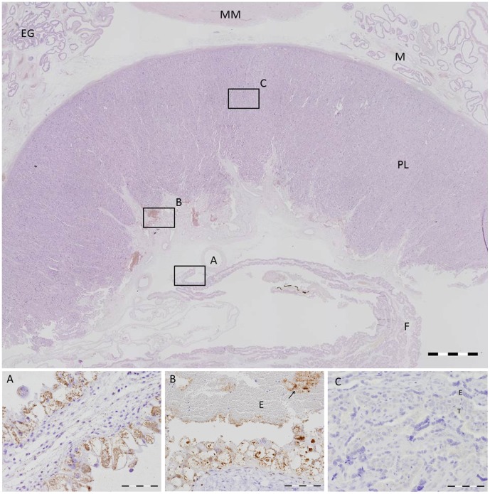
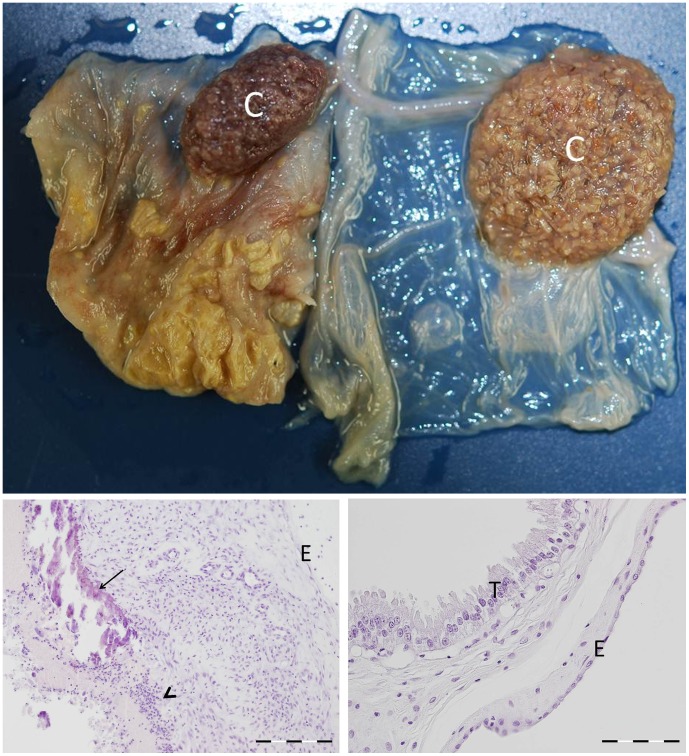
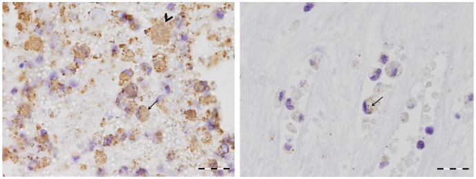
Coxiella burnetii is an intracellular bacterial pathogen that causes Q fever. Infected pregnant goats are a major source of human infection. However, the tissue dissemination and excretion pathway of the pathogen in goats are still poorly understood. To better understand Q fever pathogenesis, we inoculated groups of pregnant goats via the intranasal route with a recent Dutch outbreak C. burnetii isolate. Tissue dissemination and excretion of the pathogen were followed for up to 95 days after parturition. Goats were successfully infected via the intranasal route. PCR and immunohistochemistry showed strong tropism of C. burnetii towards the placenta at two to four weeks after inoculation. Bacterial replication seemed to occur predominantly in the trophoblasts of the placenta and not in other organs of goats and kids. The amount of C. burnetii DNA in the organs of goats and kids increased towards parturition. After parturition it decreased to undetectable levels: after 81 days post-parturition in goats and after 28 days post-parturition in kids. Infected goats gave birth to live or dead kids. High numbers of C. burnetii were excreted during abortion, but also during parturition of liveborn kids. C. burnetii was not detected in faeces or vaginal mucus before parturition. Our results are the first to demonstrate that pregnant goats can be infected via the intranasal route. C. burnetii has a strong tropism for the trophoblasts of the placenta and is not excreted before parturition; pathogen excretion occurs during birth of dead as well as healthy animals. Besides abortions, normal deliveries in C. burnetii-infected goats should be considered as a major zoonotic risk for Q fever in humans.


Conflict of interest statement

Competing Interests: The authors have declared that no competing interests exist.

Figures

Figure 1
Figure 1. Overview of the placentome of a Coxiella-inoculated goat.
Top: Scanned haematoxylin and eosin stained section of the placentome of a goat necropsied at 56 dpi (day 131 of pregnancy). No inflammatory reaction in the maternal endometrium (M), the placentome (PL) or the foetal allantochorion (F). MM = myometrium, EG = endometrial glands. Bar = 1 mm. Bottom: Higher magnification of areas A, B and C depicted by the rectangles in the overview. Serial section immunostained for the presence of Coxiella burnetii antigen (brownish colour). A. Foetal allantochorion showing severe swelling of the trophoblast cells caused by the formation of large intracytoplasmic vacuoles. The vacuoles are filled with numerous C. burnetii bacteria. Bar = 200 µm. B. Erythrophagous zone showing similar vacuolation and swelling of the trophoblasts at the base of the foetal villi. Numerous C. burnetii bacteria are seen within the vacuoles of the trophoblasts and in the blood-filled lacuna between the foetal and maternal epithelium (arrow). E = erythrocytes. Bar = 200 µm. C. Placentome. Absence of C. burnetii bacteria in the synepitheliochorial placenta. The maternal epithelium (E) and foetal trophoblasts (T) show no morphological alterations. Bar = 100 µm.
Figure 2
Figure 2. Comparison of a C. burnetii positive placenta and a C. burnetii negative placenta at parturition.
Top. Formalin-fixed allantochorion of an aborted kid (left) and the allantochorion of a kid from a control goat (right). The intercotyledonary allantochorion of the aborted kid is severely thickened and dull with a yellow-brownish exudate, while the cotyledon (C) shows no obvious macroscopic changes. The allantochorion of the control kid on the right is thin, glistening and transparent. Bottom. Left. Haematoxylin- and eosin-stained section of the intercotyledonary allantochorion of an aborted kid. Notice the severe inflammatory changes in the stroma of the allantochorion. The trophoblast layer is lost with dystrophic calcification of necrotic tissue (arrow) and a purulent exudate (arrowhead). Bar = 200 µm. Right. Histological appearance of a normal allantochorion of a kid from a control goat. The trophoblast layer is intact with a normal appearance of the trophoblast cells (T). Low cellularity in the stroma. E = endothelium of the allantochorion. Bar = 100 µm.
Figure 3
Figure 3. Close up of the allantochorion of an aborted kid due to C. burnetii infection.
The allantochorion of an aborted kid is immunostained to detect the presence of C. burnetii antigen. On the left: Numerous macrophages are present in the necropurulent exudate that covers the allantochorion. Both macrophages (arrow) and sloughed trophoblasts (arrow head) are filled with C. burnetii. On the right: macrophages in the stroma of the allantochorion have phagocytised C. burnetii bacteria (arrow). Bar = 20 µm.
Figure 4
Figure 4. Detection of C. burnetii DNA in the faeces of Coxiella-inoculated goats over time.
Detection of C. burnetii DNA in the faeces of four challenged goats of which a complete sampling sequence was present from inoculation until 119, 126, 140 and 141 days post inoculation (dpi). Goat ID 35: ⧫, 36: ▪, 38: ▴, 40: •. Faecal samples were taken at the indicated dpi and C. burnetii DNA was measured by PCR. Ct value of 40 is negative, Ct value <40 is positive. Parturition days are indicated as open symbols. Up until 42 dpi and on 56 dpi the four goats were negative. At 56 dpi three other goats of the group were positive before their parturition (data not shown). Data indicate that C. burnetii was detected in the faeces after parturition.
Figure 5
Figure 5. Detection of C. burnetii DNA in vaginal mucus of Coxiella-inoculated goats over time.
Detection of C. burnetii DNA in vaginal mucus of four challenged goats of which a complete sampling sequence was present from inoculation until 119, 126, 140 and 141 days post inoculation (dpi). Goat ID 35: ⧫, 36: ▪, 38: ▴, 40: •. Vaginal mucus samples were taken at the indicated dpi and C. burnetii DNA was measured by PCR. Ct value of 40 is negative, Ct value <40 is positive. Parturition days are indicated as open symbols. Until 42 dpi the four goats were negative. Data indicate that C. burnetii was detected in the vaginal mucus after the first parturition in the group.
Figure 6
Figure 6. Detection of C. burnetii DNA in milk of Coxiella-inoculated goats over time.
Detection of C. burnetii DNA in milk of four challenged goats of which a complete sampling sequence was present from inoculation until 119, 126, 140 and 141 days post inoculation (dpi). Goat ID 35: ⧫, 36: ▪, 38: ▴, 40: •. Milk samples were taken at the indicated days post-partum and C. burnetii DNA was measured by PCR. Ct value of 40 is negative, Ct value <40 is positive. After parturition C. burnetii DNA was detected in the milk until 32 days post-partum.
Figure 7
Figure 7. C. burnetii-specific PCR results of samples from the environment of Coxiella-inoculated goats.
C. burnetii PCR results of samples from the environment of the challenged goats measured at three time points after inoculation. ⧫: clean wood shavings stored in the facility, ▪: used wood shavings, ▴: swab from the slip mat, ×: swab from the air, ?: swab from the water, •: air filter. Ct value of 40 is negative, Ct value <40 is positive. Results indicate an environmental contamination of C. burnetii DNA at the end of the experiment.

Similar articles

Cited by

References

    1. Babudieri B (1959) Q fever: a zoonosis. Adv Vet Sci: 81–154.
    1. Greenslade E, Beasley R, Jennings L, Woodward A, Weinstein P (2003) Has Coxiella burnetii (Q fever) been introduced into New Zealand? Emerg Infect Dis 9: 138–140. - PMC - PubMed
    1. Oyston PC, Davies C (2011) Q fever: the neglected biothreat agent. J Med Microbiol 60: 9–21. - PubMed
    1. Raoult D, Marrie TJ, Mege JL (2005) Natural history and pathophysiology of Q fever. Lancet Infect Dis 5: 219–226. - PubMed
    1. Angelakis E, Raoult D (2010) Q Fever. Vet Microbiol 140: 297–309. - PubMed

Publication types

MeSH terms