. 2012;7(11):e49022.
doi: 10.1371/journal.pone.0049022. Epub 2012 Nov 12.

IL-12p40 deficiency leads to uncontrolled Trypanosoma cruzi dissemination in the spinal cord resulting in neuronal death and motor dysfunction

Affiliations

IL-12p40 deficiency leads to uncontrolled Trypanosoma cruzi dissemination in the spinal cord resulting in neuronal death and motor dysfunction

André Luis Bombeiro et al. PLoS One. 2012.

Abstract

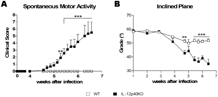
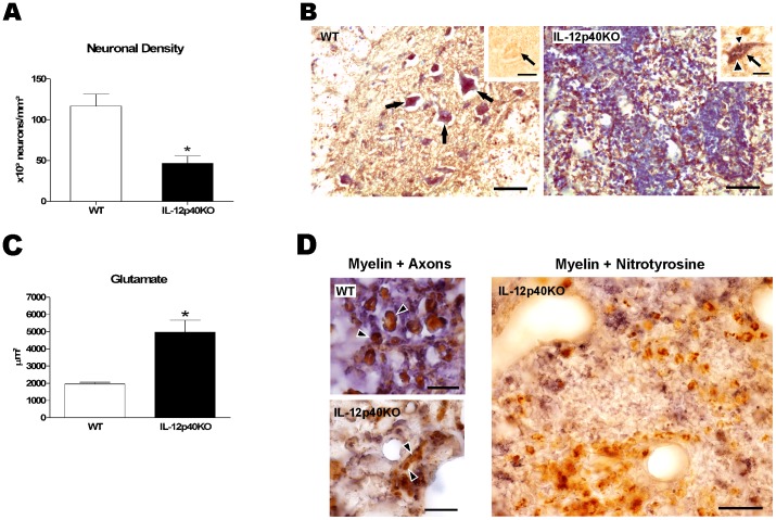
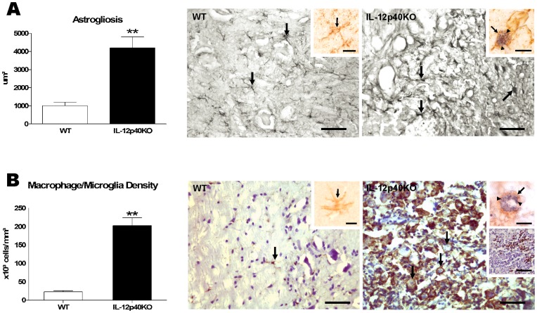
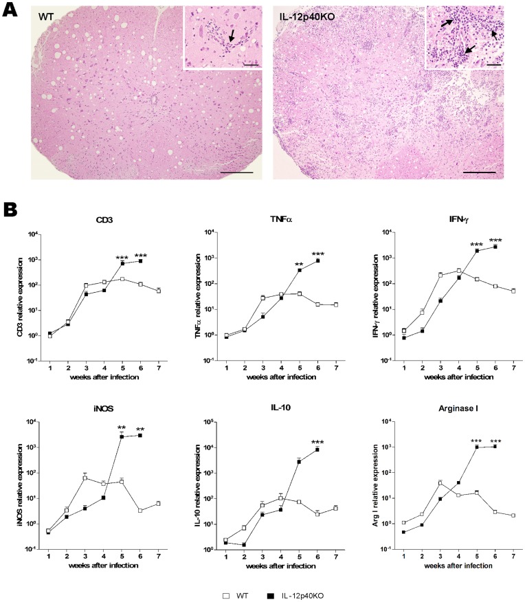
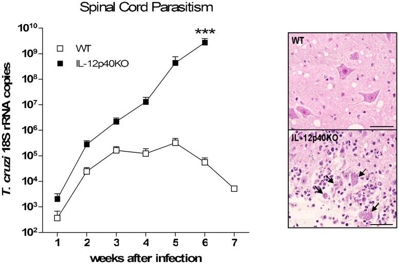
Chagas' disease is a protozoosis caused by Trypanosoma cruzi that frequently shows severe chronic clinical complications of the heart or digestive system. Neurological disorders due to T. cruzi infection are also described in children and immunosuppressed hosts. We have previously reported that IL-12p40 knockout (KO) mice infected with the T. cruzi strain Sylvio X10/4 develop spinal cord neurodegenerative disease. Here, we further characterized neuropathology, parasite burden and inflammatory component associated to the fatal neurological disorder occurring in this mouse model. Forelimb paralysis in infected IL-12p40KO mice was associated with 60% (p<0.05) decrease in spinal cord neuronal density, glutamate accumulation (153%, p<0.05) and strong demyelization in lesion areas, mostly in those showing heavy protein nitrosylation, all denoting a neurotoxic degenerative profile. Quantification of T. cruzi 18S rRNA showed that parasite burden was controlled in the spinal cord of WT mice, decreasing from the fifth week after infection, but progressive parasite dissemination was observed in IL-12p40KO cords concurrent with significant accumulation of the astrocytic marker GFAP (317.0%, p<0.01) and 8-fold increase in macrophages/microglia (p<0.01), 36.3% (p<0.01) of which were infected. Similarly, mRNA levels for CD3, TNF-α, IFN-γ, iNOS, IL-10 and arginase I declined in WT spinal cords about the fourth or fifth week after infection, but kept increasing in IL-12p40KO mice. Interestingly, compared to WT tissue, lower mRNA levels for IFN-γ were observed in the IL-12p40KO spinal cords up to the fourth week of infection. Together the data suggest that impairments of parasite clearance mechanisms in IL-12p40KO mice elicit prolonged spinal cord inflammation that in turn leads to irreversible neurodegenerative lesions.


Conflict of interest statement

Competing Interests: The authors have declared that no competing interests exist.

Figures

Figure 1
Figure 1. Neurological scoring after T. cruzi infection.
A) T. cruzi Sylvio X10/4-infected WT (n = 14) and IL-12p40KO (n = 14) mice were placed in an open field, evaluated by eye for spontaneous motor activity in a timed course and scored according to Table 1 (from 0, no paralysis, to 8, complete forelimb paralysis). While WT mice presented no sign of paralysis, IL-12p40KO mice displayed an ascending paralysis from the tail to the forelimbs. B) Infected mice were submitted to the inclined plane test (WT, n = 6; IL-12p40KO, n = 8) to assess motor function, wherein healthy mice are able to stay on a plane with a slope varying from 50 to 60 degrees. Data show that IL-12p40KO mice manifested impaired motor function, whereas WT mice did not. Mean ± SEM. **p<0.01; ***p<0.001 when comparing paired WT and IL-12p40KO groups according to Bonferroni post-tests.
Figure 2
Figure 2. Neurodegenerative aspects of IL-12p40KO spinal cords.
A) Paralyzed T. cruzi-infected IL-12p40KO mice exhibited a decreased neuronal density in the spinal cord, when compared to the WT counterparts. B) Intact WT spinal cord section (left) with healthy neurons (arrows) and a morphologically disrupted IL-12p40KO spinal cord section (right), which is rich in infiltrating cells. Sections were immunolabeled for NF-200 (a neuronal marker, in brown) and counterstained with Giemsa (for total nuclei, in blue). T. cruzi amastigote forms were not seen in the WT neurons (left panel inset, arrow points a neuron, in brown) but occurred in a few IL-12p40KO neurons (right panel inset, arrow points a neuron, in brown, and the arrowheads point amastigotes, in purple). Scale bars: left and right pictures, 50 µm; insets, 20 µm. C) Glutamate immunolabeling area was higher in the spinal cords of paralyzed T. cruzi-infected IL-12p40KO mice than in the paired WT ones. D) Myelin sheaths remained intact in infected WT spinal cords in the end-point of the infection (upper left picture, arrow-heads show myelin wrapped axons), while they were disrupted in the IL-12p40KO damaged tissue (lower left picture, arrow-heads show myelin-free axons). In the right picture, note that in the presence of nitrotyrosine (brown), little myelin debris (blue) is observed in the damaged IL-12p40KO tissue. Sections were double-immunolabeled to myelin (purple-blue) and neuronal soma and axons (brown, left pictures) or nitrotyrosine (brown, right picture). Scale bars: left smaller pictures, 10 µm; right picture, 100 µm. Data on both graphs are presented as the mean ± SEM. *p<0.05, according to the unpaired t-test.
Figure 3
Figure 3. Astrogliosis and increased macrophage/microglia density in IL-12p40KO spinal cords.
Paralyzed infected IL-12p40KO mice presented higher immunolabeled area to the astrocytic marker GFAP (A) and increased density of macrophages/microglia (B) in the spinal cord when compared to the matched infected WT group. Pictures in Figure A show astrocytes (arrows) in immunocompetent (left panel) and IL-12p40KO (right panel) spinal cords. No T. cruzi-infected astrocyte was found in WT tissue (left panel inset, arrow points an astrocyte), whereas in IL-12p40KO tissue, only a few astrocytes containing T. cruzi amastigotes were seen (right panel inset, arrow points an astrocyte and arrowheads point parasites). Pictures in Figure B show microglial cell (arrow) with long thin processes in WT tissue (left panel) and round-shaped phagocytes (arrows point macrophages or microglia) in a lesion area of an IL-12p40KO spinal cord section (right panel). While no parasite was found in the WT tissue (right panel inset, arrow shows a microglia), more than a third of all IL-12p40KO macrophages/microglias were infected (right panel upper inset, arrow shows a macrophage/microglia and arrowheads point parasites). M2 cells were visualized in the IL-12p40-KO spinal cord only (right panel lower inset, M2 cells in brown). Sections were immunolabeled to GFAP (A) and CD11b (B, also counterstained with Giemsa), GFAP and T. cruzi (A, inset), CD11b and T. cruzi (B, upper insets) or arginase I (B, right panel lower inset, also counterstained with hematoxylin). Scale bars: A and B, 50 µm; A insets and B upper insets, 20 µm; B right panel lower inset, 50 µm. Data on both graphs are presented as the mean ± SEM. **p<0.01, according to the unpaired t-test.
Figure 4
Figure 4. Spinal cord inflammation.
A) After WT and IL-12p40KO mouse transcardiac perfusion, spinal cord upper lumbar intumescences were submitted to histopathological analysis. While WT spinal cord displayed preserved morphology (left picture), with few inflammatory cells (inset, arrow), IL-12p40KO spinal cord presented intense morphological disarrangement (right picture), due to the large amount of inflammatory foci (inset, arrows). Representative pictures of HE-stained spinal cord sections of WT and IL-12p40KO mice in the sixth week after infection. Scale bars: 100 µm; insets, 20 µm. B) Once a week during seven weeks, cDNA was obtained from the spinal cord lower lumbar segments of WT and IL-12p40KO mice and submitted to real time RT-PCR procedures, using primers for CD3, TNF-α, IFN-γ, iNOS, IL-10, arginase I and HPRT, β-actin and GAPDH as endogenous control. After the fourth week of infection, the transcription ratio of all genes analyzed tended to remission in the WT tissue, while they continued increasing in the IL-12p40KO spinal cord. Of note, also around the fourth week of infection, a switch in the transcription level of the analyzed genes was observed between the two mouse strains, when IL-12p40KO mice started expressing more RNA than WT ones. Mean ± SEM. **p<0.01; ***p<0.001 when comparing paired WT and IL-12p40KO groups according to Bonferroni post-tests. n = 4 for each strain in each time point.
Figure 5
Figure 5. Spinal cord parasitism.
After transcardiac perfusion, spinal cord lower lumbar segments were processed for RNA extraction and cDNA production. Real time RT-PCR was performed using primers for T. cruzi 18S rRNA and HPRT as endogenous control. For the absolute quantification, sample values were fitted into a standard curve, constructed from a pattern sample with known number of copies of T. cruzi 18S rRNA. IL-12p40KO mice presented an ascending parasitism while WT ones managed to constrain the infection by the third week, thus decreasing the parasitic load after the fifth week. Representative pictures of HE-stained upper lumbar spinal cord sections, obtained from WT (upper square) and IL-12p40KO mice (lower square) in the sixth week after the infection. Note the high amount of amastigote nests (arrows) in the IL-12p40KO tissue. Scale bars: 20 µm. Mean ± SEM. ***p<0.001 when comparing paired WT and IL-12p40KO groups according to Bonferroni post-tests. n = 4 for each strain in each time point.

Similar articles

Cited by

References

    1. Morel CM, Lazdins J (2003) Chagas disease. Nat Rev Microbiol 1: 14–15. - PubMed
    1. Rassi A Jr, Rassi A, Marin-Neto JA (2010) Chagas disease. Lancet 375: 1388–1402. - PubMed
    1. Junqueira C, Caetano B, Bartholomeu DC, Melo MB, Ropert C, et al. (2010) The endless race between Trypanosoma cruzi and host immunity: lessons for and beyond Chagas disease. Expert Rev Mol Med 12: e29. - PubMed
    1. Benitez-Hernandez I, Mendez-Enriquez E, Ostoa P, Fortoul T, Ramirez JA, et al. (2010) Proteolytic cleavage of chemokines by Trypanosoma cruzi’s cruzipain inhibits chemokine functions by promoting the generation of antagonists. Immunobiology 215: 413–426. - PubMed
    1. Erdmann H, Steeg C, Koch-Nolte F, Fleischer B, Jacobs T (2009) Sialylated ligands on pathogenic Trypanosoma cruzi interact with Siglec-E (sialic acid-binding Ig-like lectin-E). Cell Microbiol 11. - PubMed

Publication types

MeSH terms

Substances