. 2012;7(11):e49323.
doi: 10.1371/journal.pone.0049323. Epub 2012 Nov 13.

A Baculovirus immediate-early gene, ie1, promoter drives efficient expression of a transgene in both Drosophila melanogaster and Bombyx mori

Affiliations

A Baculovirus immediate-early gene, ie1, promoter drives efficient expression of a transgene in both Drosophila melanogaster and Bombyx mori

Mika Masumoto et al. PLoS One. 2012.

Abstract

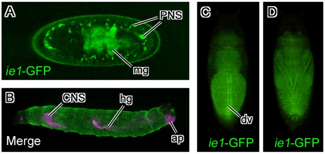
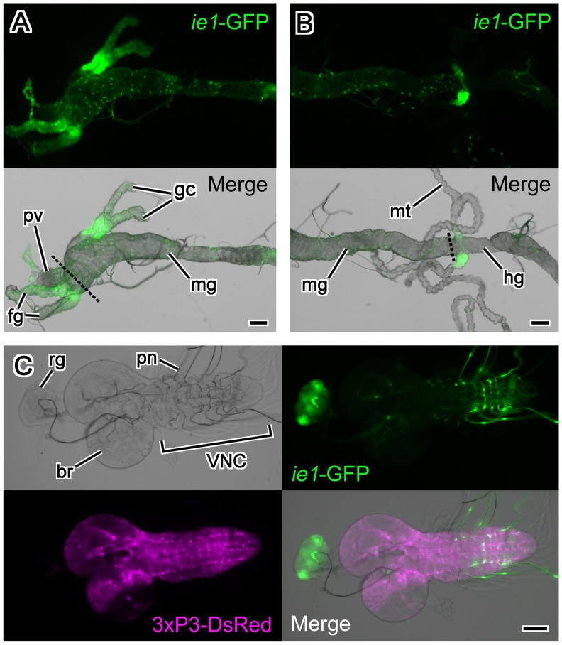
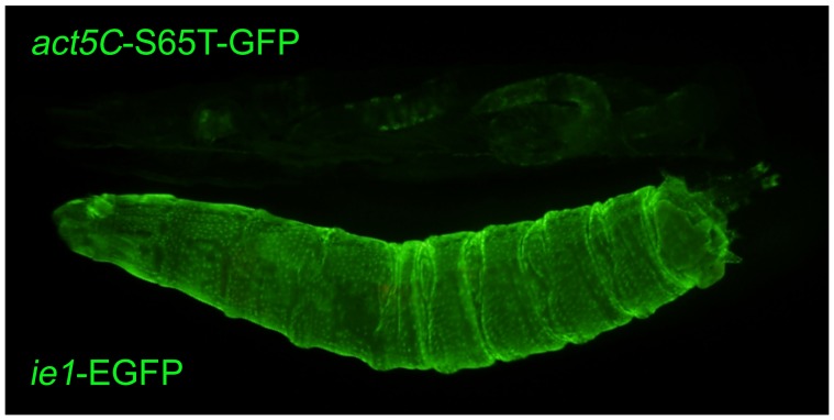
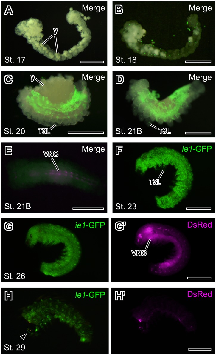
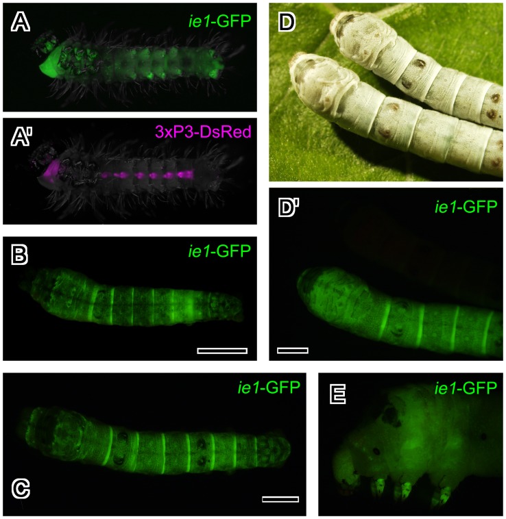
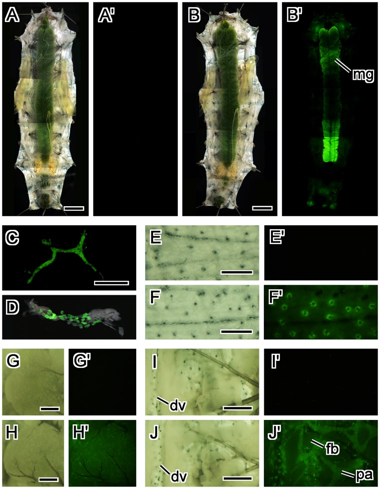
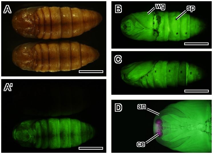
Many promoters have been used to drive expression of heterologous transgenes in insects. One major obstacle in the study of non-model insects is the dearth of useful promoters for analysis of gene function. Here, we investigated whether the promoter of the immediate-early gene, ie1, from the Bombyx mori nucleopolyhedrovirus (BmNPV) could be used to drive efficient transgene expression in a wide variety of insects. We used a piggyBac-based vector with a 3xP3-DsRed transformation marker to generate a reporter construct; this construct was used to determine the expression patterns driven by the BmNPV ie1 promoter; we performed a detailed investigation of the promoter in transgene expression pattern in Drosophila melanogaster and in B. mori. Drosophila and Bombyx belong to different insect orders (Diptera and Lepidoptera, respectively); however, and to our surprise, ie1 promoter-driven expression was evident in several tissues (e.g., prothoracic gland, midgut, and tracheole) in both insects. Furthermore, in both species, the ie1 promoter drove expression of the reporter gene from a relatively early embryonic stage, and strong ubiquitous ie1 promoter-driven expression continued throughout the larval, pupal, and adult stages by surface observation. Therefore, we suggest that the ie1 promoter can be used as an efficient expression driver in a diverse range of insect species.


Conflict of interest statement

Competing Interests: The authors have declared that no competing interests exist.

Figures

Figure 1
Figure 1. Schematic representation of the piggyBac-based BmNPV ie1 promoter reporter constructs.
This construct was designated pBac[BmNPV ie1-EGFP, 3xP3-DsRed]. A fragment containing the sequences –631 to –2 bp upstream of the codon encoding the translational start site of BmNPV ie1 was used as the BmNPV ie1 promoter and to drive expression of EGFP. DsRed was under the control of 3xP3 and was used as the transformation marker. Abbreviations: ITR, inverted terminal repeats of piggyBac; hsp70 polyA, hsp70 polyadenylation signal; SV40 polyA, SV40 polyadenylation signal.
Figure 2
Figure 2. Genomic DNA sequences surrounding piggyBac insertions.
The flanking sequences of piggyBac insertion in D. melanogaster and B. mori have 100% identity with the genome DNA sequences of chromosome 3L and Bm_scaf 21 in chromosome 17, respectively. Abbreviations: Dm, Drosophila melanogaster; Bm, Bombyx mori.
Figure 3
Figure 3. Expression pattern of the BmNPV ie1-EGFP transgene in fruit flies.
(A) Embryos at stage 16. EGFP fluorescence was primarily evident in midgut and peripheral nervous system. (B) Merged image of BmNPV ie1 promoter-driven EGFP expression and 3xP3-driven DsRed expression in a fruit fly larva. Strong EGFP expression was evident throughout the whole body. (C, D) BmNPV ie1 promoter-driven EGFP expression in a fruit fly pupa. EGFP expression was evident throughout the whole body, and strong punctate expression was evident inside the body. (C) Dorsal view. Intense EGFP fluorescence was evident along the dorsal vessel. (D) Ventral view. In (A) and (B), anterior is to the left. In (C) and (D), anterior is up. Abbreviations: ap, anal plate; CNS, central nervous system; dv, dorsal vessel; hg, hindgut; mg, midgut; PNS, peripheral nervous system.
Figure 4
Figure 4. Expression pattern of the BmNPV ie1-EGFP transgene in tissues of a 3rd instar fly larva.
(A) Foregut and midgut. EGFP expression was evident in the foregut and the gastric caecum. The dashed line indicates the boundary between the foregut and midgut. (B) Midgut and hindgut. EGFP expression was evident in the section of the Malpighian tubule that was attached to hindgut. The dashed line indicates the boundary between the midgut and hindgut. (A, B) Upper panels show EGFP fluorescence while lower panels show merged images of the transmitted light and EGFP fluorescent. EGFP was also detected along tracheoles that attached to the midgut. (C) Central nervous system and ring gland. EGFP expression was strong in the prothoracic gland region of the larval ring gland. EGFP signals were detected in the peripheral nerves emanating from the abdominal neuromere. DsRed driven by the 3xP3 promoter was expressed in the ventral nerve cord and throughout the brain except within the optic lobe. The lower right panel shows the merged images of transmitted light, EGFP fluorescence, and DsRed fluorescence. In all panels, anterior is to the left. Abbreviations: br, brain; fg, foregut; gc, gastric caecum; hg, hindgut; mg, midgut; mt, Malpighian tubule; pn, peripheral nerves; pv, proventriculus; rg, ring gland; VNC, ventral nerve cord. Scale bars = 100 µm.
Figure 5
Figure 5. Comparison of the expressions driven by the D. melanogaster actin5C and the BmNPV ie1 promoter.
The D. melanogaster actin5C promoter drove expression of S65T-GFP (upper larva), while the BmNPV ie1 promoter drove expression of EGFP (lower larva). These two larvae were examined side by side within the same field of view. Anterior is to the left.
Figure 6
Figure 6. Expression pattern of the BmNPV ie1-EGFP transgene in silkworm embryos.
(A–D) Merged images of EGFP fluorescence and epi-illumination. (A) Stage 17 embryo. EGFP expression driven by the ie1 promoter was not evident in stage 17 embryos. Most yolk had been removed. (B) Stage 18 embryo. EGFP expression was evident in some yolk cells at this stage. (C) Stage 20 embryo (dorsal closure has not complete). EGFP expression was detected in the lateral and dorsal region. (D) Stage 21B embryo. EGFP fluorescence extended to ventral region. Most yolk had been removed. (E) Ventral view of a stage 21B embryo. Merged images of DsRed fluorescence and epi-illumination. The DsRed transformation marker was evident in the developing ventral nerve cord from this stage. (F) Stage 23 embryo (dorsal closure has completed). EGFP expression was detected throughout the whole embryo at this stage. (G, G’) Stage 26 embryo. (H, H’) Stage 29 embryo. EGFP and DsRed fluorescences were obscured by the pigmentation in the cuticle. In all panels, anterior is to the left. All panels except the panel E present a lateral view. Abbreviations: T3L, the 3rd thoracic leg; VNC, ventral nerve cord; y, yolk. Arrowhead indicates stemma. Scale bars = 500 µm.
Figure 7
Figure 7. Expression pattern of the BmNPV ie1-EGFP transgene in silkworm larva at different stages.
(A, A’) First instar larva just after hatching. BmNPV ie1 promoter-driven EGFP expression did not overlap with 3xP3-driven DsRed expression in the ventral nerve cord. Ventral view. (B) Late 2nd instar larva. EGFP was expressed throughout the whole body and throughout all larval stages. Dorsal view. (C) Early 3rd instar larva. Dorsal view. (D, D’) 4th instar larvae. Dorsal view. Upper larva is a non-transgenic larva. (E) Head and thorax of 5th instar larva. Lateral view. Scale bars = 2 mm.
Figure 8
Figure 8. Expression pattern of the BmNPV ie1-EGFP transgene in tissues dissected from 5th instar silkworm larvae.
(A, A’) A dissected non-transgenic 5th instar larva. (B, B’) A dissected transgenic 5th instar larva. Intense EGFP fluorescence was evident in the anterior and posterior midgut. (C–J’) A dissected 5th instar larva. (C) Prothoracic gland. (D) Merged image of the transmitted light and EGFP fluorescence in the suboesophageal body. (E–F’) Trichogen (or trichogen and tormogen) cells in the epidermis in a non-transgenic larva (E, E’) and a transgenic larva (F, F’). (G–H’) Ovary of a non-transgenic larva (G, G’) and a transgenic larva (H, H’). EGFP was evident in tracheolar cells that were attached to the ovary. (I–J’) Tissues surrounding dorsal vessel of a non-transgenic larva (I, I’) and a transgenic larva (J, J’). EGFP was evident in pericardial cells along dorsal vessel and on the alary muscle, and peritracheal athrocytes, but not in fatbody. (A, B, E–J) White light, (A’, B’, C, E’-J’) EGFP-excitation wavelength light. The images for the comparisons of non-transgenic and transgenic larvae and tissues were obtained exactly by the same conditions. Abbreviations: dv, dorsal vessel; fb, fatbody; mg, midgut; pa, peritracheal athrocytes. Scale bars = 5 mm in (A) and (B), 500 µm in (C, E, F, G, H), 1 mm in (I, J).
Figure 9
Figure 9. Expression pattern of the BmNPV ie1-EGFP transgene in silkworm pupa.
(A–C) Two-day old pupa. EGFP expression was evident throughout the pupal body. (A, A’) Dorsal views under white light (A) and EGFP-excitation wavelength light (A’). Upper pupa is non-transgenic pupa. (B) Ventral view. (C) Lateral view. (D) A ventral view of the head and thorax of 4-day old pupa. DsRed expression was evident in the compound eyes, whereas EGFP was not. Abbreviations: an, antenna; ce, compound eye; sp, spiracle; wg, wing. Scale bar = 5 mm.

References

    1. Horn C, Schmid BGM, Pogoda FS, Wimmer EA (2002) Fluorescent transformation markers for insect transgenesis. Insect Biochem Mol Biol 32: 1221–1235. - PubMed
    1. Berghammer AJ, Klingler M, Wimmer EA (1999) A universal marker for transgenic insects. Nature 402: 370–371. - PubMed
    1. Thomas JL, Da Rocha M, Besse A, Mauchamp B, Chavancy G (2002) 3×P3-EGFP marker facilitates screening for transgenic silkworm Bombyx mori L. from the embryonic stage onwards. Insect Biochem Mol Biol 32: 247–253. - PubMed
    1. Tamura T, Thibert C, Royer C, Kanda T, Abraham E, et al. (2000) Germline transformation of the silkworm Bombyx mori L. using a piggyBac transposon-derived vector. Nat Biotechnol 18: 81–84. - PubMed
    1. Peloquin JJ, Thibault ST, Staten R, Miller TA (2000) Germ-line transformation of pink bollworm (Lepidoptera: Gelechiidae) mediated by the piggyBac transposable element. Insect Mol Biol 9: 323–333. - PubMed

Publication types