. 2012;7(11):e49496.
doi: 10.1371/journal.pone.0049496. Epub 2012 Nov 13.

17-AAG kills intracellular Leishmania amazonensis while reducing inflammatory responses in infected macrophages

Affiliations

17-AAG kills intracellular Leishmania amazonensis while reducing inflammatory responses in infected macrophages

Antonio Luis de Oliveira Almeida Petersen et al. PLoS One. 2012.

Abstract

Background: Leishmaniasis is a neglected endemic disease with a broad spectrum of clinical manifestations. Pentavalent antimonials have been the treatment of choice for the past 70 years and, due to the emergence of resistant cases, the efficacy of these drugs has come under scrutiny. Second-line drugs are less efficacious, cause a range of side effects and can be costly. The formulation of new generations of drugs, especially in developing countries, has become mandatory.

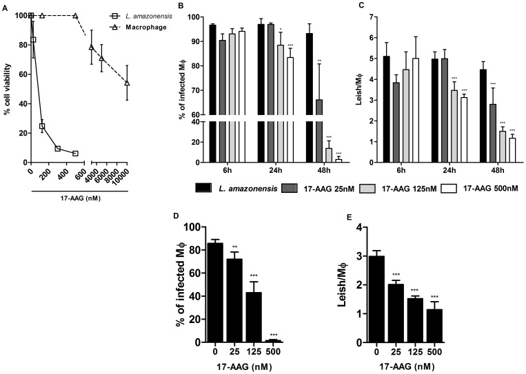
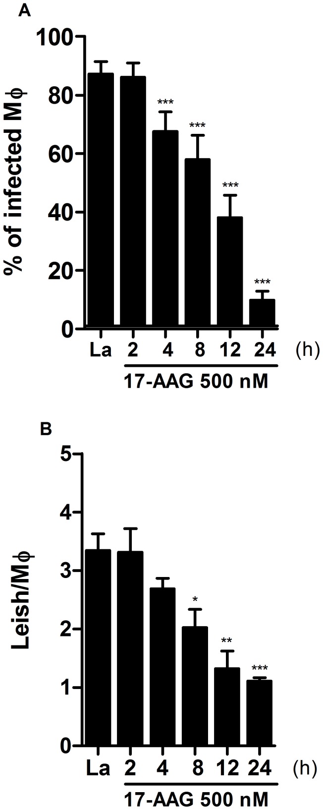
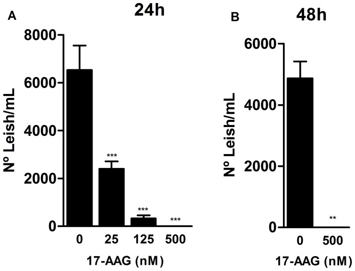
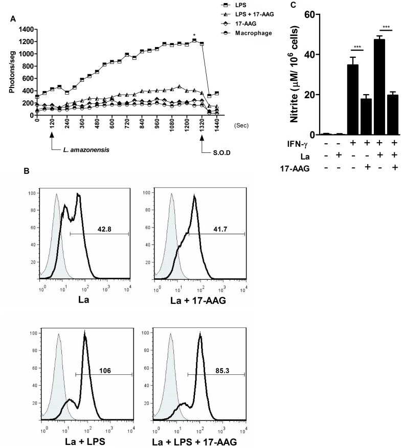
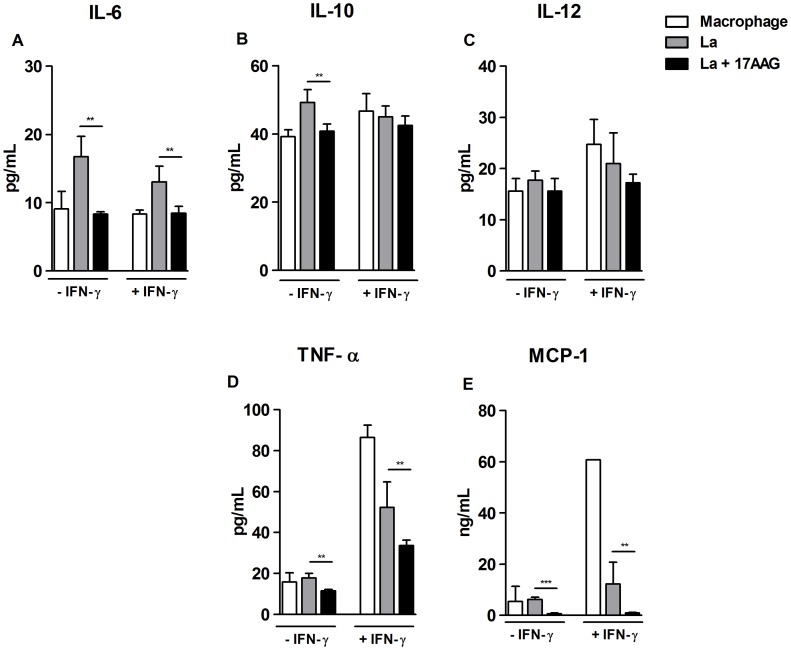
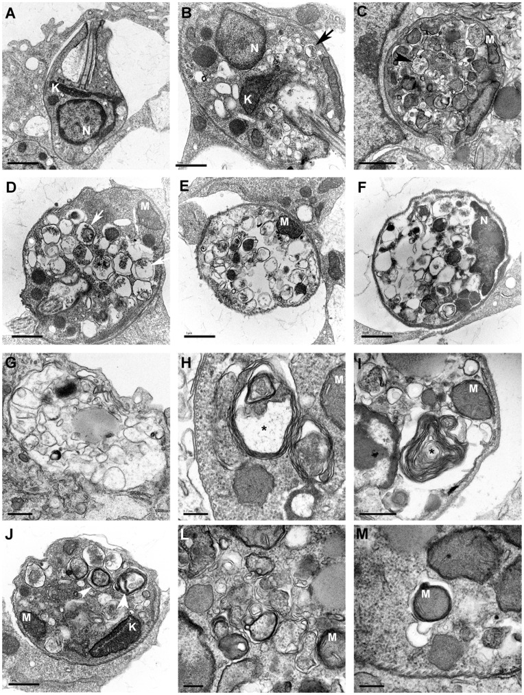
Methodology/principal findings: We investigated the anti-leishmanial effect of 17-(allylamino)-17-demethoxygeldanamycin (17-AAG), an HSP90 inhibitor, in vitro. This inhibitor is currently in clinical trials for cancer treatment; however, its effects against intracellular Leishmania remain untested. Macrophages infected with L. amazonensis were treated with 17-AAG (25-500 nM) and parasite load was quantified using optical microscopy. Parasite load declined in 17-AAG-treated macrophages in a dose- and time-dependent manner. Intracellular parasite death became irreversible after 4 h of treatment with 17-AAG, and occurred independent of nitric oxide (NO) and superoxide (O(2) (-)) production. Additionally, intracellular parasite viability was severely reduced after 48 h of treatment. Interestingly, treatment with 17-AAG reduced pro-inflammatory mediator production, including TNF-α, IL-6 and MCP-1, yet IL-12 remained unaffected. Electron microscopy revealed morphological alterations, such as double-membrane vacuoles and myelin figures at 24 and 48 h after 17-AAG treatment.

Conclusions/significance: The HSP90 inhibitor, 17-AAG, possesses high potency under low dosage and reduces both pro-inflammatory and oxidative molecule production. Therefore, further studies are warranted to investigate this inhibitor's potential in the development of new generations of anti-leishmanials.


Conflict of interest statement

Competing Interests: The authors have declared that no competing interests exist.

Figures

Figure 1
Figure 1. Inhibition of axenic growth of Leishmania and reduction in parasite load by 17-AAG.
(A) Axenic promastigotes were exposed to several concentrations of 17-AAG (25, 125, 500 nM) for 48 h and the number of viable parasites was assessed as described in Materials and Methods. Data are presented as the percentage inhibition of parasite growth related to untreated controls (4,754×107). Bars represent means ± SD of one representative out of two experiments performed in sextuplicate (one-way ANOVA, Dunnett’s Multiple Comparison Test, ***p<0.0001, post-test for linear trend, p<0.0001). (B, C) Drug effects at early times after infection. Following 6 h of incubation with parasites, macrophage cultures were treated for 6, 24 and 48 h with specific concentrations of 17-AAG (25, 125, 500 nM); (D, E) Drug effects at later stages after infection. Following 6 h of incubation with parasites, macrophage cultures were reincubated for additional 48 h then submitted to treatment with specific concentrations of 17-AAG (25, 125, 500 nM). Bars represent means ± SD of one representative experiment out of two performed in sextuplicate (one-way ANOVA, Dunnett’s Multiple Comparison Test, *p<0.05, **p<0.001, ***p<0.0001, post-test for linear trend, p<0.0001).
Figure 2
Figure 2. Irreversibility of treatment with 17-AAG on intracellular Leishmania.
To assessment the reversibility of parasite growth inhibition by treatment with 17-AAG parasite load was determined by quantifying the percentage of infected macrophages (A) and the number of parasites per macrophage (B) as described in Materials and Methods. Bars represent means ± SD of one representative experiment out of two performed in sextuplicate (one-way ANOVA, ***p<0.0001, Dunnett’s Multiple Comparison Test, *p<0.05, **p<0.001, ***p<0.0001, post-test for linear trend, p<0.0001).
Figure 3
Figure 3. Reduction of parasite intracellular viability by 17-AAG.
Treatment’s effect on parasite viability was assessed after 24 h (A) and 48 h (B) of infection, as described in Materials and Methods. Bars represent means ± SD of one representative experiment out of two performed in sextuplicate (one-way ANOVA, ***p<0.0001, Dunnett’s Multiple Comparison Test, *p<0.05, **p<0.001, ***p<0.0001, post-test for linear trend, p<0.0001; Mann Whitney test, **p<0.001).
Figure 4
Figure 4. Reduced O2
and NO production in macrophage cultures treated with 17-AAG. (A) O2 production was measured at early stages of infection using a lucigenin-derived CL method. Points on the graph represent photon emissions per second by macrophage cultures 2 min prior to the addition of L. amazonensis promastigotes, as well as throughout the incubation period of 20 min. Data are derived from one representative experiment out of four performed in uniplicate (Mann Whitney test, p = 0.028); (B) Intracellular O2 production was assessed by determining cell fluorescence in the presence of hydroethidine (5 µM) and expressed as MFI using a flow cytometer. The histogram overlay depicts the MFI of hydroethidine-labeled cells (solid lines) in comparison to unlabeled control cells (shaded areas). Data are derived from one representative experiment out of three performed in uniplicate (Mann Whitney test, p = 1). (C) NO production was measured by detecting nitrite in the supernatants of 17-AAG-treated cells, as described in Materials and Methods. Bars represent NO production measurement expressed as means ± SD of one representative experiment out of four performed in triplicate or more (Student’s t test, ***p<0.0001).
Figure 5
Figure 5. Modulation of mediator production by treatment with 17-AAG.
Mediators released by 17-AAG-treated cells were measured in cell supernatants using an inflammatory CBAKit, as described in Materials and Methods: (A) IL-6; (B) IL-10; (C) IL-12; (D) TNF-α; (E) MCP-1. Bars represent means ± SD of a single experiment performed in sextuplicate (Student’s t-test and Mann-Whitney, **p<0.001, ***p<0.0001).
Figure 6
Figure 6. Alterations suggestive of autophagy in intracellular parasites treated with 17-AAG.
Transmission electron microscopy was used to investigate ultrastructural morphological alterations in intracellular parasites inside 17-AAG-treated macrophages. (A) Control infected macrophages. After 12 h of treatment, several morphological alterations were seen in intracellular parasites, including: (B) numerous small vesicles some containing cytoplasmic material inside (black arrow), (C) vacuoles larger in size (black arrow-head), (D) with intravacuolar materials degraded (white arrow). After 24 h of treatment, the intracellular parasites presented a large number of vesicles occupying most of the cytoplasm containing well-preserved nuclei, mitochondria, and subpellicular microtubules (E–F). After 48 h of treatment, no preserved parasites inside cells were observed, yet empty vesicles, and membrane-bounded structures with an electron-density similar to parasite cytosol in parasitophorous vacuoles were seen (G). At 12 and 24 h after treatment, several alterations were also visible in parasite cytoplasm, including myelin figures (*) (H–I), vesicles with double-layered membranes (white arrow-head) (J–M), and portions of mitochondria inside membrane-bounded structures (M). The nuclei (N) and mitochondria (M) and kinetoplast (K) remained intact in all groups.

References

    1. Desjeux P (2004) Leishmaniasis: current situation and new perspectives. Comp Immunol Microbiol Infect Dis 27: 305–318. - PubMed
    1. Kedzierski L, Zhu Y, Handman E (2006) Leishmania vaccines: progress and problems. Parasitology 133 Suppl: S87–112 - PubMed
    1. Croft SL, Coombs GH (2003) Leishmaniasis–current chemotherapy and recent advances in the search for novel drugs. Trends Parasitol 19: 502–508. - PubMed
    1. Tiuman TS, Santos AO, Ueda-Nakamura T, Filho BP, Nakamura CV (2011) Recent advances in leishmaniasis treatment. Int J Infect Dis 15: e525–532. - PubMed
    1. Croft SL, Sundar S, Fairlamb AH (2006) Drug resistance in leishmaniasis. Clin Microbiol Rev 19: 111–126. - PMC - PubMed

Publication types

MeSH terms