

General anesthesia and human brain connectivity
- PMID: 23153273
- PMCID: PMC3621592
- DOI: 10.1089/brain.2012.0107
General anesthesia and human brain connectivity
Abstract
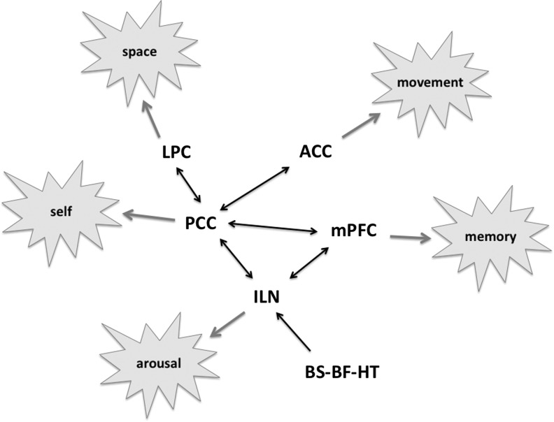
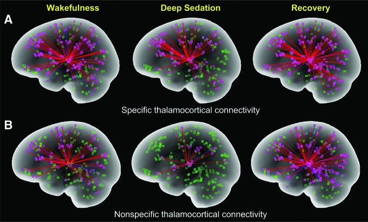
General anesthesia consists of amnesia, hypnosis, analgesia, and areflexia. Of these, the mechanism of hypnosis, or loss of consciousness, has been the most elusive, yet a fascinating problem. How anesthetic agents suppress human consciousness has been investigated with neuroimaging for two decades. Anesthetics substantially reduce the global cerebral metabolic rate and blood flow with a degree of regional heterogeneity characteristic to the anesthetic agent. The thalamus appears to be a common site of modulation by several anesthetics, but this may be secondary to cortical effects. Stimulus-dependent brain activation is preserved in primary sensory areas, suggesting that unconsciousness cannot be explained by cortical deafferentation or a diminution of cortical sensory reactivity. The effect of general anesthetics in functional and effective connectivity is varied depending on the agent, dose, and network studied. At an anesthetic depth characterized by the subjects' unresponsiveness, a partial, but not complete, reduction in connectivity is generally observed. Functional connectivity of the frontoparietal association cortex is often reduced, but a causal role of this change for the loss of consciousness remains uncertain. Functional connectivity of the nonspecific (intralaminar) thalamic nuclei is preferentially reduced by propofol. Higher-order thalamocortical connectivity is also reduced with certain anesthetics. The changes in functional connectivity during anesthesia induction and emergence do not mirror each other; the recovery from anesthesia may involve increases in functional connectivity above the normal wakeful baseline. Anesthetic loss of consciousness is not a block of corticofugal information transfer, but a disruption of higher-order cortical information integration. The prime candidates for functional networks of the forebrain that play a critical role in maintaining the state of consciousness are those based on the posterior parietal-cingulate-precuneus region and the nonspecific thalamus.
Figures
References
-
- Alkire MT. Probing the mind: anesthesia and neuroimaging. Clin Pharmacol Ther. 2008;84:149–152. - PubMed
-
- Alkire MT. Haier RJ. Barker SJ. Shah NK. Wu JC. Kao YJ. Cerebral metabolism during propofol anesthesia in humans studied with positron emission tomography. Anesthesiology. 1995;82:393–403. discussion 327A. - PubMed
-
- Alkire MT. Haier RJ. Fallon JH. Toward a unified theory of narcosis: brain imaging evidence for a thalamocortical switch as the neurophysiologic basis of anesthetic- induced unconsciousness. Conscious Cogn. 2000;9:370–386. - PubMed
-
- Alkire MT. Haier RJ. Shah NK. Anderson CT. Positron emission tomography study of regional cerebral metabolism in humans during isoflurane anesthesia. Anesthesiology. 1997;86:549–557. - PubMed
Publication types
MeSH terms
Substances
LinkOut - more resources
Full Text Sources
Medical
