Review
. 2013 Mar;24(3):137-44.
doi: 10.1016/j.tem.2012.10.002. Epub 2012 Nov 13.

Inflammation and diabetes-accelerated atherosclerosis: myeloid cell mediators

Affiliations
Review

Inflammation and diabetes-accelerated atherosclerosis: myeloid cell mediators

Jenny E Kanter et al. Trends Endocrinol Metab. 2013 Mar.

Abstract

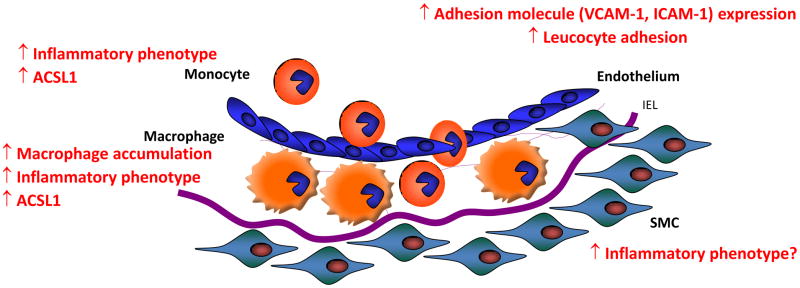
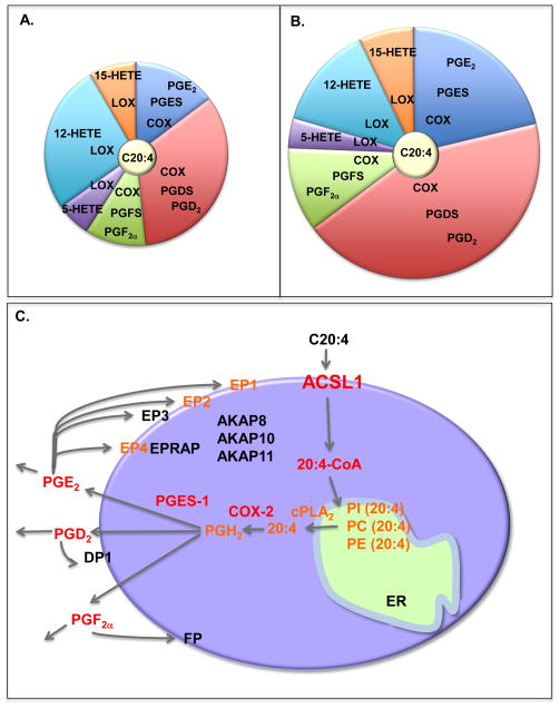
Monocytes and macrophages respond to and govern inflammation by producing a plethora of inflammatory modulators, including cytokines, chemokines, and arachidonic acid (C20:4)-derived lipid mediators. One of the most prevalent inflammatory diseases is cardiovascular disease, caused by atherosclerosis, and accelerated by diabetes. Recent research has demonstrated that monocytes/macrophages from diabetic mice and humans with type 1 diabetes show upregulation of the enzyme, acyl-CoA synthetase 1 (ACSL1), which promotes C20:4 metabolism, and that ACSL1 inhibition selectively protects these cells from the inflammatory and proatherosclerotic effects of diabetes, in mice. Increased understanding of the role of ACSL1 and other culprits in monocytes/macrophages in inflammation and diabetes-accelerated atherosclerosis offers hope for new treatment strategies to combat diabetic vascular disease.


Figures

Figure 1
Figure 1. Schematic representation of atherosclerotic lesion initiation in mouse models of T1D
Atherosclerotic lesion initiation is accelerated in mouse models of T1D in the absence of diabetes-induced hyperlipidemia. The effect of diabetes in these models is due to an increased macrophage accumulation in the arterial wall. Increased adhesion molecule expression in endothelial cells, which allows monocytes to attach and invade the arterial wall, is generally thought to mediate the increased macrophage accumulation. Recent research shows that the enzyme ACSL1 is induced in monocytes and macrophages in the setting of diabetes, and mediates the effects of diabetes on the inflammatory phenotype in these cells and on macrophage accumulation in the artery wall. It is not known if SMCs take on a more inflammatory phenotype in diabetes in vivo. Furthermore, it is unknown whether ACSL1 is induced in endothelial cells and SMCs exposed to diabetes or hyperglycemia, and the role of ACSL1 in these cells. IEL; internal elastic lamina, SMS; smooth muscle cells.
Figure 2
Figure 2. Increased ACSL1 expression and eicosanoid production in macrophages under diabetic conditions
A. Relative levels of some of the prostanoids produced by classically activated mouse bone marrow-derived macrophages differentiated in the presence of a normal (5.5 mM) glucose concentration and then stimulated with LPS and IFNγ for 48 h. B. When bone marrow-derived macrophages are differentiated in high concentration of glucose, such as that observed in diabetic mice (25 mM), the total amount of the prostanoids increases, as well as the relative levels of PGD2 and PGE2. C. A proposed role for ACSL1 in arachidonic acid handling in an inflammatory macrophage is shown. Arachidonic acid (C20:4) is taken up by macrophages through diffusion across the plasma membrane or by FA transport proteins. C20:4 is rapidly converted to arachidonoyl-CoA (20:4-CoA) by ACSL1 and perhaps other enzymes with acyl-CoA synthetase activity. This reaction traps C20:4 within the cell because of the bulky hydrophilic CoA moiety, and also allows it to be incorporated into phospholipids, such as phosphatidylinolsitol (PI), phosphatidylcholine (PC) and phosphatidylethanolamine (PE), through reacylation or through de novo synthesis. Activation of cytosolic PLA2 (cPLA2) liberates C20:4 from phospholipids and makes it available for eicosanoid production. In macrophages subjected to an inflammatory environment, such as diabetes or following TLR4 activation, production of eicosanoids is skewed toward a pathway mediated by cyclooxygenase 2 (COX-2). The COX-2 pathway results in enhanced production of prostaglandin PGE2, PGD2 and PGF, after TLR4 stimulation or in the setting of diabetes. These prostaglandins in turn act on other cells, or bind to specific receptors on the macrophage, resulting in activation of intracellular signal transduction pathways. Downstream signaling from these receptors is in turn governed by a number of modulators, such as A-kinase anchor proteins (AKAPs). Thus, induction of ACSL1 in inflammatory macrophages has the potential to enhance and fine-tune a complex network of potent modulators of the inflammatory response. Red font, mediators shown to be increased by diabetes; orange font, mediators shown to be increased by inflammatory stimuli, e.g. TLR4 activation.

References

    1. Bornfeldt KE, Tabas I. Insulin resistance, hyperglycemia, and atherosclerosis. Cell Metab. 2011;14:575–585. - PMC - PubMed
    1. Moore KJ, Tabas I. Macrophages in the pathogenesis of atherosclerosis. Cell. 2011;145:341–355. - PMC - PubMed
    1. Kanter JE, et al. Diabetes promotes an inflammatory macrophage phenotype and atherosclerosis through acyl-CoA synthetase 1. Proc Natl Acad Sci U S A. 2012;109:E715–E724. - PMC - PubMed
    1. Grundy SM. Pre-diabetes, metabolic syndrome, and cardiovascular risk. J Am Coll Cardiol. 2012;59:635–643. - PubMed
    1. Hu FB, et al. Elevated risk of cardiovascular disease prior to clinical diagnosis of type 2 diabetes. Diabetes Care. 2002;25:1129–1134. - PubMed

Publication types