. 2012 Nov 13;23(5):981-94.
doi: 10.1016/j.devcel.2012.10.013.

Sox2 in the dermal papilla niche controls hair growth by fine-tuning BMP signaling in differentiating hair shaft progenitors

Affiliations

Sox2 in the dermal papilla niche controls hair growth by fine-tuning BMP signaling in differentiating hair shaft progenitors

Carlos Clavel et al. Dev Cell. .

Abstract

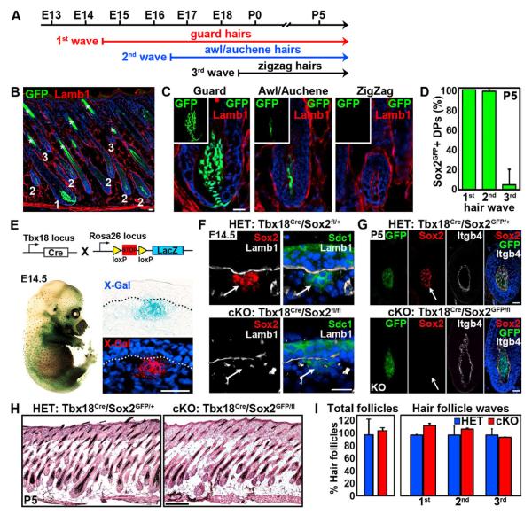
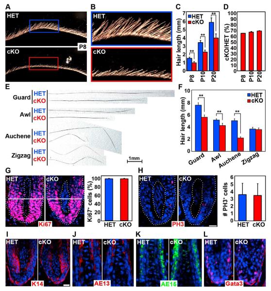
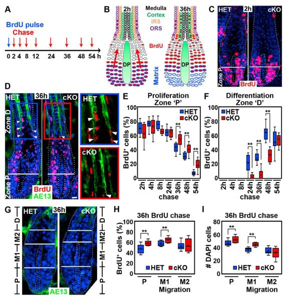
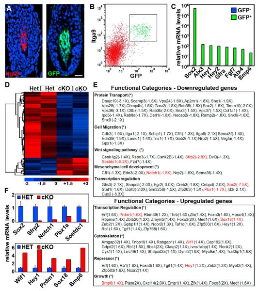
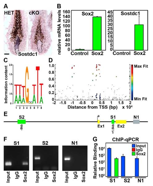
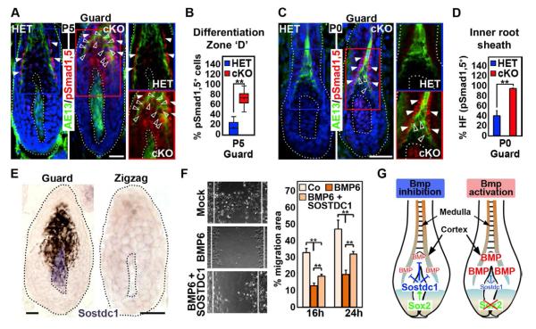
How dermal papilla (DP) niche cells regulate hair follicle progenitors to control hair growth remains unclear. Using Tbx18(Cre) to target embryonic DP precursors, we ablate the transcription factor Sox2 early and efficiently, resulting in diminished hair shaft outgrowth. We find that DP niche expression of Sox2 controls the migration speed of differentiating hair shaft progenitors. Transcriptional profiling of Sox2 null DPs reveals increased Bmp6 and decreased BMP inhibitor Sostdc1, a direct Sox2 transcriptional target. Subsequently, we identify upregulated BMP signaling in knockout hair shaft progenitors and demonstrate that Bmp6 inhibits cell migration, an effect that can be attenuated by Sostdc1. A shorter and Sox2-negative hair type lacks Sostdc1 in the DP and shows reduced migration and increased BMP activity of hair shaft progenitors. Collectively, our data identify Sox2 as a key regulator of hair growth that controls progenitor migration by fine-tuning BMP-mediated mesenchymal-epithelial crosstalk.


Figures

Figure 1
Figure 1. Sox2 ablation in DP precursors does not affect hair follicle formation
(A) Schematic of the three major hair follicle formation waves during embryonic morphogenesis. (B) Sox2 expression analysis (green) in postnatal day P5 skin of Sox2GFP knock-in reporter mice. Lamb1 stained the basement membrane (red). Nuclei are highlighted with DAPI (blue). Asterisk marks autofluorescence of hair shafts. Note GFP is expressed in DPs of 1st wave guard (1) and 2nd wave awl/auchene (2) hair follicles. DPs of 3rd wave zigzag follicles (3) do not express Sox2. (C) High magnification examples of hair follicle types with Sox2GFP positive and negative DPs. (D) Quantification of Sox2GFP positive DPs in P5 follicles of all 3 waves. n=130 hairs per mouse. Data are mean ± SD from 3 mice. (E)Targeting of DP precursor cells with Tbx18Cre. Top: Schematic of Tbx18Cre crossed with R26RLacZ reporter line. Bottom: Whole-mount X-Gal stained Tbx18Cre/R26RLacZ embryo showed robust Cre activity in a hair follicle distribution at E14.5. Histological analyses of sectioned embryos with Cre reporter activity in DP precursors. (F)Efficient ablation of Sox2 protein in Tbx18Cre/Sox2fl/fl conditional knockout (cKO) embryo. Sox2 immunofluorescence was absent in cKO dermal condensates (arrows) at E14.5. Syndecan-1 (Sdc1) labeled dermal condensates and Lamb1 marked the basement membrane. (G) Immunofluorescence staining for Sox2 at P5. Sox2 is absent in null DPs (arrow) in a Sox2GFP reporter background. (H) Hematoxylin/eosin staining of Sox2 heterozygous (HET; Tbx18Cre/Sox2GFP/+) and conditional knockout (cKO; Tbx18Cre/Sox2GFP/fl) skins at P5. (I) Quantification of total hair follicles and of follicles from the 3 waves at P5. n=75 hairs per mouse. Data are mean ± SD from 2 mice. Scale bars are 25 μm in A-F and 250 μm in G. See also Figure S1 and S2.
Figure 2
Figure 2. Sox2 ablation in the DP strongly impairs hair shaft outgrowth
(A) Side view of dorsal skin with outgrowing hair shafts at P8. Hair shafts are shorter in Sox2 cKO skin. (B) High magnification view of HET (blue frame) and cKO (red frame) inserts. 1st wave guard hair shafts are of considerable length by P8 in HET, but are much shorter in cKO. (C) Quantification of guard hair shaft lengths. n=20 guard hairs per mouse. Data are mean ± SD from 3 mice. **p < 0.01. (D) Relative guard hair shaft length reduction remains constant at all time points. (E) Examples of HET and cKO hair shafts from all 4 hair types at P20. Note that Sox2 expressing guard, awl and auchene hairs were shorter in cKO. Sox2 negative zigzag hairs were unchanged. (F) Quantification of hair shaft lengths of P20 clipped hairs. n=20 hairs per hair type. Data are mean ± SD from 2 mice. **p < 0.01. (G) Immunofluorescence staining of proliferation marker Ki67. Dotted line marks basement membrane. Right: Quantification of Ki67+ cells below Auber’s line (white). n=10 guard hairs per mouse. Data are mean ± SD from two mice. (H) Immunofluorescence staining of mitosis marker phospho-histone H3 (PH3). Right: Quantification of PH3+ cells in guard hair bulbs. n=20 guard hairs per mouse. Data are mean ± SD from 2 mice. (I-L) Immunofluorescence staining of (I) matrix marker keratin-14 and hair shaft differentiation markers (J) AE13 (hair cortex), (K) AE15 (inner root sheath) and (L) Gata3 (inner root sheath). Scale bar is 25 μm. See also Figure S3.
Figure 3
Figure 3. Sox2 ablation impairs progenitor migration during hair growth
(A) Pulse/chase BrdU labeling to track proliferating and migrating matrix cells. Timeline of BrdU injection at P5 (pulse) and chase time points for tissue harvest and BrdU label analysis (red arrows). (B) Schematic of initial BrdU uptake (2h) and of migration of matrix cells towards the hair shaft differentiation areas (36h). (C) BrdU immunofluorescence staining at 2h. Proliferation Zone “P” is below Auber’s line (white). Note robust BrdU uptake in both Sox2 HET and cKO follicles. (D) BrdU/AE13 double immunofluorescence staining at 36h. Differentiation Zone “D”: BrdU+ cells in the differentiating hair cortex labeled by AE13 (arrowheads). Note decreased BrdU+ matrix cells migrating into Zone “D” in cKO. Right: Inserts show higher magnification views of Zone “D” in HET (blue frame) and cKO (red frame). Dotted line marks basement membrane. Nuclei are counterstained with DAPI. Scale bars are 25 μm. (E,F) Quantification of BrdU+ cells in Zone “P” and “D”. Note delayed exit of Zone “P” and entry into Zone “D” of BrdU+ matrix cells in follicles with Sox2 null DPs. (G) Migration Zone “M” between Proliferation Zone “P” and Differentiation Zone “D”. Zone “M” is subdivided into proximal Zone “M1” and distal Zone “M2” at the level of the distal DP tip. AE13 staining defines the border to Differentiation Zone “D”. (H,I) Quantification of BrdU+ cells and total cell numbers (as DAPI+ nuclei) in Zone “P”, “M1” and “M2” at 36h. Note increased BrdU+ cells (H) and total cell numbers (I) in Zone “P” and “M1” in follicles with Sox2 null DPs. All box-and-whisker plots: mid-line, median; mean, plus symbol; box, 25th and 75th percentiles; whiskers, 10th and 90th percentiles. N=10 guard hairs/time point. Counts are from 3 (E,F) and 2 (H,I) independent experiments. **p < 0.01; *p < 0.05.
Figure 4
Figure 4. Sox2 ablation in the DP alters its gene expression signature
(A-C) FACS sorting strategy to isolate Sox2GFP/Integrin-alpha 9 double-positive DP cells from Sox2 HET and cKO P5 skin for transcriptional profiling. (A) Sox2GFP and Itga9+ DPs. Nuclei are counterstained with DAPI. Scale bar is 25 μm. (B) DP population is FACS isolated as Sox2GFP+/Itga9+ cells. (C) Real-Time PCR of sorted Sox2GFP-positive and -negative cells. (D) Heat map and clustering analysis of altered gene expression in microarrays from HET and cKO sorted P5 DPs. (E) Functional gene ontology categories enriched in downregulated (top) and upregulated (bottom) genes in cKO DPs. Genes highlighted in red were corroborated by Real-Time PCR. (F) Real-Time PCR verification of downregulated and upregulated genes in cKO DPs. Data are mean ± SD (n=2).
Figure 5
Figure 5. Expression of Bmp inhibitor Sostdc1 in DP cells is directly regulated by Sox2
(A) In situ hybridization for Sostdc1 mRNA expression in Sox2 HET and cKO guard hair follicle. Dotted line marks the basement membrane. Scale bar is 25 μm. (B) Real-Time PCR for Sox2 (left) and Sostdc1 (right) in isolated DP cells that lentivirally overexpress Sox2. Data are mean ± SD (n=2). (C) Predicted Sox2 binding site consensus sequence. (D) Heat map of putative Sox2 binding sites in the Sostdc1 genomic region based on consensus sequence and species conservation. (E) Schematic of genomic region displaying two putative Sox2 binding sites, “S1” and “S2”. “N1” is used as control site. (F) ChIP-PCR to detect Sox2 binding to conserved Sox2 binding sites “S1”, “S2”. “N1” is a control intronic fragment with no predicted site. Shown is 1 of 2 representative experiments. (G) ChIP-qPCR analyses to assess relative binding of Sox2 to “S1”, “S2” sites. Fold-enrichments of immunoprecipitated DNA fragments are compared to control “N1” and presented relative to IgG. Data are mean ± SD of 2 independent experiments.
Figure 6
Figure 6. Increased Bmp signaling in differentiating hair shaft TIP lineages in Sox2 ablated guard hair follicles
(A) Immunofluorescence for pSmad1,5 in P5 Sox2GFP HET and cKO guard hair follicles.RAE13 highlights differentiating hair cortex (green). GFPC signal in the DP is from Sox2GFP. Normal Bmp signaling in inner root sheath (arrowheads). Bmp signaling in hair cortex progenitors is upregulated in cKO MANUS (open arrowheads). Nuclei are counterstained with DAPI. Right: blue (HET) and red (cKO) inserts without DAPI. (B) Quantification of pSmad1,5-positive cells within the AE13 domain of P5 guard hair follicles. (C) pSmad1,5 immunofluorescence TED at P0. Precocious Bmp signaling in inner root sheath (arrowheads) and hair cortex (open arrowheads) in cKO follicles. Bmp activity is also increased in upper DP. (D) Quantification of hair follicles (HF) with pSmad1,5-positive inner root sheath/hair cortex progenitors in guard hair follicles at P0. (E) In situ hybridization for Sostdc1 mRNA expression in Sox2 guard and zigzag hair follicle. Dotted line marks the basement membrane. (F) In vitro scratch migration assay. BMP6 (500 ng/ml) decreases migration of skin epithelial cells, which is alleviated by SOSTDC1 (300 ng/ml). Right: Quantification of migrated area. Data are mean ± SEM from 2 independent experiments. **p < 0.01. (G) Model of fine-tuning Bmp signaling in hair shaft progenitors. Sox2 in the DP niche drives the expression of Bmp inhibitor Sostdc1, counteracting Bmps produced by matrix cells at the base and by the DP to fine-tune Bmp signaling in differentiating hair shaft progenitors. In follicles with Sox2 null DPs, the equilibrium is unbalanced leading to increased Bmp signaling and decreased migration during hair shaft differentiation. All box-and-whisker plots: mid-line, median; mean, plus symbol; box, 25th and 75th percentiles; whiskers, 10th and 90th percentiles. N=10 guard and zigzag hairs. Counts are from 2 independent experiments. **p < 0.01. All scale bars are 25 μm. See also Figure S4.

Comment in

References

    1. Ahmed MI, Mardaryev AN, Lewis CJ, Sharov AA, Botchkareva NV. MicroRNA-21 is an important downstream component of BMP signalling in epidermal keratinocytes. J Cell Sci. 2011;124:3399–3404. - PMC - PubMed
    1. Alonso L, Okada H, Pasolli HA, Wakeham A, You-Ten AI, Mak TW, Fuchs E. Sgk3 links growth factor signaling to maintenance of progenitor cells in the hair follicle. J Cell Biol. 2005;170:559–570. - PMC - PubMed
    1. Andl T, Ahn K, Kairo A, Chu EY, Wine-Lee L, Reddy ST, Croft NJ, Cebra-Thomas JA, Metzger D, Chambon P, et al. Epithelial Bmpr1a regulates differentiation and proliferation in postnatal hair follicles and is essential for tooth development. Development. 2004;131:2257–2268. - PubMed
    1. Arnold K, Sarkar A, Yram MA, Polo JM, Bronson R, Sengupta S, Seandel M, Geijsen N, Hochedlinger K. Sox2(+) adult stem and progenitor cells are important for tissue regeneration and survival of mice. Cell Stem Cell. 2011;9:317–329. - PMC - PubMed
    1. Biernaskie J, Paris M, Morozova O, Fagan BM, Marra M, Pevny L, Miller FD. SKPs derive from hair follicle precursors and exhibit properties of adult dermal stem cells. Cell Stem Cell. 2009;5:610–623. - PMC - PubMed

Publication types

MeSH terms

Substances