. 2012 Nov 13;22(5):668-82.
doi: 10.1016/j.ccr.2012.10.009.

Relief of profound feedback inhibition of mitogenic signaling by RAF inhibitors attenuates their activity in BRAFV600E melanomas

Affiliations

Relief of profound feedback inhibition of mitogenic signaling by RAF inhibitors attenuates their activity in BRAFV600E melanomas

Piro Lito et al. Cancer Cell. .

Abstract

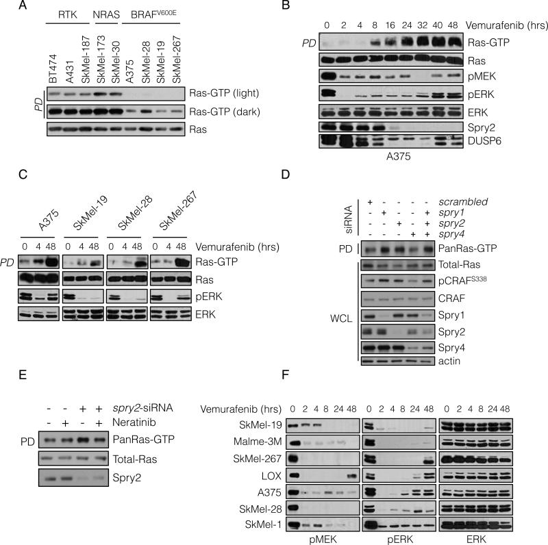
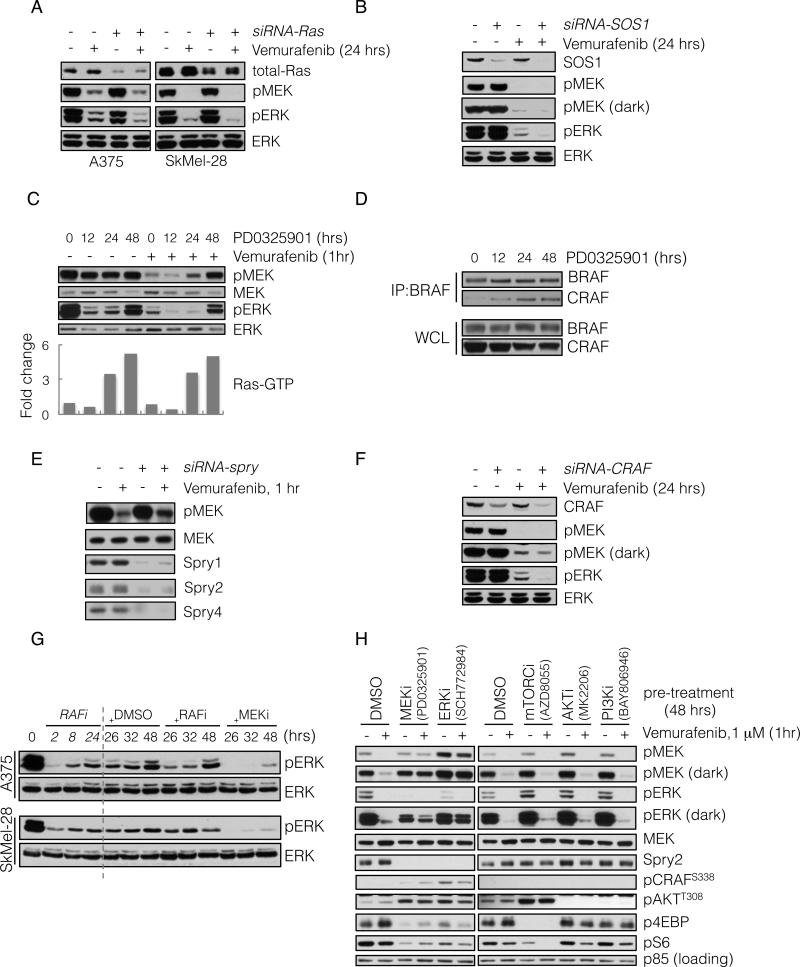
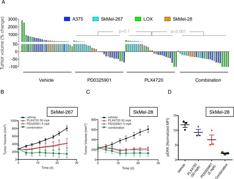
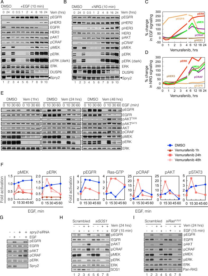
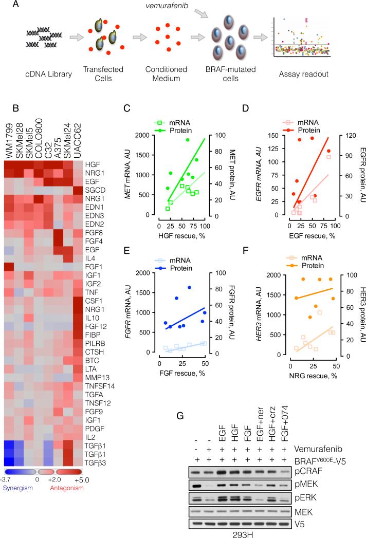
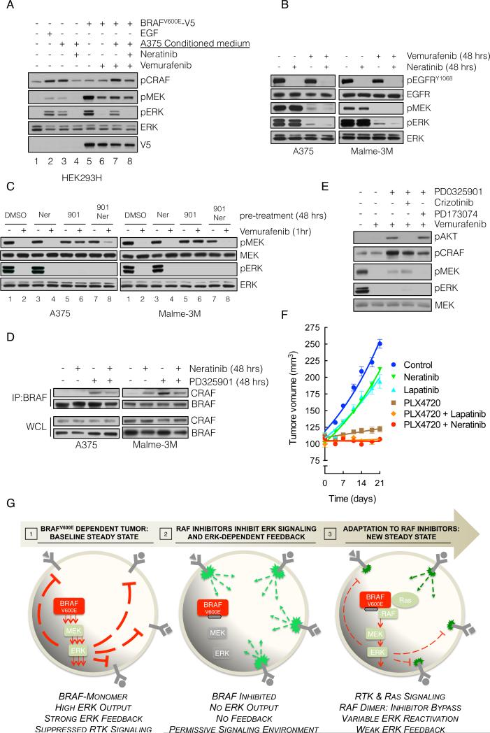
BRAF(V600E) drives tumors by dysregulating ERK signaling. In these tumors, we show that high levels of ERK-dependent negative feedback potently suppress ligand-dependent mitogenic signaling and Ras function. BRAF(V600E) activation is Ras independent and it signals as a RAF-inhibitor-sensitive monomer. RAF inhibitors potently inhibit RAF monomers and ERK signaling, causing relief of ERK-dependent feedback, reactivation of ligand-dependent signal transduction, increased Ras-GTP, and generation of RAF-inhibitor-resistant RAF dimers. This results in a rebound in ERK activity and culminates in a new steady state, wherein ERK signaling is elevated compared to its initial nadir after RAF inhibition. In this state, ERK signaling is RAF inhibitor resistant, and MEK inhibitor sensitive, and combined inhibition results in enhancement of ERK pathway inhibition and antitumor activity.


Figures

Figure 1
Figure 1. BRAFV600E melanomas maintain a state of low Ras-GTP through negative feedback regulation
(A) Whole cell lysates (WCL) from the indicated cell lines were subjected to pull-down (PD) assays with GST-bound CRAF Ras-binding domain (RBD). WCL and PD products were immunoblotted (IB) with a pan-Ras antibody. (B, C) BRAF-mutated melanoma cell lines were treated with vemurafenib (2 μM) for the indicated times. Ras-GTP was detected as in A. Phospho- and total levels of ERK pathway components were assayed by IB. (D) A375 cells (BRAFV600E) were transfected with siRNA pools targeting the indicated Spry isoforms or scrambled oligonucleotides. WCL were subjected to GST-RBD PD and analyzed by IB for the indicated proteins. (E) A375 cells were transfected with spry2 siRNA and 48 hrs after transfection they were treated with neratinib (1μM) for 1 hr. Ras-GTP levels were determined as above. (F) BRAFV600E melanoma cell lines were treated with vemurafenib (2 μM) for various times. The effect on ERK signaling is shown. See also Figure S1.
Figure 2
Figure 2. ERK rebound is dependent on formation of CRAF-containing dimers that are resistant to RAF inhibition
(A) BRAFV600E expressing A375 and SkMel-28 cells were transfected with siRNA pools targeting all three Ras isoforms (+) or scrambled oligonucleotides (-). 48 hours after transfection the cells were treated with vemurafenib (2 μM) for 24 hours and analyzed by immunoblotting (IB) to detect the indicated proteins. (B) A375 cells were transfected with siRNA pools targeting SOS1. 48 hours after transfection the cells were treated with vemurafenib (2 μM) for 24 hours, and analyzed by IB as above. (C) A375 cells were untreated or initially pre-treated with MEK inhibitor PD0325901 (50 nM) for the times shown. Subsequently, vemurafenib (1 μM) was added for 1 hour. WCL were assayed to determine changes in pMEK and pERK. WCLs were also analyzed with a GST-RBD Elisa assay. The fold change in the amount of GTP-bound Ras, as compared to untreated cells, is shown. (D) BRAFV600E expressing Malme-3M cells were treated with PD0325901 (50 nM) for various times. To assess BRAF-CRAF dimerization, WCL were subjected to immunoprecipitation (IP) with a BRAF-specific antibody and then IB for CRAF. (E) A375 cells were transfected with siRNA pools targeting spry1-4 genes. After 48 hrs they were treated with vemurafenib (1 μM) for 1 hr. WCL were analyzed to determine changes in inhibition of MEK phosphorylation. (F) A375 cells were transfected with CRAF siRNAs and subsequently treated with vemurafenib for 24 hours. Changes in phospho- and total ERK are shown. (G) BRAFV600E expressing cell lines were initially treated with vemurafenib (1 μM) for 2, 8 and 24 hours. After 24 hours of treatment, DMSO, vemurafenib (1 μM, RAFi) or PD0325901 (5 nM, MEKi) was added for an additional 2, 8 and 24 hours respectively. The total treatment time is indicated. The effect on ERK signaling is shown. (H) A375 cells were pre-treated with inhibitors targeting the indicated kinases for 48 hrs, followed by treatment with vemurafenib for 1 hr. WCL were analyzed to detect the ability of vemurafenib to inhibit MEK phosphorylation by RAF, as well as the ability of the indicated compounds to inhibit their targets. Their effects on Spry2 and pCRAF are also shown. See also Figure S2.
Figure 3
Figure 3. Combination of RAF and MEK inhibitors results in improved tumor growth inhibition in vivo
(A) Mice bearing xenografts from four different BRAFV600E melanoma cell lines were treated with vehicle, PD0325901 (2 mg/kg), PLX4720 (12.5 mg/kg) or their combination for four weeks. A waterfall representation of the best response for each tumor is shown. (B, C) Mice bearing SkMel-267 (B) and SkMel-28 (C) xenografts were treated with PLX4720 (50 mg/kg) alone or in combination with PD0325901 (5 mg/kg) for the indicated times. The tumor volumes (and SEM) are shown as a function of time after treatment. (D) Cells derived from SkMel-28 xenografts treated as shown for 48 hrs were subjected to flow-cytometric analysis to measure levels of pERK in isolated human melanoma cells. See also Figure S3.
Figure 4
Figure 4. Relief of ERK-dependent feedback potentiates receptor signaling
(A-D) BRAFV600E expressing A375 cells were treated with vemurafenib (vem, 2 μM) for various times. At the indicated times the cells were stimulated with either EGF (100 ng/mL, A) or NRG (100 ng/mL, B) for 10 min. WCL were analyzed by immunoblotting (IB) to assay downstream signaling. The percent change in ligand induced signaling (C, D) was determined by densitometric analysis of the bands in A and B, respectively. (E, F) A375 cells were untreated (DMSO) or treated with vemurafenib for 1, 24 or 48 hrs in serum free media and then stimulated with EGF for the indicated times. EGF-induced changes in phosphorylation of signaling intermediates were determined by IB (E) and quantified by densitometry (F). Changes in Ras-GTP were measured with an ELISA based GST-RBD assay (shown) and confirmed with a traditional pulldown assay (data not shown). (G) A375 cells were transfected with spry2 siRNAs, followed by serum starvation for 24 hrs and EGF stimulation for 10 min. WCL were IB with the indicated antibodies. (H, I) A375 cells were transfected with siRNAs targeting either SOS1 (H) or the three Ras isoforms (I) or scrambled siRNAs. Then, the cells were treated with vemurafenib for 24 hrs in serum-free media, followed by stimulation with EGF for 10 min. The phosphorylation of various signaling intermediates is shown. See also Figure S4.
Figure 5
Figure 5. Secreted exogenous ligands reduce the effectiveness of RAF inhibitors
(A) Schematic representation of the secretome assay. 293T cells grown in 384-well plates were transfected with a cDNA library encoding 220 unique secreted and single pass transmembrane proteins. Secreted ligands were collected in the conditioned medium, which was combined with vemurafenib (1 μM) and added to melanoma cell lines to assay the effect on proliferation. (B) Heat map representation of the effect of secreted ligands in the ability of vemurafenib to inhibit growth in a panel of eight BRAFV600E melanomas (some ligands were encoded by multiple cDNAs). Ligands with most potent effects are shown. (C-D) The percent rescue by the indicated growth factors was plotted as a function of the mRNA and protein level of the respective RTK. The mRNA level was obtained from expression profiling of the indicated lines, whereas the protein level was quantified by immunoblotting (shown in Figure S5C) and densitometry. (G) 293H cells were transfected with V5-tagged BRAFV600E, followed by treatment with vemurafenib (1 μM, 15 min), alone, and in combination with the indicated ligands and inhibitors (1 μM each) of HER kinases (neratinib), MET (crizotinib) or FGFR (PD173074). The ability of vemurafenib to inhibit MEK phosphorylation is shown. See also Figure S5.
Figure 6
Figure 6. Targeting HER receptor signaling restores sensitivity to RAF inhibition
(A) BRAFV600E-expressing A375 cells were grown in serum-free medium for 24 hrs and the medium was collected. This conditioned medium (CM) was added to 293H cells (lanes 1-4) or to 293H cells expressing exogenous V5 tagged BRAFV600E (lanes 5-8). The cells were also treated with the indicated combinations of CM, vemurafenib and neratinib in order to assay the effect on ERK singaling. (B) A375 and Malme3M cells were treated with vemurafenib (1 μM) alone or in combination with neratinib (1 μM) for 48 hours, and the effect on EGFR and downstream signaling was analyzed. (C) The cells were pre-treated with MEK inhibitor PD0325901 (901, 50 nM), and HER-kinase inhibitor neratinib (Ner, 1 μM), alone or in combination for 48 hours, followed by vemurafenib (1 μM) for 1 hour. Phospho- and total MEK and ERK are determined by IB. (D) The indicated cell lines were treated with PD0325901 and/or neratinib. WCL were subjected to IP with a BRAF-specific antibody and IB for CRAF. The level of BRAF and CRAF in the WCL is shown. (E) A375 cells were treated as in C but PD0325901 was combined with crizotinib, and PD173074 (1 μM each), targeting MET and FGFR, respectively. (F) SkMel-28-derived xenografts were treated with combinations of PLX4720 (50 mg/kg) neratinib (20 mg/kg) and lapatinib (150 mg/kg). Tumor growth (and SEM) as a function of time is shown. (G) Graphical representation of BRAFV600E melanomas adapting to RAF inhibitors. High levels of ERK-dependent feedback suppress RTK signaling and maintain mutant BRAF in a monomeric, drug-sensitive state. Inhibition of ERK signaling inactivates feedback, and restores RTK signaling to Ras. The resulting RAF dimers are resistant to RAF inhibitors, leading to bypass of inhibition and reactivation of ERK signaling.

References

    1. Avraham R, Yarden Y. Feedback regulation of EGFR signalling: decision making by early and delayed loops. Nature reviews Molecular cell biology. 2011;12:104–117. - PubMed
    1. Badalian-Very G, Vergilio JA, Degar BA, MacConaill LE, Brandner B, Calicchio ML, Kuo FC, Ligon AH, Stevenson KE, Kehoe SM, et al. Recurrent BRAF mutations in Langerhans cell histiocytosis. Blood. 2010;116:1919–1923. - PMC - PubMed
    1. Barretina J, Caponigro G, Stransky N, Venkatesan K, Margolin AA, Kim S, Wilson CJ, Lehar J, Kryukov GV, Sonkin D, et al. The Cancer Cell Line Encyclopedia enables predictive modelling of anticancer drug sensitivity. Nature. 2012;483:603–607. - PMC - PubMed
    1. Basso AD, Solit DB, Chiosis G, Giri B, Tsichlis P, Rosen N. Akt forms an intracellular complex with heat shock protein 90 (Hsp90) and Cdc37 and is destabilized by inhibitors of Hsp90 function. J Biol Chem. 2002;277:39858–39866. - PubMed
    1. Bollag G, Hirth P, Tsai J, Zhang J, Ibrahim PN, Cho H, Spevak W, Zhang C, Zhang Y, Habets G, et al. Clinical efficacy of a RAF inhibitor needs broad target blockade in BRAF-mutant melanoma. Nature. 467:596–599. - PMC - PubMed

Publication types

MeSH terms