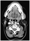
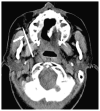
Review
. 2012 Dec;45(6):1293-310.
doi: 10.1016/j.otc.2012.08.004.

Retropharyngeal and prevertebral spaces: anatomic imaging and diagnosis

Affiliations
Review

Retropharyngeal and prevertebral spaces: anatomic imaging and diagnosis

J Matthew Debnam et al. Otolaryngol Clin North Am. 2012 Dec.

Abstract

Cross-sectional imaging plays an important role in the evaluation of the retropharyngeal space (RPS) and the prevertebral space (PVS). Because of their deep location within the neck, lesions arising within these spaces are difficult, if not impossible, to evaluate on clinical examination. This article details the cross-sectional anatomy and imaging appearances of primary and secondary diseases involving the RPS and PVS, including metastasis and spread from adjacent spaces. The role of image-guided biopsy is also discussed.


Conflict of interest statement

The authors have no financial information or potential conflicts of interest to disclose.

Figures
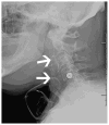
Fig. 1
Fig. 1
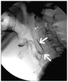
Thickening of the PVS. Lateral plane radiograph in a patient after total laryngectomy shows thickening of the RPS and PVS (arrows) secondary to reconstruction of the larynx and pharynx.
Fig. 2
Fig. 2
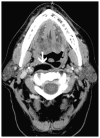
Fistulous tract between the aerodigestive tract and the RPS. Gastrografin contrast fluoroscopy image in a patient after chemotherapy and radiation therapy for base of tongue carcinoma shows a fistulous tract containing contrast (large arrows) between the aerodigestive tract and the RPS. Note presence of air (small arrow) superiorly in the RPS.
Fig. 3
Fig. 3
Metastatic melanoma. (A) Axial PET/CT scan shows a fluorodeoxyglucose-avid right lateral retropharyngeal node (large arrow) and bilateral neck nodes (small arrows). (B) PET image shows diffuse metastatic disease.
Fig. 4
Fig. 4
(A) Axial diagram of the retropharyngeal, danger, and prevertebral spaces with associated fascial planes. (B) The relationship of the retropharyngeal, danger, and prevertebral spaces.
Fig. 5
Fig. 5
Lipoma. Axial contrast-enhanced neck CT scan shows fat density (−20 Hounsfield units) in a right RPS lipoma (arrow).
Fig. 6
Fig. 6
PVS air collection. (A) Axial contrast-enhanced neck CTscan shows air (−920 Hounsfield units) (arrow) in the PVS. (B) Axial contrast-enhanced neck CT scan shows placement of a tracheostomy cannula (large arrow) in the soft tissues anterior to the trachea (small arrow).
Fig. 7
Fig. 7
NPC. (A) Sagittal T1 postcontrast MRI scan shows inferior extension of tumor (large arrows) into the RPS and PVS. Note the clival involvement (small arrow). (B) Axial PET/CT scan shows FDG-avid disease (arrow) in the RPS and PVS.
Fig. 8
Fig. 8
Chordoma. (A) Axial T1 postcontrast and (B) axial fast spin echo T2 MRI scans show a heterogeneously enhancing, T2-hyperintense PVS mass (large arrow) that is anteriorly displacing the prevertebral musculature (small arrow).
Fig. 9
Fig. 9
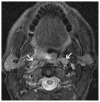
Retropharyngeal nodal metastasis. Axial T1 postcontrast MRI scan shows enlarged bilateral retropharyngeal nodes (arrows).
Fig. 10
Fig. 10
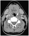
Retropharyngeal nodal metastasis. Axial contrast-enhanced neck CT scan shows a nonpathologically enlarged, centrally necrotic, right lateral retropharyngeal node (arrow).
Fig. 11
Fig. 11
Retropharyngeal cyst. Axial contrast-enhanced neck CT scan shows a branchial cleft cyst in the left RPS (arrow).
Fig. 12
Fig. 12
Schwannoma. Axial T1 postcontrast MRI scan with fat saturation shows a presumed right retropharyngeal schwannoma (large arrow) in a patient with neurofibromatosis 1. Nerve sheath tumor is seen extending out of the right neural foramen (small arrow).
Fig. 13
Fig. 13
Retropharyngeal fluid. Axial contrast-enhanced neck CT scan shows fluid in the retropharynx following radiation therapy (large arrow). A fat graft is noted in the oral cavity following total glossectomy (small arrow).
Fig. 14
Fig. 14
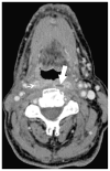
Retropharyngeal abscess. Axial contrast-enhanced neck CT scan shows a peripherally enhancing left RPS fluid collection (large arrow) under tension. Heterogeneous enhancement is present in the right RPS (small arrow) consistent with inflammatory change.
Fig. 15
Fig. 15
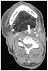
Soft tissue ulceration. Axial contrast-enhanced neck CT scan shows a radiation-associated ulceration (arrow) extending from the pharyngeal wall through the RPS and PVS to the anterior margin of the exposed vertebral body.
Fig. 16
Fig. 16
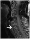
Anterior disk extrusion. Sagittal T1 postcontrast MRI scan with fat saturation shows an incidentally discovered anterior disk extrusion (arrow).
Fig. 17
Fig. 17
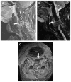
Prevertebral abscess/osteomyelitis. (A) Sagittal T1 postcontrast MRI scan, (B) sagittal fast spin echo T2 scan, and (C) axial T1 postcontrast MRI scan show infection of the vertebral column, epidural space, and the RPS and PVS (small arrows) characterized by soft tissue enhancement and a prevertebral abscess (large arrows).
Fig. 18
Fig. 18
Axial contrast-enhanced CT scan shows medial positioning of the internal carotid arteries (arrows) into the RPS.
Fig. 19
Fig. 19
CT-guided transfacial biopsy. Axial non–contrast-enhanced CT scan shows the biopsy needle (arrow) extending toward the right RPS.
Fig. 20
Fig. 20
Ultrasound-guided transoral FNA biopsy. Sonogram shows the biopsy needle (large arrow) extending into a right retropharyngeal lymph node (small arrows).

References

    1. Nyberg DA, Jeffrey RB, Brant-Zawadzki M, et al. Computed tomography of cervical infections. J Comput Assist Tomogr. 1985;9(2):288–96. - PubMed
    1. Wong YK, Novotny GM. Retropharyngeal space - a review of anatomy, pathology, and clinical presentation. J Otolaryngol. 1978;7(6):528–36. - PubMed
    1. Warner WC. Rockwood and Wilkins’ fractures in children. In: Beaty JH, Kasser JR, editors. Cervical spine injuries in children. Baltimore (MD): Lippincott Williams & Wilkins; 2001. pp. 809–84.
    1. Branstetter BF, 4th, Weissman JL. Normal anatomy of the neck with CT and MR imaging correlation. Radiol Clin North Am. 2000;38(5):925–40. - PubMed
    1. Chong VF, Fan YF. Radiology of the retropharyngeal space. Clin Radiol. 2000;55(10):740–8. - PubMed

Publication types

MeSH terms