

The high mobility group box: the ultimate utility player of a cell
- PMID: 23153957
- PMCID: PMC4437563
- DOI: 10.1016/j.tibs.2012.09.003
The high mobility group box: the ultimate utility player of a cell
Abstract
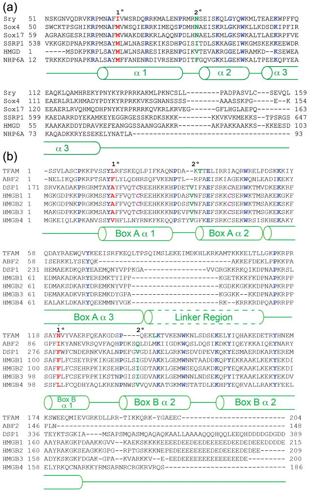
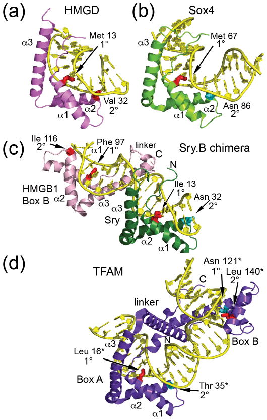
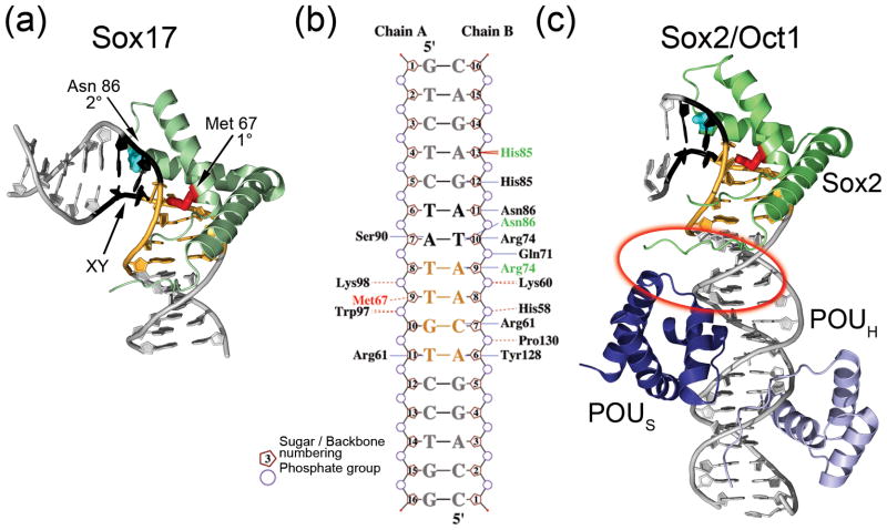
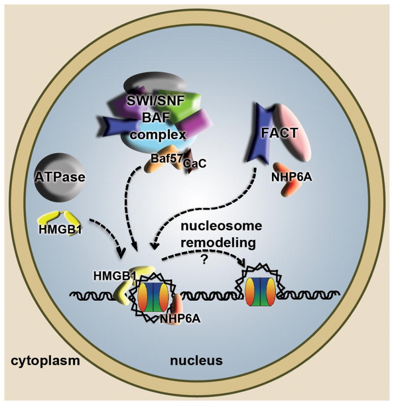
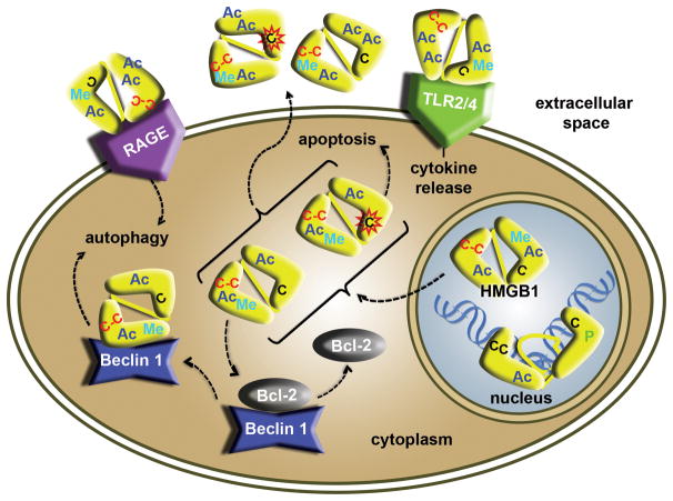
High mobility group (HMG) box proteins are abundant and ubiquitous DNA binding proteins with a remarkable array of functions throughout the cell. The structure of the HMG box DNA binding domain and general mechanisms of DNA binding and bending have been known for more than a decade. However, new mechanisms that regulate HMG box protein intracellular translocation, and by which HMG box proteins recognize DNA with and without sequence specificity, have only recently been uncovered. This review focuses primarily on the Sry-like HMG box family, HMGB1, and mitochondrial transcription factor A. For these proteins, structural and biochemical studies have shown that HMG box protein modularity, interactions with other DNA binding proteins and cellular receptors, and post-translational modifications are key regulators of their diverse functions.
Copyright © 2012 Elsevier Ltd. All rights reserved.
Figures
Similar articles
-
Differences in the DNA-binding properties of the HMG-box domains of HMG1 and the sex-determining factor SRY.Eur J Biochem. 1995 Jun 15;230(3):943-50. doi: 10.1111/j.1432-1033.1995.tb20640.x. Eur J Biochem. 1995. PMID: 7601157
-
Solution structure of the sequence-specific HMG box of the lymphocyte transcriptional activator Sox-4.J Biol Chem. 1995 Dec 22;270(51):30516-24. doi: 10.1074/jbc.270.51.30516. J Biol Chem. 1995. PMID: 8530483
-
Evolutionary conservation in the DNA-binding and -bending properties of HMG-boxes from SRY proteins of primates.Gene. 1995 Mar 10;154(2):277-80. doi: 10.1016/0378-1119(94)00853-k. Gene. 1995. PMID: 7890177
-
Floppy SOX: mutual induced fit in hmg (high-mobility group) box-DNA recognition.Mol Endocrinol. 2001 Mar;15(3):353-62. doi: 10.1210/mend.15.3.0617. Mol Endocrinol. 2001. PMID: 11222737 Review.
-
HMG1 and 2, and related 'architectural' DNA-binding proteins.Trends Biochem Sci. 2001 Mar;26(3):167-74. doi: 10.1016/s0968-0004(01)01801-1. Trends Biochem Sci. 2001. PMID: 11246022 Review.
Cited by
-
SOXE transcription factors form selective dimers on non-compact DNA motifs through multifaceted interactions between dimerization and high-mobility group domains.Sci Rep. 2015 May 27;5:10398. doi: 10.1038/srep10398. Sci Rep. 2015. PMID: 26013289 Free PMC article.
-
Epigenetic Modulation of Chromatin States and Gene Expression by G-Quadruplex Structures.Int J Mol Sci. 2020 Jun 11;21(11):4172. doi: 10.3390/ijms21114172. Int J Mol Sci. 2020. PMID: 32545267 Free PMC article. Review.
-
Spätzle processing enzyme is required to activate dorsal switch protein 1 induced Toll immune signalling pathway in Tenebrio molitor.PLoS One. 2023 Sep 21;18(9):e0291976. doi: 10.1371/journal.pone.0291976. eCollection 2023. PLoS One. 2023. PMID: 37733725 Free PMC article.
-
The Catalytic-Dependent and -Independent Roles of Lsd1 and Lsd2 Lysine Demethylases in Heterochromatin Formation in Schizosaccharomyces pombe.Cells. 2020 Apr 13;9(4):955. doi: 10.3390/cells9040955. Cells. 2020. PMID: 32295063 Free PMC article.
-
Functional roles of the DNA-binding HMGB domain in the histone chaperone FACT in nucleosome reorganization.J Biol Chem. 2018 Apr 20;293(16):6121-6133. doi: 10.1074/jbc.RA117.000199. Epub 2018 Mar 7. J Biol Chem. 2018. PMID: 29514976 Free PMC article.
References
-
- Goodwin GH, et al. A new group of chromatin-associated proteins with a high content of acidic and basic amino acids. Eur J Biochem. 1973;38:14–19. - PubMed
-
- Bustin M. Revised nomenclature for high mobility group (HMG) chromosomal proteins. Trends Biochem Sci. 2001;26:152–153. - PubMed
-
- Sgarra R, et al. HMGA molecular network: From transcriptional regulation to chromatin remodeling. Biochim Biophys Acta. 2010;1799:37–47. - PubMed
Publication types
MeSH terms
Substances
Grants and funding
LinkOut - more resources
Full Text Sources
