Review
. 2012 Dec;12(12):845-57.
doi: 10.1038/nri3328. Epub 2012 Nov 16.

Recognition of CD1d-restricted antigens by natural killer T cells

Affiliations
Review

Recognition of CD1d-restricted antigens by natural killer T cells

Jamie Rossjohn et al. Nat Rev Immunol. 2012 Dec.

Abstract

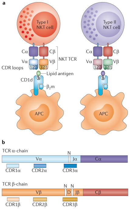
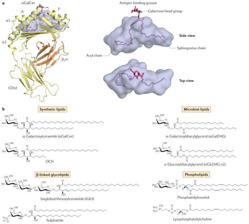
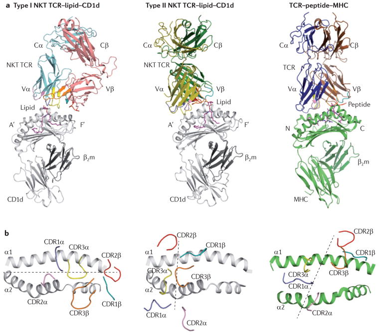
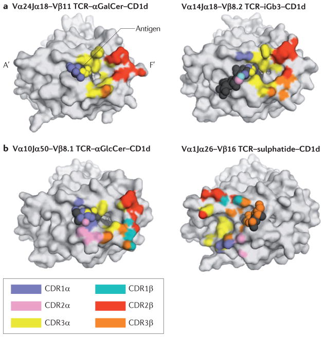
Natural killer T (NKT) cells are innate-like T cells that rapidly produce a variety of cytokines following T cell receptor (TCR) activation and can shape the immune response in many different settings. There are two main NKT cell subsets: type I NKT cells are typically characterized by the expression of a semi-invariant TCR, whereas the TCRs expressed by type II NKT cells are more diverse. This Review focuses on the defining features and emerging generalities regarding how NKT cells specifically recognize self, microbial and synthetic lipid-based antigens that are presented by CD1d. Such information is vitally important to better understand, and fully harness, the therapeutic potential of NKT cells.


Conflict of interest statement

Competing interests statement

The authors declare no competing financial interests.

Figures

Figure 1
Figure 1. NKT cells
a | The figure shows a schematic representation of type I and type II natural killer T (NKT) cells. These two subsets use different variable (V) region gene segments in the α- and β-chains of their T cell receptors (TCRs), and they recognize different CD1d-restricted antigens. b | The αβ TCR is composed of two chains, with the V domains containing the complementarity-determining region (CDR) loops. The CDR3 loops are encoded by multiple gene segments and also contain non-templated (N) regions, which add further diversity to the TCR repertoire. The colour coding is the same as that used for the type I NKT TCR in part a. β2m, β2-microglobulin; APC, antigen-presenting cell; C, constant; D, diversity; J, joining.
Figure 2
Figure 2. CD1d-mediated antigen presentation
a | The figure shows the structure of human CD1d bound to α-galactosylceramide (αGalCer) (PDB code 1ZT4). αGalCer is positioned within the CD1d antigen-binding groove, which is characterized by two main pockets: the A′-pocket and the F′-pocket. The galactose head group is surface exposed, whereas the lipid tails are buried within the cavity. b | The figure shows the chemical structures of various lipid antigens that bind to CD1d. These include examples of synthetic lipids (αGalCer and OCH), microbial lipids (α-galactosyldiacylglycerol from Borrelia burgdorferi and α-glucosyldiacylcerol from Streptococcus pneumoniae), β-linked glycolipids (isoglobotrihexosylceramide and sulphatide) and phospholipids (phosphatidylinositol and lysophosphatidylcholine). β2m, β2-microglobulin.
Figure 3
Figure 3. Structural comparison between NKT TCR–lipid–CD1d complexes and the conventional TCR–peptide–MHC complex
a | The figure shows the docking mode of the T cell receptor (TCR) in a type I natural killer T (NKT) cell TCR–lipid–CD1d complex (left; PDB code 2PO6), a type II NKT TCR–lipid–CD1d complex (middle; PDB code 4EI5) and a TCR–peptide–MHC complex (right; PDB code 3SJV). The CD1d antigen-binding pockets are labelled A′ and F′, and the amino and carboxyl termini of the peptide are labelled N and C, respectively. b | The figure shows the view looking down into the antigen-binding groove of the three complexes showing the parallel docking mode in the type I NKT–lipid–CD1d complex (left), the orthogonal docking mode in the type II NKT–lipid–CD1d complex (middle) and the diagonal docking mode in the TCR–peptide–MHC complex (right). Dashed lines represent the docking mode. β2m, β2-microglobulin.
Figure 4
Figure 4. The footprint of contact made by NKT TCRs on the surface of CD1d. a
| The image on the left shows the footprint of the human type I natural killer T (NKT) cell Vα24Jα18–Vβ11 T cell receptor (TCR) on the surface of human CD1d, which is presenting an α-linked glycolipid, α-galactosylceramide (αGalCer) (PDB code 2PO6). The image on the right shows the footprint of the mouse type I NKT cell Vα14Jα18–Vβ8.2 TCR on the surface of mouse CD1d, which is presenting a β-linked glycolipid, isoglobotrihexosyl-ceramide (iGb3) (PDB code 3SCM). b | On the left is the footprint of the type I NKT cell Vα10Jα50–Vβ8.1 TCR on the surface of mouse CD1d, which is presenting α-glucosylceramide (αGlcCer) (PDB code 3RUG). On the right is the footprint of the mouse type II NKT cell Vα1Jα26–Vβ16 TCR on the surface of mouse CD1d, which is presenting a β-linked self glycolipid, sulphatide (PDB code 4EI5).
Figure 5
Figure 5. Changes in the conformation of the lipid or CD1d following binding to NKT TCRs
a | The image on the left shows a surface representation of the α-galactosylceramide analogue OCH presented by CD1d, showing the A′ and F′ pockets (PDB code 3G08). The image on the right shows the closing of the F′-pocket roof in the CD1d–OCH complex following binding to the T cell receptor (TCR) (PDB code 3ARB), which is caused by movement of the side chains of Leu84, Vαl149 and Leu150 in CD1d (these residues are shown in blue). b | On the left is the structure of the β-linked glycolipid isoglobotrihexosylceramide (iGb3) presented by CD1d (PDB code 2Q7Y). The terminal sugar for iGb3 is modelled and shown in yellow. The image on the right shows the extensive interactions between the three sugars of the iGb3 head group and the residues of the CD1d α2-helix within type I natural killer T (NKT) cell TCR–iGb3–CD1d complex (PDB code 3SCM).
Figure 6
Figure 6. Modes of NKT cell activation
a | In the steady state, antigen-presenting cells (APCs) present non-agonist self lipids on CD1d molecules that do not promote the activation of the natural killer T (NKT) cell T cell receptor (TCR).b | APCs can present non-self lipids derived from bacteria or environmental allergens on CD1d molecules. The direct recognition of foreign lipid antigens from these sources can promote NKT cell activation. c | Activation of Toll-like receptors (TLRs) on APCs can induce the production of self lipid antigens (such as β-galactosylceramide) that can serve as agonists for the NKT TCR. Recognition of these CD1d-presented self lipid agonists, in conjunction with exposure to TLR-induced inflammatory cytokines, leads to NKT cell activation. IL-12, interleukin-12; IL-12R, IL-12 receptor.

Similar articles

Cited by

References

    1. Brigl M, Brenner MB. CD1: antigen presentation and T cell function. Annu Rev Immunol. 2004;22:817–890. - PubMed
    1. Mori L, De Libero G. T cells specific for lipid antigens. Immunol Res. 2012;53:191–199. - PubMed
    1. Bendelac A, Savage PB, Teyton L. The biology of NKT cells. Annu Rev Immunol. 2007;25:297–336. - PubMed
    1. Godfrey DI, MacDonald HR, Kronenberg M, Smyth MJ, Van Kaer L. NKT cells: what’s in a name? Nature Rev Immunol. 2004;4:231–237. - PubMed
    1. Godfrey DI, Kronenberg M. Going both ways: immune regulation via CD1d-dependent NKT cells. J Clin Invest. 2004;114:1379–1388. - PMC - PubMed

Publication types