Review
. 2012 Dec;10(12):852-67.
doi: 10.1038/nrmicro2911.

Animal models for HIV/AIDS research

Affiliations
Review

Animal models for HIV/AIDS research

Theodora Hatziioannou et al. Nat Rev Microbiol. 2012 Dec.

Abstract

The AIDS pandemic continues to present us with unique scientific and public health challenges. Although the development of effective antiretroviral therapy has been a major triumph, the emergence of drug resistance requires active management of treatment regimens and the continued development of new antiretroviral drugs. Moreover, despite nearly 30 years of intensive investigation, we still lack the basic scientific knowledge necessary to produce a safe and effective vaccine against HIV-1. Animal models offer obvious advantages in the study of HIV/AIDS, allowing for a more invasive investigation of the disease and for preclinical testing of drugs and vaccines. Advances in humanized mouse models, non-human primate immunogenetics and recombinant challenge viruses have greatly increased the number and sophistication of available mouse and simian models. Understanding the advantages and limitations of each of these models is essential for the design of animal studies to guide the development of vaccines and antiretroviral therapies for the prevention and treatment of HIV-1 infection.


Conflict of interest statement

Competing interests statement

The authors declare no competing financial interests.

Figures
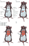
Figure 1
Figure 1. ‘Humanized’ mouse models used in HIV-1 research
ad | The different mouse models that are used for HIV-1 research are depicted here, grouped according to the presence of human immune cells in mouse tissues, and of HIV-1 infection, as reported in the literature (indicated in red). Peyer’s patches and the appendix represent the GALT (gut-associated lymphoid tissue). For simplicity, not all lymphoid organs are shown. The conjoint human thymus–liver organ formed following injection of human fetal liver and thymus cells into the mouse renal capsule is shown in parts a and d. For details of each mouse model, see main text. BLT, bone marrow–liver–thymus; Il2rg, interleukin-2 receptor common γ-chain gene; NOD, non-obese diabetic; NSG, NOD scid gamma; PBL, peripheral blood lymphocyte; Rag2, V(D)J recombination-activating gene 2; scid, severe combined immunodeficiency.
Figure 2
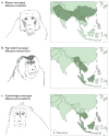
Figure 2. Macaque species commonly used in AIDS research, and their geographical ranges
a | The geographical range of the rhesus macaque (Macaca mulatta) exceeds that of all other primate species except humans, extending from western India and Pakistan across China. Distinct populations of rhesus macaques can be differentiated on the basis of mitochondrial DNA sequences or SNPs,. Captive-breeding programmes in the United States were initially established using animals imported from India, contributing to the widespread use of Indian-origin rhesus macaques in AIDS research. However, owing to the increasing demand for rhesus macaques and an embargo on the exportation of these animals from India since 1978, there has been a substantial decline in their availability and a sharp increase in their cost. This has led to greater dependence on rhesus macaques imported from China and Burma. b | The pig-tailed macaque (Macaca nemestrina) is native to Southeast Asia, Malaysia and Indonesia, and last shared a common ancestor with rhesus macaques approximately 3.5 million years ago. c | The cynomolgus macaque (Macaca fascicularis), also known as the long-tailed or crab-eating macaque, is native to regions of Indochina, Malaysia, Indonesia and the Philippines. Genetic evidence suggests that cynomolgus and rhesus macaques diverged from a common ancestor approximately 1.9 million years ago,. Distribution data from REF. .
Figure 3
Figure 3. Restriction factor polymorphism and the effects on viral replication in macaques
a | Numerous alleles encoding tripartite-motif-containing protein 5α (TRIM5α) and TRIMCyp have been identified in macaques. Although several amino acid differences have been found throughout the proteins, the encoding alleles can be grouped into two main variants depending on the amino acids at position 339–341 (TFP or ΔΔQ) in TRIM5α and at position 369 (N or D) in TRIMCyp. These crucial residues determine the specificity of these variants against lentiviruses. b | A summary of the restriction factors of macaques and the effects of the different variants on viral replication, as demonstrated in vivo for the simian immunodeficiency virus (SIV) strain SIVsm-E543-3 and in vitro for HIV-1. The description of TRIM5α polymorphism in cynomolgus macaques is very recent. Although the alleles described so far belong to group 2 and are not predicted to restrict SIV or HIV-1, amino acid differences at other locations in these proteins might affect restriction activity. There are also allelic variations for each APOBEC3 gene family member in macaques, but these are not described here. The extent of tetherin polymorphism in macaques is presently unclear. The number of variants listed is based on samples from 1–2 animals or cell lines (it is sometimes unclear whether cell lines were derived from animals of Indian or Chinese origin). The single tetherin variants reported for Chinese rhesus macaques and cynomolgus macaques is predicted to restrict HIV-1, but this has not been tested. +, restriction of replication; −, no significant restriction of replication (less than twofold); CypA, cyclophilin A-like.
Figure 4
Figure 4. Derivation of common simian immunodeficiency virus and simian–human immunodeficiency virus strains
a | Monkey cartoons indicate animals from which simian immunodeficiency virus (SIV) isolates were derived. Green boxes represent uncloned biological isolates, and yellow boxes represent infectious molecular clones. Solid arrows represent direct viral transmission or isolation steps, and dotted arrows indicate passage and/or cloning steps omitted for simplicity. b | Schematic representation of HIV-1, SIV and chimeric viral genomes. Maroon and blue boxes indicate HIV-1- and SIV-derived sequences, respectively. Ca, Cercocebus atys (sooty mangabey); CNPRC, California National Primate Research Center (Davis, California, USA); Env, envelope glycoprotein; LTR, long terminal repeat; Mm, Macaca mulatta (rhesus macaque); Mn, Macaca nemestrina (pig-tailed macaque); SHIV, simian–human immunodeficiency virus; SIVmac, macaque SIV; SIVmne, pig-tailed macaque SIV; SIVsmm, sooty mangabey SIV; stHIV-1, simian-tropic HIV-1; RT, reverse transcriptase.

References

    1. Gao F, et al. Origin of HIV-1 in the chimpanzee Pan troglodytes troglodytes. Nature. 1999;397:436–441. - PubMed
    1. Keele BF, et al. Chimpanzee reservoirs of pandemic and nonpandemic HIV-1. Science. 2006;313:523–526. - PMC - PubMed
    1. Keele BF, et al. Increased mortality and AIDS-like immunopathology in wild chimpanzees infected with SIVcpz. Nature. 2009;460:515–519. - PMC - PubMed
    1. Alter HJ, et al. Transmission of HTLV-III infection from human plasma to chimpanzees: an animal model for AIDS. Science. 1984;226:549–552. - PubMed
    1. O’Neil SP, et al. Progressive infection in a subset of HIV-1-positive chimpanzees. J Infect Dis. 2000;182:1051–1062. - PubMed

Publication types

MeSH terms