. 2013 Apr:67:78-87.
doi: 10.1016/j.neuropharm.2012.10.004. Epub 2012 Nov 12.

Combination treatment with progesterone and vitamin D hormone is more effective than monotherapy in ischemic stroke: the role of BDNF/TrkB/Erk1/2 signaling in neuroprotection

Affiliations

Combination treatment with progesterone and vitamin D hormone is more effective than monotherapy in ischemic stroke: the role of BDNF/TrkB/Erk1/2 signaling in neuroprotection

Fahim Atif et al. Neuropharmacology. 2013 Apr.

Abstract

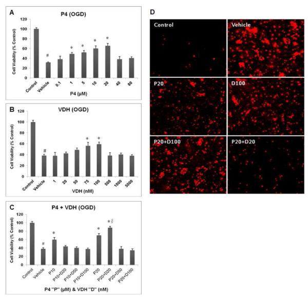
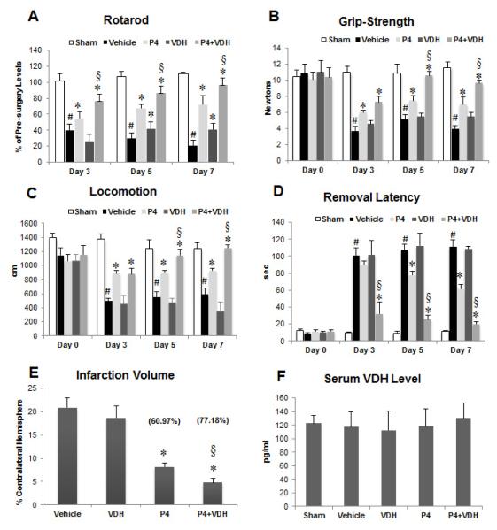
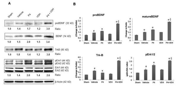
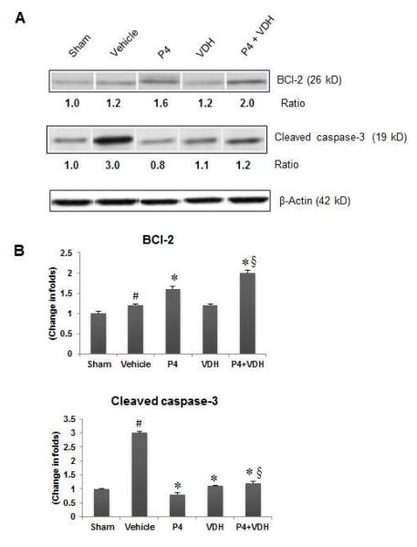
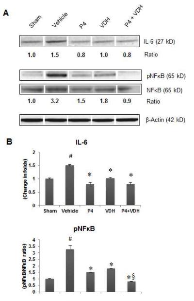
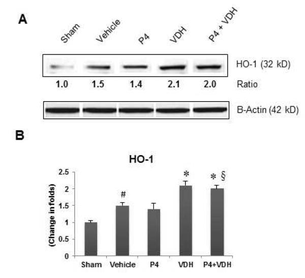
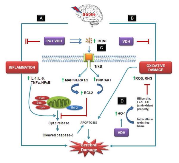
We investigated whether combinatorial post-injury treatment with progesterone (P4) and vitamin D hormone (VDH) would reduce ischemic injury more effectively than P4 alone in an oxygen glucose deprivation (OGD) model in primary cortical neurons and in a transient middle cerebral artery occlusion (tMCAO) model in rats. In the OGD model, P4 and VDH each showed neuroprotection individually, but combination of the "best" doses did not show substantial efficacy; instead, the lower dose of VDH in combination with P4 was the most effective. In the tMCAO model, P4 and VDH were given alone or in combination at different times post-occlusion for 7 days. In vivo data confirmed the in vitro findings and showed better infarct reduction at day 7 and functional outcomes (at 3, 5 and 7 days post-occlusion) after combinatorial treatment than when either agent was given alone. VDH, but not P4, upregulated heme oxygenase-1, suggesting a pathway for the neuroprotective effects of VDH differing from that of P4. The combination of P4 and VDH activated brain-derived neurotrophic factor and its specific receptor, tyrosine kinase receptor-B. Under specific conditions VDH potentiates P4's neuroprotective efficacy and should be considered as a potential partner of P4 in a low-cost, safe and effective combinatorial treatment for stroke.


Figures

Fig. 1
Fig. 1
Concurrent treatments of P4 (A) and VDH (B) and their combinations (C) on the viability of primary cortical neurons 24 h post-OGD. Panel (D) shows representative photomicrographs of cell death by PI staining 24 h post-OGD. Values are expressed as means±SEM of three experiments. Significant difference #P<0.05 compared to sham; *P<0.05 compared to vehicle; and §P<0.05 compared to P4.
Fig. 2
Fig. 2
Functional outcomes on days 3, 5 and 7 post-injury using rotarod (A), grip strength (B), locomotor activity (C), and sticky tape removal (D) tests; and effect of P4 and VDH on infarct volume (E) and serum VDH level (F) following tMCAO. Values are expressed as means ± SEM. Significant difference #P<0.05 compared to sham; *P<0.05 compared to vehicle; and §P<0.05 compared to P4. Vaues in parentheses represent percent reduction in infarct volume.
Fig. 3
Fig. 3
P4 and VDH effects on expression of proBDNF and mature BDNF (BDNF), its receptor TrkB, and downstream Erk1/2 phosphorylation in rat brain 24 h post-injury. (A) Representative protein bands of Western data. (B) Densitometry data. β-actin was used as the loading control. Values are expressed as means ± SEM. Significant difference #P<0.05 compared to sham; *P<0.05 compared to vehicle; and §P<0.05 compared to P4. Combination treatment with P4 and VDH triggered BDNF/TrkB/Erk1/2 signaling.
Fig. 4
Fig. 4
B>. P4 and VDH effects on expression of anti-apoptotic protein BCl-2 and cleaved caspase-3 24 h post-injury. (A) Representative protein bands of Western data. (B) Densitometry data. β-actin was used as the loading control. Values are expressed as means ± SEM. Significant difference #P < 0.05 compared to sham; *P < 0.05 compared to vehicle; §P < 0.05 compared to P4. The P4 and VDH combination showed a synergistic effect on BCl-2 expression.
Fig. 5
Fig. 5
P4 and VDH effects on expression of inflammatory markers IL-6 and NFκB 24 h post-injury. (A) Representative protein bands of Western data. (B) Densitometry data. β-actin was used as the loading control. Values are expressed as means ± SEM. Significant difference #P<0.05 compared to sham; *P<0.05 compared to vehicle; §P<0.05 compared to P4. VDH enhanced the anti-inflammatory efficacy of P4 in combination.
Fig. 6
Fig. 6
P4 and VDH effects on the expression of HO-1, a marker of neuronal oxidative damage, 24 h post-injury. (A) Representative protein bands of Western data. (B) Densitometry data. β-actin was used as the loading control. Values are expressed as means ± SEM. Significant difference #P<0.05 compared to sham; *P<0.05 compared to vehicle; §P<0.05 compared to P4. Data indicate that VDH, but not P4, increased the expression of HO-1, suggesting a neuroprotective response against oxidative damage.
Fig. 7
Fig. 7
Hypothetical presentation of some possible mechanisms of action of P4 and VDH against ischemic stroke. We target two important detrimental pathways of ischemic injury: (A) inflammation, and (B) oxidative damage, and their modulation by P4 and VDH alone and in combination. P4 and VDH inhibited inflammatory response synergistically by suppressing IL-6 and NFκB. Both inflammation and oxidative damage cause mitochondrial damage. As a result, damaged mitochondria release cytochrome c (cyt c) into the cytosol, which further activates caspases and associated apoptosis. (C) Growth factor signaling (BDNF/TrkB/Erk1/2) is one of the neuroprotective mechanisms of P4 and VDH. BDNF promotes cell survival through activation of TrkB, a high-affinity receptor for BDNF. Upon activation, TrkB receptor signaling activates several signaling proteins and pathways regulated by MAP kinase (MAPK/Erk1/2) and PI3 kinase (PI3K/Akt). These pathways further activate the anti-apoptotic protein BCl-2, which prevents mitochondrial damage and cyt-c release-mediated caspase activation and apoptosis, thus promoting neuronal survival. Both P4 and VDH synergistically activate BDNF, TrkB, Erk1/2 signaling, and BCl-2, and suppress cleaved caspase-3 activation. VDH alone showed modulatory effects on oxidative damage as evidenced by the expression of HO-1. HO-1 is a rapidly induced heat shock protein which catabolizes free heme to biliverdin, CO, and Fe2+. These byproducts of HO-1 activity have potential antioxidant activity and provide cytoprotection (D).

Similar articles

Cited by

References

    1. Abe K, Hayashi T. Expression of the glial cell line-derived neurotrophic factor gene in rat brain after transient MCA occlusion. Brain Res. 1997;776:230–234. - PubMed
    1. Atif F, Sayeed I, Ishrat T, Stein DG. Progesterone with vitamin D affords better neuroprotection against excitotoxicity in cultured cortical neurons than progesterone alone. Mol. Med. 2009;15:328–336. - PMC - PubMed
    1. Balden R, Selvamani A, Sohrabji F. Vitamin D deficiency exacerbates experimental stroke injury and dysregulates ischemia-induced inflammation in adult rats. Endocrinol. 2012;153:2420–2435. - PMC - PubMed
    1. Cekic M, Cutler SM, VanLandingham JW, Stein DG. Vitamin D deficiency reduces the benefits of progesterone treatment after brain injury in aged rats. Neurobiol. Aging. 2011;32:864–874. - PMC - PubMed
    1. Cekic M, Sayeed I, Stein DG. Combination treatment with progesterone and vitamin D hormone may be more effective than monotherapy for nervous system injury and disease. Front. Neuroendocrinol. 2009;30:158–172. - PMC - PubMed

Publication types

MeSH terms