. 2013 Mar;20(3):456-64.
doi: 10.1038/cdd.2012.141. Epub 2012 Nov 16.

Recruitment of Grb2 and SHIP1 by the ITT-like motif of TIGIT suppresses granule polarization and cytotoxicity of NK cells

Affiliations

Recruitment of Grb2 and SHIP1 by the ITT-like motif of TIGIT suppresses granule polarization and cytotoxicity of NK cells

S Liu et al. Cell Death Differ. 2013 Mar.

Abstract

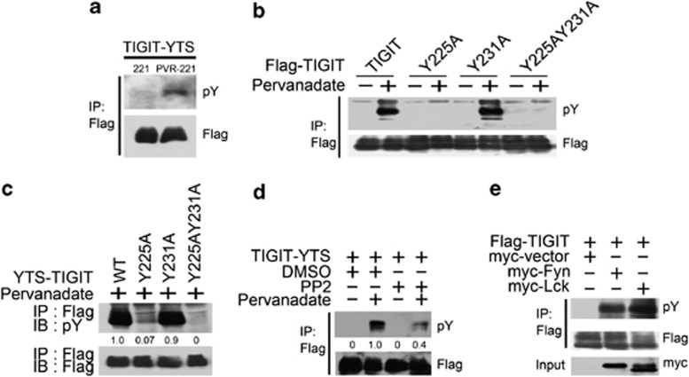
Activating and inhibitory receptors control natural killer (NK) cell activity. T-cell immunoglobulin and ITIM (immunoreceptor tyrosine-based inhibition motif) domain (TIGIT) was recently identified as a new inhibitory receptor on T and NK cells that suppressed their effector functions. TIGIT harbors the immunoreceptor tail tyrosine (ITT)-like and ITIM motifs in its cytoplasmic tail. However, how its ITT-like motif functions in TIGIT-mediated negative signaling is still unclear. Here, we show that TIGIT/PVR (poliovirus receptor) engagement disrupts granule polarization leading to loss of killing activity of NK cells. The ITT-like motif of TIGIT has a major role in its negative signaling. After TIGIT/PVR ligation, the ITT-like motif is phosphorylated at Tyr225 and binds to cytosolic adapter Grb2, which can recruit SHIP1 to prematurely terminate phosphatidylinositol 3-kinase (PI3K) and MAPK signaling, leading to downregulation of NK cell function. In support of this, Tyr225 or Asn227 mutation leads to restoration of TIGIT/PVR-mediated cytotoxicity, and SHIP1 silencing can dramatically abolish TIGIT/PVR-mediated killing inhibition.


Figures

Figure 1
Figure 1
TIGIT/PVR engagement disrupts granule polarization and cytotoxicity of YTS cells. (a) TIGIT/PVR engagement disrupts granule and MTOC polarization. PVR-221 or 221 target cells were labeled by CellTracker Blue and then incubated with TIGIT-YTS or YTS cells at 37 °C for 30 min. Treated cells were stained with anti-Flag and anti-perforin antibody (upper panel) or anti-β-tubulin antibody (lower panel) followed by confocal microscopy. Blue fluorescence for target cells is shown in the first lane, green for TIGIT in the second lane, red for perforin or β-tubulin in the third lane, the merged image in the fourth lane and differential interference control (DIC) in the last. Granule polarization (middle) was counted at least 100 conjugates in each experiment. Inhibition rates (right) of YTS and TIGIT-YTS were calculated as the polarization rate against 221 cells minus the polarization rate against PVR-221 cells. These data are representative of at least six separate experiments shown as means±S.D. The Student's t test was used for statistical analysis. **P<0.01. CTB: CellTracker Blue; 221: 721.221 cells. (b) TIGIT/PVR engagement suppresses YTS cell-mediated cytolysis. PVR-221 (right) or 221 (left) target cells were labeled with 51Cr and incubated with TIGIT-YTS or YTS cells at the indicated E:T ratios followed by a 51Cr-release assay. Data are representative of at least three independent experiments as means±S.D. The color representation of this figure is available at the Cell Death and Differentiation Journal online
Figure 2
Figure 2
TIGIT is phosphorylated at its cytoplasmic tail after its ligation with PVR. (a) TIGIT is phosphorylated once engaged with PVR. TIGIT-YTS cells were incubated with 221 or PVR-221 cells at E:T ratio of 5 : 1 at 37 °C for 5 min. Treated cells were lysed and Flag-tagged TIGIT was immunoprecipitated with anti-Flag beads followed by immunoblotting with anti-pY antibody. The same blot was stripped and reprobed with anti-Flag antibody. IP: immunoprecipitation; pY: phosphorylated tyrosine. (b) Tyr225A mutation abolishes tyrosine phosphorylation of TIGIT in 293A cells. Plasmids of 3 × Flag-tagged TIGIT and its mutants (Y225A, Y231A or Y225AY231A) were transfected into 293A cells for 48 h and treated with or without pervanadate for immunoblotting with anti-pY antibody. The same blot was stripped and reprobed with anti-Flag antibody. Y225A: Tyr225Ala mutation; Y231A: Tyr231Ala mutation; Y225AY231A: Tyr225AlaTyr231Ala mutation. (c) Tyr225A mutation abrogates tyrosine phosphorylation of TIGIT in YTS cells. Different mutants of TIGIT-YTS cells were treated with pervanadate for 10 min and detected as above. (d) PP2 can inhibit TIGIT phosphorylation. TIGIT-YTS cells were pretreated with a Src family inhibitor PP2 for 6 h, and then treated with or without pervanadate for 10 min followed by immunoblotting as above. (e) Fyn and Lck can phosphorylate TIGIT. 3 × Flag-TIGIT vector was co-transfected with myc-tagged Fyn or myc-tagged Lck vector into 293A cells for 48 h and then lysed for immunoblotting. All above data are representative of at least three independent experiments. The numbers in Figure 2c and Figure 2d show relative amount of the indicated proteins
Figure 3
Figure 3
pTIGIT can recruit the adapter molecule Grb2 by its ITT-like motif. (a) pTIGIT associates with Grb2. Plasmids of 3 × Flag-tagged TIGIT and myc-tagged Grb2 were co-transfected into 293A cells for 48 h. Transfected cells were lysed with or without pervanadate treatment and immunoprecipitated with anti-Flag beads followed by immunoblotting. myc-Grb2 in total lysates was probed for myc as a loading control. (b) pTIGIT can precipitate Grb2 in TIGIT-YTS cells. TIGIT-YTS or YTS cell lysates with or without pervanadate treatment were immunoprecipitated with anti-Flag beads and blotted with anti-Grb2 or anti-Flag antibody. Grb2 in the lysates was probed as a loading control. (c) pTIGIT can directly bind to rGrb2. Non-phosphorylated TIGIT (TIGIT) or pTIGIT was coincubated with recombinant His-Grb2 for a GST-pulldown assay. Precipitates were detected by immunoblotting with anti-Grb2 or anti-His antibody. Grb2 expression in the inputs served as a loading control. (d) Tyr225 mutation can not interact with Grb2 in 293A cells. Flag-tagged TIGIT mutant (Y225A, Y231A or Y225AY231A) and myc-Grb2 vectors were co-transfected into 293A for 48 h and lysed with or without pervanadate treatment for immunoprecipitation. Myc-Grb2 expression in the inputs was used as a loading control. (e) Tyr225 mutation fails to bind Grb2 in YTS cells. Tyr225, Tyr231 or both mutated TIGIT-YTS cells with or without pervanadate treatment were lysed and immunoprecipitated with anti-Flag beads for immunoblotting. Grb2 in the inputs was used as a loading control. (f) Asn227 mutation abolishes the association of TIGIT with Grb2. TIGIT-YTS and TIGIT-N227Q-YTS (N227Q) cells were lysed with or without pervanadate treatment followed by immunoprecipitation. Grb2 in lysates was probed as a loading control. All above data represent at least three separate experiments. (g) Tyr225 mutation dramatically loses TIGIT/PVR-mediated killing inhibition. Tyr225 or Tyr231-mutated TIGIT-YTS cells were incubated with 51Cr-labeled 221 (left) or PVR-221 (right) target cells at the indicated E:T ratios followed by a 51Cr-release assay. (h) Asn227 mutation abrogates TIGIT/PVR-mediated killing inhibition against PVR-221 cells. TIGIT-N227Q-YTS (N227Q) or TIGIT-YTS cells were incubated with 51Cr-labeled PVR-221 or 221 cells as detected above. Data are representative of at least four independent experiments as means±S.D. *P<0.05; **P<0.01. The numbers in Figures 3d, e and f show relative amount of the indicated proteins
Figure 4
Figure 4
Grb2 facilitates SHIP1 recruitment to TIGIT. (a and b) pTIGIT mainly interacts with SHIP1. TIGIT-YTS (a) or primary NK cell (b) lysates with or without pervanadate treatment were immunoprecipitated by anti-Flag beads and immunoblotted with anti-SHIP1, anti-SHP2, anti-SHP1, anti-pY, or anti-Grb2 antibody. The same blots for testing pY were stripped and reprobed with anti-Flag antibody. (c) Tyr225 mutation fails to recruit SHIP1. TIGIT-YTS mutant (Y225A, Y231A or Y225AY231A) was transduced into YTS cells and lysed with or without pervanadate treatment for immunoprecipitation. SHIP1 in YTS lysates was used as a loading control. (d) Asn227 mutation can not associate with SHIP1. Recombinant non-phosphorylated TIGIT (TIGIT), pTIGIT, or phosphorylated TIGIT-Asn227Gln (pN227Q) were coincubated with YTS lysates and then immunoblotted with anti-SHIP1 or anti-His antibody. SHIP1 in the inputs was used as a loading control. (e) Asn227 mutation of TIGIT remarkably diminishes its recruitment of SHIP1. N227Q-YTS or TIGIT-YTS cell lysates were immunoprecipitated by anti-Flag beads and immunoblotted with anti-SHIP1 or anti-Flag antibody. SHIP1 in lysates was used as a loading control. (f) Grb2 silencing dramatically reduces SHIP1 recruitment. TIGIT-YTS cells were transfected with siRNA duplexes either for Grb2 (siGrb2) or for scrambled siRNA control (siCtrl) for 48 h. Lysates were immunoprecipitated with anti-Flag beads and immunoblotted for SHIP1, SHP2, Grb2 or Flag. SHIP1, SHP2 and β-actin in the inputs were used as loading controls. All above data are representative of at least three independent experiments. The numbers in Figures 4c–f show relative amount of the indicated proteins
Figure 5
Figure 5
TIGIT engagement prematurely terminates PI3K and MAPK activation signaling in YTS cells. (a and b) TIGIT/PVR ligation prematurely terminates phosphorylation of Akt or of Erk and MEK. TIGIT-YTS cells were stimulated with 221 or PVR-221 cells followed by immunoblotting with anti-p-Akt antibody (a, upper) and the same blot was reprobed with anti-Akt antibody (a, lower). p-Erk (b, upper panel) and p-MEK (b, lower panel) were detected as above. (c) TIGIT/PVR engagement inhibits Erk phosphorylation by confocal microscopy. TIGIT-YTS cells were incubated with CellTracker Blue-labeled 221 (upper) or PVR-221 (lower) cells for 45 min and stained for p-Erk followed by confocal microscopy. Blue fluorescence for target cells is shown in the first lane, red for p-Erk in the second lane, the merged image in the third lane, differential interference control (DIC) in the last lane. CTB: CellTracker Blue. (d) Tyr225 mutation disrupts TIGIT/PVR-mediated premature termination of Erk phosphorylation. TIGIT-Y225A-YTS (Y225A-YTS), TIGIT-Y231A-YTS (Y231A-YTS), TIGIT-Y225AY231A-YTS (Y225AY231A-YTS) cells were incubated with PVR-221 or 221 cells and immunoblotted with anti-p-Erk or anti-Erk antibody. (e) TIGIT/PVR ligation attenuates phosphorylation of Erk in primary NK cells. Human primary NK cells were incubated with PVR-221 or 221 cells and immunoblotted with anti-p-Erk or anti-Erk antibody. (f) Blockade of TIGIT disrupts the premature termination of Erk phosphorylation. TIGIT-YTS cells were preincubated with anti-TIGIT (40 μg/ml) or isotype control mIgG for 30 min on ice, then were incubated with PVR-221 or 221 cells for the indicated times. All above data represent at least three separate experiments. The numbers in Figures 5a and b and Figures 5d–f show relative amount of the indicated proteins. The color representation of this figure is available at the Cell Death and Differentiation Journal online
Figure 6
Figure 6
SHIP1 silencing opposes TIGIT/PVR-mediated inhibitory signaling and restores cytotoxicity of YTS cells. (a) SHIP1 expression was silenced in TIGIT-YTS cells. TIGIT-YTS cells were transfected with siRNA duplexes for SHIP1 (siSHIP1), SHP2 (siSHP2) or scrambled siRNA control (siCtrl) for 48 h and confirmed by immunoblotting. β-Actin was probed as a negative control. (b) SHIP1 depletion rescues Erk phosphorylation. SHIP1- or SHP2-silenced TIGIT-YTS cells were incubated with PVR-221 or 221 cells and detected for Erk phosphorylation. (c) SHIP1 knockdown restores TIGIT-YTS-mediated cytolysis against PVR-221 cells. SHIP1- or SHP2-silenced TIGIT-YTS cells were incubated with 51Cr-labeled PVR-221 or 221 cells at different E:T ratios for a 51Cr-release assay. Data are representative of at least four independent experiments as means±S.D. **P<0.01. The numbers in Figures 6a and b show relative amount of the indicated proteins

References

    1. Vivier E, Tomasello E, Baratin M, Walzer T, Ugolini S. Functions of natural killer cells. Nat Immunol. 2008;9:503–510. - PubMed
    1. Fan Z, Yu P, Wang Y, Fu ML, Liu W, Sun Y, et al. NK-cell activation by LIGHT triggers tumor-specific CD8+ T-cell immunity to reject established tumors. Blood. 2006;107:1342–1351. - PMC - PubMed
    1. Zhong C, Li C, Wang X, Toyoda T, Gao G, Fan Z. Granzyme K inhibits replication of influenza virus through cleaving the nuclear transport complex importin alpha1/beta dimer of infected host cells. Cell Death Differ. 2011;19:882–890. - PMC - PubMed
    1. Tang H, Li C, Wang L, Zhang H, Fan Z. Granzyme H of cytotoxic lymphocytes is required for clearance of the hepatitis B virus through cleavage of the hepatitis B virus X protein. J Immunol. 2012;188:824–831. - PubMed
    1. Orr MT, Lanier LL. Natural killer cell education and tolerance. Cell. 2010;142:847–856. - PMC - PubMed

Publication types

MeSH terms

Substances