Review
. 2013 Jan-Feb;7(1):64-71.
doi: 10.4161/cam.22479. Epub 2012 Nov 15.

Protein composition and biomechanical properties of in vivo-derived basement membranes

Affiliations
Review

Protein composition and biomechanical properties of in vivo-derived basement membranes

Willi Halfter et al. Cell Adh Migr. 2013 Jan-Feb.

Abstract

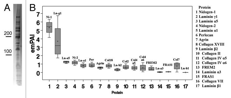
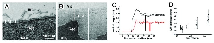
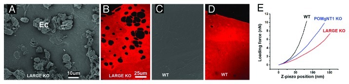
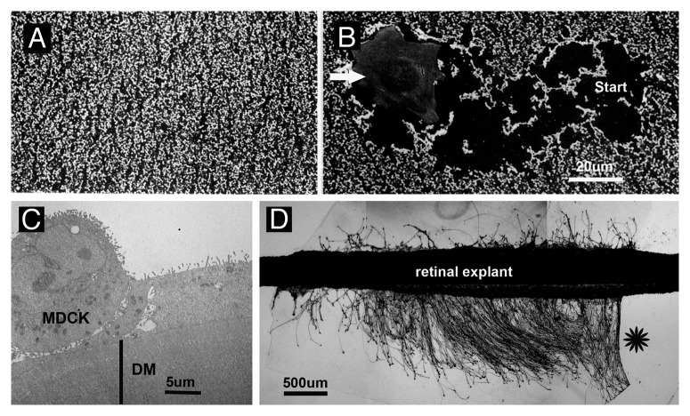
Basement membranes (BMs) evolved together with the first metazoan species approximately 500 million years ago. Main functions of BMs are stabilizing epithelial cell layers and connecting different types of tissues to functional, multicellular organisms. Mutations of BM proteins from worms to humans are either embryonic lethal or result in severe diseases, including muscular dystrophy, blindness, deafness, kidney defects, cardio-vascular abnormalities or retinal and cortical malformations. In vivo-derived BMs are difficult to come by; they are very thin and sticky and, therefore, difficult to handle and probe. In addition, BMs are difficult to solubilize complicating their biochemical analysis. For these reasons, most of our knowledge of BM biology is based on studies of the BM-like extracellular matrix (ECM) of mouse yolk sac tumors or from studies of the lens capsule, an unusually thick BM. Recently, isolation procedures for a variety of BMs have been described, and new techniques have been developed to directly analyze the protein compositions, the biomechanical properties and the biological functions of BMs. New findings show that native BMs consist of approximately 20 proteins. BMs are four times thicker than previously recorded, and proteoglycans are mainly responsible to determine the thickness of BMs by binding large quantities of water to the matrix. The mechanical stiffness of BMs is similar to that of articular cartilage. In mice with mutation of BM proteins, the stiffness of BMs is often reduced. As a consequence, these BMs rupture due to mechanical instability explaining many of the pathological phenotypes. Finally, the morphology and protein composition of human BMs changes with age, thus BMs are dynamic in their structure, composition and biomechanical properties.


Figures

None
Figure 1. BMs are detected in tissue sections of human skin by immunostaining with antibodies to BM-specific proteins, such as collagen IV (A) (red), or by TEM (B). The epidermal BM (BM) of the skin is located between the epidermis (Ep) and the underlying dermal stroma (St). The vascular BMs are detected along the endothelial lining of blood vessels (BV). When the skin is experimentally split into the epidermal (C) and the dermal layers (D), the epidermal BM consistently stays with the dermal connective tissue, as shown by anti-collagen IV staining (C and D). It is inseparable from the dermal connective tissue, making a clean preparation of this BM impossible (D). The sections were counter-stained with SytoxGreen. Bars: (A, C and D), 100 µm; (B) 500 nm.
None
Figure 2. Isolation of BMs and the gross morphology of isolated BMs. The DM is peeled off the inner surface of a detergent-treated human cornea (A). The BM is completely transparent. When imaged by SEM, the DM appears as clean ECM sheet without cellular contamination (B) (St, stromal surface; Ep, epithelial surface). Sheets of isolated ILM and retinal vascular BMs are shown in (C and D). The micrographs show the appearance of the BMs under a dissecting microscope using dark field. The vascular BM sheet (D) was from the foveal area of the retina. Note the foveal avascular zone in the middle of the sample. Bars: (B), 100 µm; (C and D), 500 µm.
None
Figure 3. Proteome analysis of a BM. A Coomassie-stained SDS PAGE of chick ILM proteins is shown in (A). Note that BM proteins typically have molecular weights between 150 kD and 1,000 kD. Semi-quantitative mass spectrometry analysis of the E10 chick ILM proteome based on emPAI values (B) shows that laminin and nidogens are the most prevalent proteins. The collagen IV members represent only a minor portion of the ILM proteome.
None
Figure 4. The thickness of human BMs. TEM images of a fetal (A) and an 83-year-old retina (B). White bars indicate the height of the ILMs. The ILM increases in thickness by a factor of 20 from 100 nm at fetal stages (A) to 1.5 µm at old age (B). In contrast to the even, regular and text-book-like fetal ILM (A), the retinal surface (Ret) of the aged ILM is highly irregular, whereas the vitreal side (Vit) is even and smooth (B). Representative AFM height measurements from a 44- and 88-y-old ILM confirm that the thickness of the ILMs increases with age (C). The scans extend from the flat glass surface on the left to the convoluted edge and further deeper into the BM sheet on the right. The thickness is measured by comparing the height difference of the glass surface and deep averaged ILM surface. The graph in (D) shows the age-related increase in ILM thickness as measured by AFM. The thickness of the ILMs, as measured by AFM, ranges from 0.4–4 µm, on average four-times greater than measured by TEM. Bar: (A and B), 500 nm.
None
Figure 5. Stiffness measurements of mutant and wild type mouse ILMs. SEM images of the vitreal surfaces of an intact retina from a LARGE mutant (LARGE KO) and of a wild type mouse (WT). The images show numerous ectopias (Ec) in the retina of the mutant mouse (A) and a continuous, smooth surface of the retina from the control mouse (C). ILMs isolated from the mutant mice have numerous perforations (B), while wild type mice have a continuous ILM (D); the ILM flat mounts had been stained for collagen IV. Representative force curves of ILMs from two different mutant mouse lines (LARGE and POMgNT1) show a much shallower slope than the force curves of ILMs from wild type mice (E) indicating a lower Young’s modulus. LARGE and POMgNT1 mutations affect the glycosylation of dystroglycan, one of the laminin receptors that are essential for BM assembly. Bars: (A and C), 10 µm; (B and D), 25 µm.
None
Figure 6. Cell adhesion/migration and axonal growth on embryonic chick ILM (A and B) and adult human DM (C and D). SEM micrograph showing an ILM whole mount from the embryonic chick retina. The BM is covered by a dense monolayer of neuroepithelial endfeet (A). When cells (Bowes human melanoma cells, arrow) are plated at low density onto this BM, the cells adhere and migrate on the BM by displacing the endfeet, and the cells leave behind a track as a record of their migratory behavior (B; start: site, where the cell adhered and started migration for 10 h). TEM micrograph showing a crossection of MDCK cells on a human DM after 4 d of culture. The cells attached to the BM and formed microvilli at the apical surface (C). When chick embryonic retina strips are cultured on an adult human DM flat mount, numerous axons grow out from the explants during 24 h in vitro. The neurites were visualized by anti-tubulin staining. Note, neurite outgrowth was restricted to the BM substrate; there was no neurite outgrowth on the poly-lysine coated plastic (star). Bars: (A and B), 20 µm; (C), 5 µm; (D) 500 µm.

References

    1. Yurchenco PD, Patton BL. Developmental and pathogenic mechanisms of basement membrane assembly. Curr Pharm Des. 2009;15:1277–94. doi: 10.2174/138161209787846766. - DOI - PMC - PubMed
    1. Candiello J, Cole GJ, Halfter W. Age-dependent changes in the structure, composition and biophysical properties of a human basement membrane. Matrix Biol. 2010;29:402–10. doi: 10.1016/j.matbio.2010.03.004. - DOI - PubMed
    1. Timpl R, Brown JC. Supramolecular assembly of basement membranes. Bioessays. 1996;18:123–32. doi: 10.1002/bies.950180208. - DOI - PubMed
    1. Chung AE. Embryonal carcinoma and the basement membrane glycoproteins laminin and entactin. Int J Dev Biol. 1993;37:141–50. - PubMed
    1. Stephens LE, Sutherland AE, Klimanskaya IV, Andrieux A, Meneses J, Pedersen RA, et al. Deletion of beta 1 integrins in mice results in inner cell mass failure and peri-implantation lethality. Genes Dev. 1995;9:1883–95. doi: 10.1101/gad.9.15.1883. - DOI - PubMed

Publication types

LinkOut - more resources