Distinct requirements for wnt9a and irf6 in extension and integration mechanisms during zebrafish palate morphogenesis
- PMID: 23154410
- PMCID: PMC6514306
- DOI: 10.1242/dev.080473
Distinct requirements for wnt9a and irf6 in extension and integration mechanisms during zebrafish palate morphogenesis
Abstract
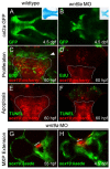
Development of the palate in vertebrates involves cranial neural crest migration, convergence of facial prominences and extension of the cartilaginous framework. Dysregulation of palatogenesis results in orofacial clefts, which represent the most common structural birth defects. Detailed analysis of zebrafish palatogenesis revealed distinct mechanisms of palatal morphogenesis: extension, proliferation and integration. We show that wnt9a is required for palatal extension, wherein the chondrocytes form a proliferative front, undergo morphological change and intercalate to form the ethmoid plate. Meanwhile, irf6 is required specifically for integration of facial prominences along a V-shaped seam. This work presents a mechanistic analysis of palate morphogenesis in a clinically relevant context.
Figures


Similar articles
-
Requirement for frzb and fzd7a in cranial neural crest convergence and extension mechanisms during zebrafish palate and jaw morphogenesis.Dev Biol. 2013 Sep 15;381(2):423-33. doi: 10.1016/j.ydbio.2013.06.012. Epub 2013 Jun 25. Dev Biol. 2013. PMID: 23806211
-
Zebrafish wnt9a is expressed in pharyngeal ectoderm and is required for palate and lower jaw development.Mech Dev. 2011 Jan-Feb;128(1-2):104-15. doi: 10.1016/j.mod.2010.11.003. Epub 2010 Nov 18. Mech Dev. 2011. PMID: 21093584
-
Roles of Wnt pathway genes wls, wnt9a, wnt5b, frzb and gpc4 in regulating convergent-extension during zebrafish palate morphogenesis.Development. 2016 Jul 15;143(14):2541-7. doi: 10.1242/dev.137000. Epub 2016 Jun 10. Development. 2016. PMID: 27287801 Free PMC article.
-
Closing the Gap: Mouse Models to Study Adhesion in Secondary Palatogenesis.J Dent Res. 2017 Oct;96(11):1210-1220. doi: 10.1177/0022034517726284. Epub 2017 Aug 17. J Dent Res. 2017. PMID: 28817360 Free PMC article. Review.
-
Extracellular Matrix in Secondary Palate Development.Anat Rec (Hoboken). 2020 Jun;303(6):1543-1556. doi: 10.1002/ar.24263. Epub 2019 Oct 30. Anat Rec (Hoboken). 2020. PMID: 31513730 Review.
Cited by
-
mir152 hypomethylation as a mechanism for non-syndromic cleft lip and palate.Epigenetics. 2022 Dec;17(13):2278-2295. doi: 10.1080/15592294.2022.2115606. Epub 2022 Sep 1. Epigenetics. 2022. PMID: 36047706 Free PMC article.
-
An Fgf-Shh signaling hierarchy regulates early specification of the zebrafish skull.Dev Biol. 2016 Jul 15;415(2):261-277. doi: 10.1016/j.ydbio.2016.04.005. Epub 2016 Apr 7. Dev Biol. 2016. PMID: 27060628 Free PMC article.
-
Orofacial Cleft and Mandibular Prognathism-Human Genetics and Animal Models.Int J Mol Sci. 2022 Jan 16;23(2):953. doi: 10.3390/ijms23020953. Int J Mol Sci. 2022. PMID: 35055138 Free PMC article. Review.
-
Wnt9a deficiency discloses a repressive role of Tcf7l2 on endocrine differentiation in the embryonic pancreas.Sci Rep. 2016 Jan 14;6:19223. doi: 10.1038/srep19223. Sci Rep. 2016. PMID: 26771085 Free PMC article.
-
Zebrafish Models of Craniofacial Malformations: Interactions of Environmental Factors.Front Cell Dev Biol. 2020 Nov 16;8:600926. doi: 10.3389/fcell.2020.600926. eCollection 2020. Front Cell Dev Biol. 2020. PMID: 33304906 Free PMC article. Review.
References
-
- Beaty T. H., Murray J. C., Marazita M. L., Munger R. G., Ruczinski I., Hetmanski J. B., Liang K. Y., Wu T., Murray T., Fallin M. D., et al. (2010). A genome-wide association study of cleft lip with and without cleft palate identifies risk variants near MAFB and ABCA4. Nat. Genet. 42, 525–529. - PMC - PubMed
-
- Ben J., Jabs E. W., Chong S. S. (2005). Genomic, cDNA and embryonic expression analysis of zebrafish IRF6, the gene mutated in the human oral clefting disorders Van der Woude and popliteal pterygium syndromes. Gene Expr. Patterns 5, 629–638. - PubMed
-
- Brugmann S. A., Goodnough L. H., Gregorieff A., Leucht P., ten Berge D., Fuerer C., Clevers H., Nusse R., Helms J. A. (2007). Wnt signaling mediates regional specification in the vertebrate face. Development 134, 3283–3295. - PubMed
Publication types
MeSH terms
Substances
Grants and funding
LinkOut - more resources
Full Text Sources
Molecular Biology Databases

