. 2013 Jan 1;140(1):136-46.
doi: 10.1242/dev.087791. Epub 2012 Nov 15.

Genetic elevation of sphingosine 1-phosphate suppresses dystrophic muscle phenotypes in Drosophila

Affiliations

Genetic elevation of sphingosine 1-phosphate suppresses dystrophic muscle phenotypes in Drosophila

Mario Pantoja et al. Development. .

Abstract

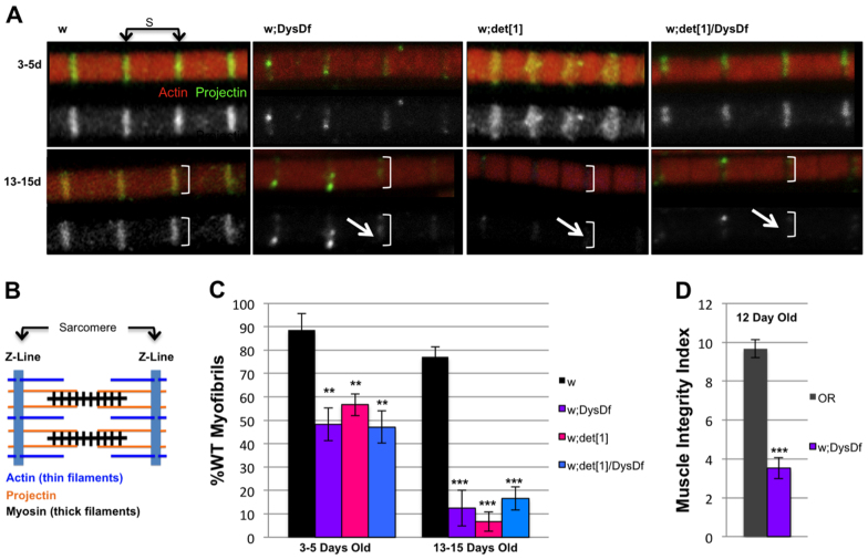
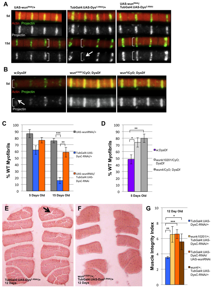
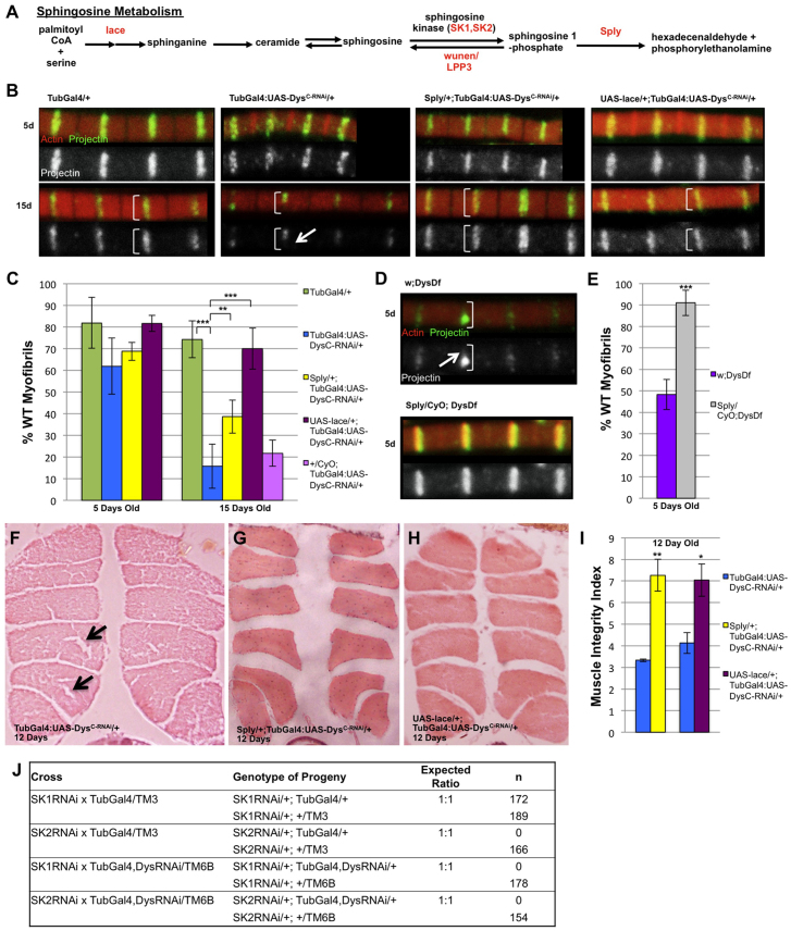
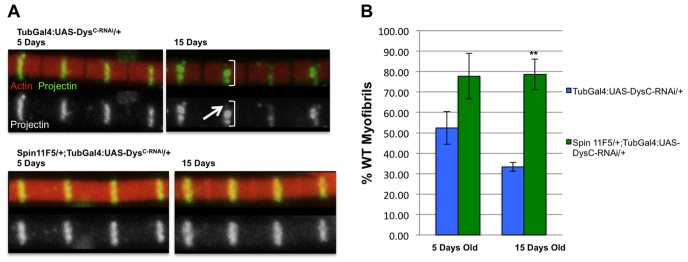
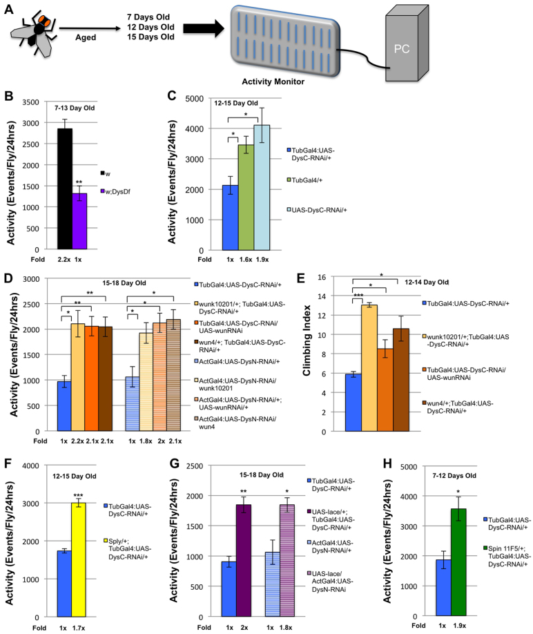
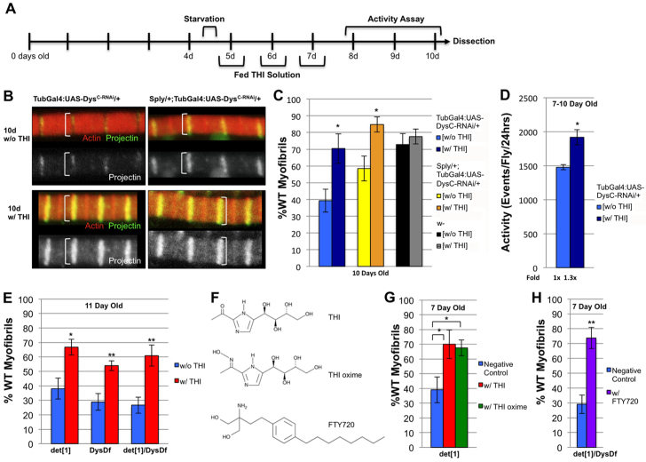
Duchenne muscular dystrophy is a lethal genetic disease characterized by the loss of muscle integrity and function over time. Using Drosophila, we show that dystrophic muscle phenotypes can be significantly suppressed by a reduction of wunen, a homolog of lipid phosphate phosphatase 3, which in higher animals can dephosphorylate a range of phospholipids. Our suppression analyses include assessing the localization of Projectin protein, a titin homolog, in sarcomeres as well as muscle morphology and functional movement assays. We hypothesize that wunen-based suppression is through the elevation of the bioactive lipid Sphingosine 1-phosphate (S1P), which promotes cell proliferation and differentiation in many tissues, including muscle. We confirm the role of S1P in suppression by genetically altering S1P levels via reduction of S1P lyase (Sply) and by upregulating the serine palmitoyl-CoA transferase catalytic subunit gene lace, the first gene in the de novo sphingolipid biosynthetic pathway and find that these manipulations also reduce muscle degeneration. Furthermore, we show that reduction of spinster (which encodes a major facilitator family transporter, homologs of which in higher animals have been shown to transport S1P) can also suppress dystrophic muscle degeneration. Finally, administration to adult flies of pharmacological agents reported to elevate S1P signaling significantly suppresses dystrophic muscle phenotypes. Our data suggest that localized intracellular S1P elevation promotes the suppression of muscle wasting in flies.


Figures

Fig. 1.
Fig. 1.
Age-dependent dystrophic myofibril phenotypes. (A) Confocal images of individual IFM myofibrils of 3- to 5-day-old and 13- to 15-day-old flies of wild type (w), w;DysDf, w;Dysdet1 and w;DysDf/Dysdet1. Actin (red) and Projectin (green, grayscale). White brackets indicate width of the myofibril. Arrows indicate a representative defective Projectin pattern. S marks the length of one sarcomere. (B) Schematic representation of a sarcomere. (C) Percentage of wild-type IFM myofibrils from each genotype shown in A at 3-5 days and 13-15 days. (D) Quantification of histological section data. Muscle integrity index is a weighted average of scored sections measuring the intactness of the IFMs (see Materials and methods). Ten flies were scored for each experiment; each experiment was repeated three times. **P<0.01, ***P<0.001. Error bars represent s.e.m.
Fig. 2.
Fig. 2.
Dystrophic flies with reduced wunen have less age-dependent degeneration over time. (A) Confocal images of individual IFM myofibrils of 5- and 15-day-old flies of the following genotypes (from left to right): undriven UAS-wunRNAi/+, tubulin-Gal4-driven Dys RNAi (TubGal4:UAS-DysC-RNAi/+) and tubulin-Gal4-driven wun RNAi, Dys RNAi double knockdown (UAS-wunRNAi/TubGal4:UAS-DysC-RNAi). (B) Confocal images of individual IFM myofibrils of 5-day-old flies of the following genotypes (from left to right): w;DysDf and the same Dys mutant with reduced wunen (wunk10201 and wun4). (C) Percentage of wild-type IFM myofibrils from each genotype shown in A at 5 and 15 days including UAS-wunRNAi/+, tubulin-Gal4-driven Dys RNAi (TubGal4:UAS-DysC-RNAi/+) and tubulin-Gal4-driven wun RNAi in Dys RNAi background (UAS-wunRNAi/TubGal4:UAS-DysC-RNAi). (D) Percentage of wild-type IFM myofibrils from each genotype shown in B at 5 days. (E) Tubulin-Gal4-driven Dys RNAi mutant transverse section (H&E stained) from a 12-day-old fly. Black arrow shows a hole in the flight muscle indicative of severe degeneration. (F) Transverse section (H&E stained) of a 12-day-old tubulin-Gal4-driven Dys RNAi mutant with reduced wunen (wunk10201) in the background. At least nine flies of each genotype were used per experiment. Each experiment was repeated three times. (G) Muscle integrity index of the tubulin-Gal4-driven Dys RNAi mutant alone and with reduced wunen (wunk10201, wunRNAi, wun4). In A and B, white brackets indicate width of the myofibril; white arrows indicate a representative defective Projectin pattern; Actin (red) and Projectin (green, grayscale). *P<0.05, **P<0.01, ***P<0.001. Error bars represent s.e.m.
Fig. 3.
Fig. 3.
Dystrophic muscle phenotypes are suppressed with altered sphingolipid metabolism gene dosage. (A) De novo sphingolipid synthesis pathway. (B) Confocal images of individual IFM myofibrils of 5- and 15-day-old flies of the following genotypes (from left to right): tubulin-Gal4 (TubGal4/+), tubulin-Gal4-driven Dys RNAi (TubGal4:UAS-DysC-RNAi/+), Sply, Dys double heterozygote (Sply/+; TubGal4:UAS-DysC-RNAi/+), and the Dys mutant with overexpressed lace (UAS-lace/+;TubGal4:UAS-DysC-RNAi/+). Actin (red) and Projectin (green, grayscale). White brackets indicate width of the myofibril. White arrows indicate a representative defective Projectin pattern. (C) Percentage of wild-type IFM myofibrils from each genotype shown in B at 5 and 15 days as well as from flies with tubulin-Gal4-driven Dys RNAi with the second chromosome balancer CyO (+/CyO;TubGal4:UAS-DysC-RNAi/+). (D) Confocal images of individual IFM myofibrils of 5-day-old flies of the following genotypes: w;DysDf and w; Sply/+; DysDf. (E) Graph quantifying the percentage of wild-type IFM myofibrils from each genotype shown in D at 5 days. (F) Transverse section (H&E stained) of a tubulin-Gal4-driven Dys RNAi 12-day-old mutant fly (TubGal4:UAS-DysC-RNAi/+). Black arrows indicate holes in the flight muscle indicative of the most severe degeneration. (G) Transverse section (H&E stained) of a tubulin-Gal4-driven Dys RNAi 12-day-old mutant with reduced Sply (Sply/+; TubGal4:UAS-DysC-RNAi/+). (H) Transverse section (H&E stained) of a tubulin-Gal4-driven Dys RNAi 12-day-old mutant with lace overexpressed (UAS-lace/+;TubGal4:UAS-DysC-RNAi/+). (I) Graph comparing the muscle integrity index of the tubulin-Gal4-driven Dys RNAi mutant alone (TubGal4:UAS-DysC-RNAi/+), with reduced Sply (Sply/+; TubGal4:UAS-DysC-RNAi/+), and with overexpressed lace (UAS-lace/+;TubGal4:UAS-DysC-RNAi/+). Eight flies were sectioned per experiment; each experiment was repeated three times; total n=24 for each genotype. (J) Table of viability of globally reducing the sphingosine kinases (SK1 and SK2) by expressing RNAi transgenes using the tubulin-Gal4 driver in the presence and absence of Dys RNAi. **P<0.01, ***P<0.001. Error bars represent s.e.m.
Fig. 4.
Fig. 4.
Suppression of the dystrophic myofibril phenotype by genetic reduction of Spinster, a putative S1P transporter. (A) Confocal images of individual IFM myofibrils of 5- and 15-day-old flies of the following genotypes: tubulin-Gal4-driven Dys RNAi (TubGal4:UAS-DysC-RNAi/+) and the spin11F5, Dys double heterozygote (spin11F5/+; TubGal4:UAS-DysC-RNAi/+). Actin (red) and Projectin (green, grayscale). White brackets indicate width of the myofibril. Arrow indicates a representative defective Projectin pattern. (B) Percentage of wild-type IFM myofibrils from each genotype shown in A at 5 and 15 days. **P<0.01. Error bars represent s.e.m.
Fig. 5.
Fig. 5.
Dystrophic movement phenotypes suppressed with genetic elevation of S1P. (A) Experimental flow for automated activity monitoring. (B) Activity analysis of 7-day-old w;DysDf and w control flies over 144 hours. (C) Activity analysis of 12-day-old tubulin-Gal4-driven Dys RNAi mutant and transgene control flies TubGal4/+ and UAS-DysC-RNAi/+ monitored over 72 hours. (D) Activity analysis of 15-day-old tubulin- or actin-Gal4-driven Dys RNAi mutants alone and combined with different wunen alleles (wunk10201, wunRNAi, wun4) over 72 hours. (E) Climbing index comparing 12- to 14-day-old dystrophic flies (TubGal4:UAS-DysC-RNAi/+) alone and with reduced wunen (wunk10201, wunRNAi, wun4). Twenty flies were used per experiment; each experiment was repeated three times. (F) Activity analysis of the 12-day-old tubulin-Gal4-driven Dys RNAi (TubGal4:UAS-DysC-RNAi/+) mutant alone and with reduced Sply (Sply/+; TubGal4:UAS-DysC-RNAi/+). (G) Activity analyses of 15-day-old tubulin- and actin-Gal4 Dys RNAi mutants alone and with overexpressed lace (TubGal4:UAS-DysC-RNAi/+, UAS-lace/+;TubGal4:UAS-DysC-RNAi/+, and ActGal4:UAS-DysN-RNAi/+ and UAS-lace/ActGal4:UAS-DysN-RNAi). (H) Activity analysis of the 7-day-old tubulin-Gal4-driven Dys RNAi (TubGal4:UAS-DysC-RNAi/+) mutant alone and with reduced spin (spin11F5/+; TubGal4:UAS-DysC-RNAi/+). *P<0.05, **P<0.01, ***P<0.001. Error bars represent s.e.m.
Fig. 6.
Fig. 6.
Dystrophic phenotypes suppressed with THI treatment during adulthood. (A) Experimental timeline of THI administration to Drosophila. (B) Confocal images of individual IFM myofibrils isolated at 10 days of tubulin-Gal4-driven Dys RNAi (TubGal4:UAS-DysC-RNAi/+) mutants alone and with reduced Sply (Sply/+; TubGal4:UAS-DysC-RNAi/+) that were fed a corn syrup solution with and without THI. Actin (red) and Projectin (green, grayscale). White brackets indicate the width of the myofibril. Arrow indicates reduction/loss of Projectin staining at the Z-band. (C) Percentage of wild-type myofibrils from tubulin-Gal4-driven Dys RNAi (TubGal4:UAS-DysC-RNAi/+), Sply, Dys double heterozygote (Sply/+; TubGal4:UAS-DysC-RNAi/+) flies fed a corn syrup solution with and without THI. (D) Activity analysis of tubulin-Gal4-driven Dys RNAi (TubGal4:UAS-DysC-RNAi/+) flies fed a corn syrup solution with and without THI. (E) Percentage of wild-type myofibrils from 11-day-old Dys mutants (Dysdet1, DysDf and Dysdet1/DysDf) fed a corn syrup solution with and without THI. (F) Chemical structures of THI, THI oxime and FTY720. (G) Percentage of wild-type myofibrils from 7-day-old Dysdet1 flies fed a corn syrup solution with and without THI and THI oxime. (H) Percentage of wild-type myofibrils from 7-day-old Dysdet1/DysDf flies fed a corn syrup solution with and without FTY720. For activity assays, six flies of each genotype listed on the x-axis were analyzed per experiment; each experiment was repeated three times. Total time per experiment was 72 hours; total n=18. *P<0.05. Error bars represent s.e.m.

Similar articles

Cited by

References

    1. Adachi-Yamada T., Gotoh T., Sugimura I., Tateno M., Nishida Y., Onuki T., Date H. (1999). De novo synthesis of sphingolipids is required for cell survival by down-regulating c-Jun N-terminal kinase in Drosophila imaginal discs. Mol. Cell. Biol. 19, 7276–7286 - PMC - PubMed
    1. Ayme-Southgate A., Saide J., Southgate R., Bounaix C., Cammarato A., Patel S., Wussler C. (2005). In indirect flight muscles Drosophila projectin has a short PEVK domain, and its NH2-terminus is embedded at the Z-band. J. Muscle Res. Cell Motil. 26, 467–477 - PubMed
    1. Bagdanoff J. T., Donoviel M. S., Nouraldeen A., Tarver J., Fu Q., Carlsen M., Jessop T. C., Zhang H., Hazelwood J., Nguyen H., et al. (2009). Inhibition of sphingosine-1-phosphate lyase for the treatment of autoimmune disorders. J. Med. Chem. 52, 3941–3953 - PubMed
    1. Bagdanoff J. T., Donoviel M. S., Nouraldeen A., Carlsen M., Jessop T. C., Tarver J., Aleem S., Dong L., Zhang H., Boteju L., et al. (2010). Inhibition of sphingosine 1-phosphate lyase for the treatment of rheumatoid arthritis: discovery of (E)-1-(4-((1R,2S,3R)-1,2,3,4-tetrahydroxybutyl)-1H-imidazol-2-yl)ethanone oxime (LX2931) and (1R,2S,3R)-1-(2-(isoxazol-3-yl)-1H-imidazol-4-yl)butane-1,2,3,4-tetraol (LX2932). J. Med. Chem. 53, 8650–8662 - PubMed
    1. Benzer S. (1967). Behavioural mutants of Drosophila isolated by countercurrent distribution. Proc. Natl. Acad. Sci. USA 58, 1112–1119 - PMC - PubMed

Publication types

MeSH terms