

Residual malignant and normal plasma cells shortly after high dose melphalan and stem cell transplantation. Highlight of a putative therapeutic window in Multiple Myeloma?
- PMID: 23154454
- PMCID: PMC4539173
- DOI: 10.18632/oncotarget.650
Residual malignant and normal plasma cells shortly after high dose melphalan and stem cell transplantation. Highlight of a putative therapeutic window in Multiple Myeloma?
Abstract
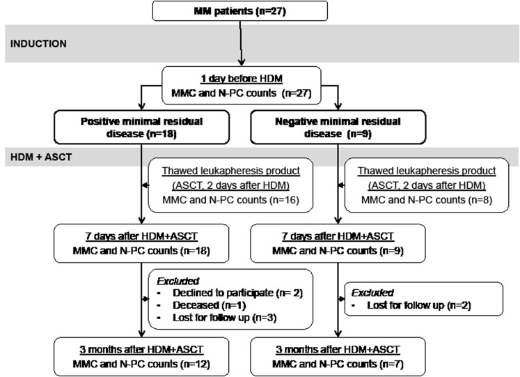
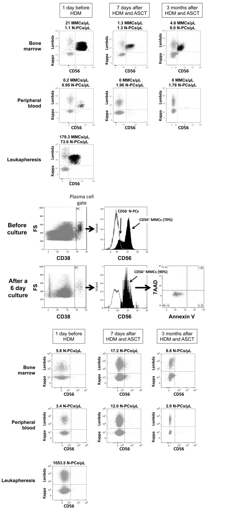
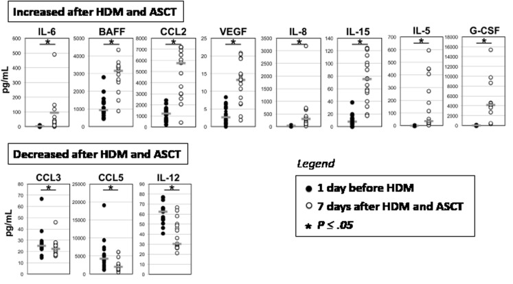
Multiple Myeloma (MM) is an incurable malignant plasma cell disorder. We have evaluated the counts of Multiple Myeloma Cells (MMCs) and normal plasma cells (N-PCs), seven days after high-dose melphalan (HDM) and autologous stem transplantation (ASCT). Two third of patients had detectable minimal residual disease (MRD+) (71.7 MMCs/µL) after induction treatment with dexamethasone and proteasome inhibitor. MMC counts were reduced by 92% (P ≤ .05) but not eradicated 7 days after HDM+ASCT. Post-HDM+ASCT MMCs were viable and bathed in a burst of MMC growth factors, linked with post-HDM aplasia. In one third of patients (MRD- patients), MMCs were not detectable after induction treatment and remained undetectable after HDM+ASCT. Major difference between MRD- and MRD+ patients is that N-PC counts were increased 3 fold (P〈.05) by HDM+ASCT in MRD- patients, but were unaffected in MRD+ patients. Possible explanation could be that clearance of MMCs in MRD- patients makes more niches available for N-PCs. Thus, MMCs are not fully eradicated shortly after HDM, are bathed in high concentrations of MMC growth factors in an almost desert BM, are viable in short-term culture, which suggests providing additional therapies shortly after HDM to kill resistant MMCs before full repair of lesions.
Conflict of interest statement
The authors reported no potential conflicts of interest.
Figures
Similar articles
-
Prognostic value of sequencing-based minimal residual disease detection in patients with multiple myeloma who underwent autologous stem-cell transplantation.Ann Oncol. 2017 Oct 1;28(10):2503-2510. doi: 10.1093/annonc/mdx340. Ann Oncol. 2017. PMID: 28945825 Free PMC article.
-
The role of high-dose melphalan with autologous stem-cell transplant in multiple myeloma: is it time for a paradigm shift?Br J Haematol. 2020 Dec;191(5):692-703. doi: 10.1111/bjh.16764. Epub 2020 Jun 5. Br J Haematol. 2020. PMID: 32501533 Free PMC article. Review.
-
The changing role of high dose melphalan with stem cell rescue in the treatment of newly diagnosed multiple myeloma in the era of modern therapies-back to the future!Best Pract Res Clin Haematol. 2020 Mar;33(1):101150. doi: 10.1016/j.beha.2020.101150. Epub 2020 Jan 17. Best Pract Res Clin Haematol. 2020. PMID: 32139015 Review.
-
Single versus tandem high-dose melphalan followed by autologous blood stem cell transplantation in multiple myeloma: long-term results from the phase III GMMG-HD2 trial.Br J Haematol. 2016 Jun;173(5):731-41. doi: 10.1111/bjh.13994. Epub 2016 Mar 17. Br J Haematol. 2016. PMID: 26990892 Clinical Trial.
-
The role of high-dose melphalan and autologous stem cell transplant in the rapidly evolving era of modern multiple myeloma therapy.Clin Adv Hematol Oncol. 2016 Sep;14(9):719-28. Clin Adv Hematol Oncol. 2016. PMID: 27673290 Review.
Cited by
-
Inhibiting the anaphase promoting complex/cyclosome induces a metaphase arrest and cell death in multiple myeloma cells.Oncotarget. 2016 Jan 26;7(4):4062-76. doi: 10.18632/oncotarget.6768. Oncotarget. 2016. PMID: 26716651 Free PMC article.
-
Treatment Strategies Considering Micro-Environment and Clonal Evolution in Multiple Myeloma.Cancers (Basel). 2021 Jan 8;13(2):215. doi: 10.3390/cancers13020215. Cancers (Basel). 2021. PMID: 33435539 Free PMC article. Review.
-
The role of DNA damage and repair in decitabine-mediated apoptosis in multiple myeloma.Oncotarget. 2014 May 30;5(10):3115-29. doi: 10.18632/oncotarget.1821. Oncotarget. 2014. PMID: 24833108 Free PMC article.
-
Targeted Therapies in Adult B-Cell Malignancies.Biomed Res Int. 2015;2015:217593. doi: 10.1155/2015/217593. Epub 2015 Sep 6. Biomed Res Int. 2015. PMID: 26425544 Free PMC article. Review.
-
Antioxidant Defenses Confer Resistance to High Dose Melphalan in Multiple Myeloma Cells.Cancers (Basel). 2019 Mar 28;11(4):439. doi: 10.3390/cancers11040439. Cancers (Basel). 2019. PMID: 30925767 Free PMC article.
References
-
- Becker N. Epidemiology of multiple myeloma. Recent Results Cancer Res. 2011;183:25–35. - PubMed
-
- Cavo M, Tacchetti P, Patriarca F, Petrucci MT, Pantani L, Galli M, Di Raimondo F, Crippa C, Zamagni E, Palumbo A, Offidani M, Corradini P, Narni F, Spadano A, Pescosta N, Deliliers GL, et al. Bortezomib with thalidomide plus dexamethasone compared with thalidomide plus dexamethasone as induction therapy before, and consolidation therapy after, double autologous stem-cell transplantation in newly diagnosed multiple myeloma: a randomised phase 3 study. Lancet. 2010;376(9758):2075–2085. - PubMed
-
- Paiva B, Almeida J, Perez-Andres M, Mateo G, Lopez A, Rasillo A, Vidriales MB, Lopez-Berges MC, Miguel JF, Orfao A. Utility of flow cytometry immunophenotyping in multiple myeloma and other clonal plasma cell-related disorders. Cytometry B Clin Cytom. 2010;78(4):239–252. - PubMed
Publication types
MeSH terms
Substances
LinkOut - more resources
Full Text Sources
Other Literature Sources
Medical
