. 2013 Jan 4;288(1):633-43.
doi: 10.1074/jbc.M112.347682. Epub 2012 Nov 15.

ras-Induced up-regulation of CTP:phosphocholine cytidylyltransferase α contributes to malignant transformation of intestinal epithelial cells

Affiliations

ras-Induced up-regulation of CTP:phosphocholine cytidylyltransferase α contributes to malignant transformation of intestinal epithelial cells

Daniel J Arsenault et al. J Biol Chem. .

Abstract

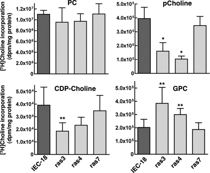
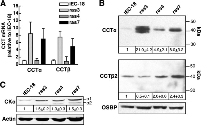
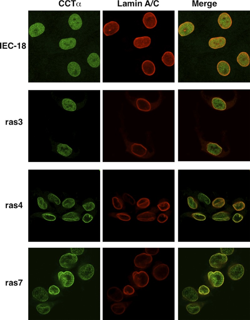
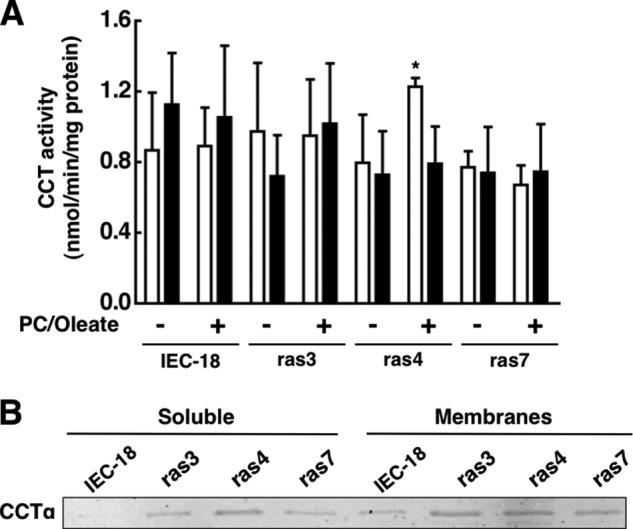
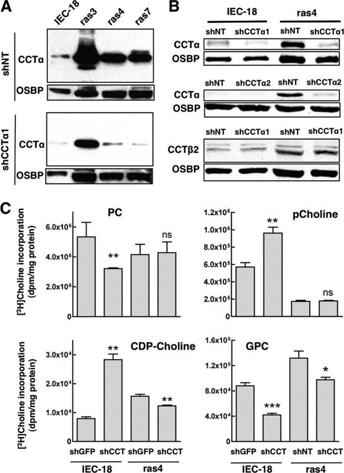
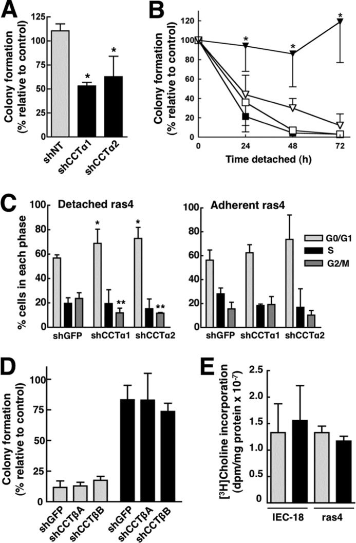
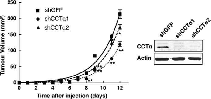
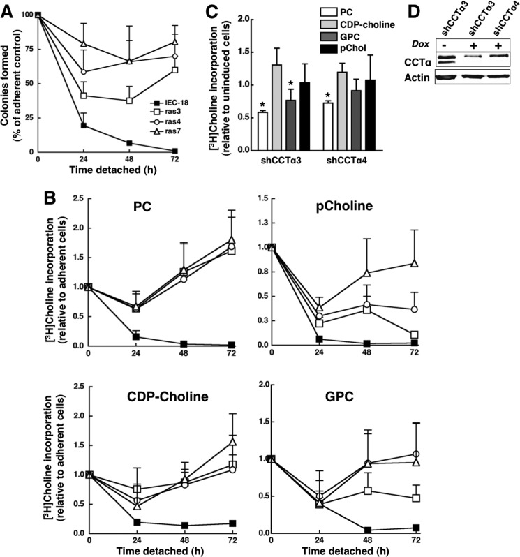
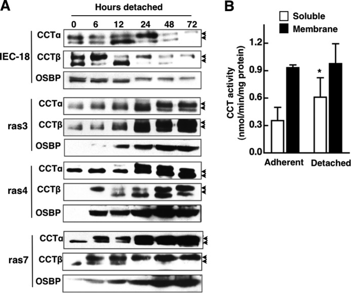
Cancer cells have enhanced lipogenic capacity characterized by increased synthesis of fatty acids and complex lipids, including phosphatidylcholine (PC). As the rate-limiting enzyme in the CDP-choline pathway for PC synthesis, CTP:phosphocholine cytidylyltransferase α (CCTα) is implicated in the provision of membranes and bioactive lipids necessary of cell proliferation. In this study, we assessed the role of CCTα in malignant intestinal epithelial cells transformed with activated H-ras (IEC-ras). Three IEC-ras clones had significant up-regulation CCTα expression, but PC synthesis and in vitro activity of CCTα were similar to control IEC. RNA interference of CCTα in adherent IEC-ras did not affect PC synthesis, confirming that the enzyme was relatively inactive. However, CCTα silencing in ras-transformed IEC reduced anchorage-independent growth, a criterion for malignant transformation, as well as tumorigenicity in mice. Relative to their adherent counterparts, detached IEC-ras had increased PC synthesis that was attenuated by inducible CCTα silencing. Detachment of IEC-ras was accompanied by increased CCTα phosphorylation and cytosolic enzyme activity. We conclude that the expanded pool of CCTα in IEC-ras is activated by detachment. This provides the increased PC biosynthetic capacity that contributes to malignant transformation of intestinal epithelial cells when detached from the extracellular matrix.


Figures

FIGURE 1.
FIGURE 1.
[3H]Choline incorporation into PC and choline metabolites of IEC-18 and IEC-ras cells. IEC-18, ras3, ras4, and ras7 cells were incubated with [3H]choline for 3 h, and incorporation into PC, pCholine, GPC, and CDP-choline was measured as described under “Experimental Procedures.” Results are the mean and S.D. (error bars) of three independent experiments. *, p < 0.01; **, p < 0.05 compared with IEC.
FIGURE 2.
FIGURE 2.
CCTα expression in adherent and detached IEC-18 and IEC-ras cells. A, CCTα and CCTβ mRNA were quantified by qPCR, normalized to GAPDH expression, and expressed relative to IEC-18. Results are the mean and S.D. (error bars) of 4–6 experiments. B, total cell lysates prepared from IEC-18 and IEC-ras cells cultured on plastic dishes were resolved by SDS-8% PAGE and immunoblotted for CCTα, CCTβ, and oxysterol-binding protein (OSBP), as described under “Experimental Procedures.” CCTα and CCTβ expression was normalized to the load control (oxysterol-binding protein) and expressed relative to IEC-18 (mean and S.D.). C, cell lysates prepared as described above were immunoblotted for the α1 and α2 isoforms of CK, and total CK was quantified relative to actin. Results are the mean and S.D. of four experiments.
FIGURE 3.
FIGURE 3.
CCTα is localized to the nucleoplasm and nuclear envelope in IEC-ras cells. IEC-18, ras3, ras4, and ras7 cells cultured on glass coverslips were immunostained with the following primary and secondary antibodies: rabbit anti-CCTα and AlexaFluor488-conjugated goat anti-rabbit; goat anti-lamin A/C and AlexaFluor594-conjugated donkey anti-goat. Images are 1-μm optical sections through the mid-nuclei captured using a Zeiss LSM520 Meta confocal microscope.
FIGURE 4.
FIGURE 4.
CCTα activity in cytosol and membranes of IEC-18 and IEC-ras cells. A, CCT activity was measured in the soluble (open bars) and membrane particulate fractions (black bars) from IEC-18 and IEC-ras in the presence or absence of PC/oleate vesicles as described under “Experimental Procedures.” Results are the mean and S.D. (error bars) of three experiments. B, the soluble and membrane particulate fraction (25 μg) of IEC-18 and IEC-ras cells were separated by SDS-8% PAGE and immunoblotted for CCTα. *, p < 0.05 compared with IEC-18 activity assayed under the same conditions.
FIGURE 5.
FIGURE 5.
Effect of CCTα silencing on PC metabolism in IEC-18 and IEC-ras cells. A, lentivirus encoding an shRNA against CCTα (shCCTα1) or a non-targeting shRNA control (shNT) were expressed in IEC-18 and IEC-ras cells and selected with puromycin for 24 h. Total cell lysates were resolved by SDS-8% PAGE and immunoblotted for CCTα and oxysterol-binding protein (OSBP). B, knockdown of CCTα by lentiviral shCCTα1 and shCCTα2 was determined in IEC-18 and ras4 cells. CCTβ2 expression was determined in shCCTα1-transduced cells. C, IEC-18 and ras4 cells transduced with shGFP or shCCTα1 were pulse-labeled with [3H]choline (2 μCi/ml) for 3 h. Cells were harvested, and [3H]choline incorporation into PC, pCholine, CDP-choline, and GPC was measured as described under “Experimental Procedures.” Results are the mean and S.D. (error bars) of three independent experiments. *, p < 0.1; **, p < 0.05; ***, p > 0.01; ns, not significant compared with shGFP controls.
FIGURE 6.
FIGURE 6.
Knockdown of CCTα, but not CCTβ, sensitizes ras4 cells to anoikis. A, lentiviral shCCTα1, shCCTα2, or a non-targeting control (shNT) were transduced into ras4 cells and selected with puromycin for 48 h. Cells (500) were suspended in 0.3% agar, and colonies were quantified after 5 days in culture. Results are the mean and S.D. of three experiments. *, p < 0.05 compared with shNT ras4 cells. B, IEC-18 (squares) and ras4 cells (triangles) transduced with shNT (■ and ▾) or shCCTα1 (□ and ▿) were cultured on SeaPlaque agarose for the indicated times and transferred to plastic dishes, and colonies were quantified after 3–4 days as described under “Experimental Procedures.” Results are the mean and S.D. (error bars) of 5–6 experiments. *, p < 0.05 compared with shNT ras4 cells. C, the cell cycle distribution of shGFP-transduced (control), shCCTα1-transduced, or shCCTα2-transduced ras4 cells was determined under adherent and 24-h detached conditions as described under “Experimental Procedures.” Results are the mean and S.D. of three experiments. *, p < 0.05; **, p < 0.005, compared with shGFP control. D, IEC-18 (gray bars) and ras4 cells (black bars) were transduced with lentiviral shGFP, shCCTβ1, or shCCTβ2 and cultured on SeaPlaque agarose for 48 h prior to seeding on plastic dishes for 3–4 days as described in B. Results are the mean and S.D. of 3–6 experiments. E, [3H]choline incorporation into PC was measured in adherent IEC-18 and ras4 cells transduced with shGFP (gray bars) or shCCTβ2 (black bars) as described in the legend to Fig. 6. Results are the mean and S.D. of three experiments.
FIGURE 7.
FIGURE 7.
Activation and/or overexpression of CCTα does not grant anchorage-independent growth to IEC-18. After transfection with pCCTα-GFP or empty vector for 24 h, IEC-18 (500 cells) were cultured on 60-mm dishes coated with SeaPlaque agarose in media supplemented without or with 300 μm oleate complexed to BSA for 72 h. Cells were then seeded on plastic dishes, and colony formation was determined. CCTα-GFP overexpression relative to endogenous CCTα was determined after SDS-8% PAGE and immunoblotting using a CCTα antibody. Results are the means and S.D. (error bars) of three separate experiments.
FIGURE 8.
FIGURE 8.
Tumor growth of ras4 cells is reduced by CCTα silencing. ras4 cells (2 × 10⁵) expressing shCCTα1, shCCTα2, or shGFP were resuspended in sterile PBS and injected subcutaneously into the right flank of athymic mice. Tumor growth was measured daily using a skin fold caliper. Results are the mean and S.D. (error bars) of three separate experiments involving 8–12 mice. *, p < 0.05; **, p < 0.005 compared with shGFP. The inset shows a representative immunoblot of CCTα expression in ras4 cells prior to injection into mice.
FIGURE 9.
FIGURE 9.
Detached IEC-ras cells have sustained growth and PC synthesis. A, IEC, ras3, ras4, and ras7 cells were assayed for viability using a colonogenicity assay as described under “Experimental Procedures.” Results are the mean and S.D. of three experiments. B, IEC-18 (■), ras3 (□), ras4 (○), and ras7 (▵) cells were cultured on plastic dishes or dishes coated with 1% SeaPlaque agarose in MEM containing [3H]choline (2 μCi/ml) for 0–72 h. At the indicated times, cells were harvested, and [3H]choline incorporation into PC, pCholine, GPC, and CDP-choline was determined and quantified relative to cellular protein. The ratio of specific incorporation into PC and choline metabolites of detached versus adherent cells was determined as an estimate of relative biosynthetic rates. Results are the mean and S.D. (error bars) of five separate experiments. C, ras4 cells were transduced with TripZ lentiviral shCCTα3 or shCCTα4 and selected under adherent conditions. Cells were then cultured on agarose for 24 h, followed by induction of shRNAs with 1 μg/ml doxycycline for 48 h, at which time [3H]choline (1 μCi/ml) incorporation into PC and water-soluble metabolites was measured by a 3-h pulse. Results are expressed relative to cells that were not induced with doxycycline and are the mean and S.D. of four experiments. *, p < 0.05 compared with uninduced cells. D, lysates from ras4 cells cutured on agarose for 48 h in the absence or presence or doxycycline were immunoblotted for CCTα.
FIGURE 10.
FIGURE 10.
Effect of anchorage-independent growth on CCTα expression and activity. A, CCTα and CCTβ expression in cell lysates from IEC-18 and IEC-ras cells cultured on agarose for 0–72 h was analyzed by immunoblotting. Because of the high level of CCTα overexpression in IEC-ras cells, 4 times more IEC lysate was loaded, and exposure times were adjusted to improve resolution and comparison. The arrows denote the upper hyperphosphorylated and lower hypophosphorylated forms of CCTα. B, CCT activity in the soluble and membrane (particulate fractions) of adherent and 72-h detached ras4 cells. Results are the mean and S.D. (error bars) of three separate experiments. *, p < 0.05 compared with activity in the soluble fraction from adherent cells.

References

    1. Hsu P. P., Sabatini D. M. (2008) Cancer cell metabolism. Warburg and beyond. Cell 134, 703–707 - PubMed
    1. Menendez J. A., Lupu R. (2007) Fatty acid synthase and the lipogenic phenotype in cancer pathogenesis. Nat. Rev. Cancer 7, 763–777 - PubMed
    1. Bauer D. E., Hatzivassiliou G., Zhao F., Andreadis C., Thompson C. B. (2005) ATP citrate lyase is an important component of cell growth and transformation. Oncogene 24, 6314–6322 - PubMed
    1. Hatzivassiliou G., Zhao F., Bauer D. E., Andreadis C., Shaw A. N., Dhanak D., Hingorani S. R., Tuveson D. A., Thompson C. B. (2005) ATP citrate lyase inhibition can suppress tumor cell growth. Cancer Cell 8, 311–321 - PubMed
    1. Brusselmans K., De Schrijver E., Verhoeven G., Swinnen J. V. (2005) RNA interference-mediated silencing of the acetyl-CoA-carboxylase-α gene induces growth inhibition and apoptosis of prostate cancer cells. Cancer Res. 65, 6719–6725 - PubMed

Publication types

Substances

LinkOut - more resources