

Enterolithiasis-associated ileus in Crohn's disease
- PMID: 23155347
- PMCID: PMC3496895
- DOI: 10.3748/wjg.v18.i42.6160
Enterolithiasis-associated ileus in Crohn's disease
Abstract
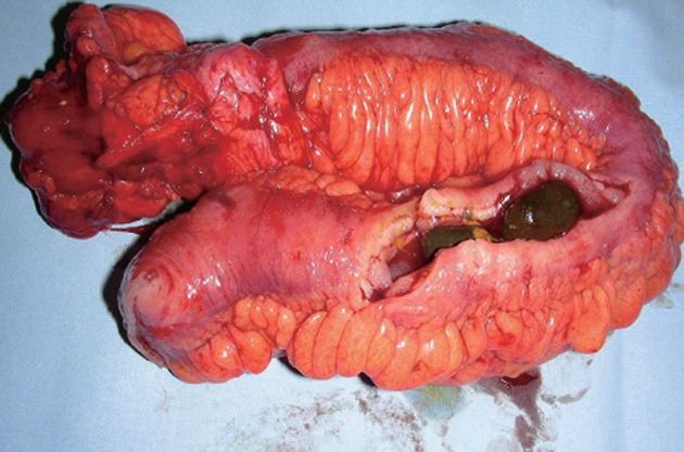
Stasis of the flow of the intestinal contents, ingested material and unfavorable composition of the chylus can lead to the formation of enteroliths inside the bowel. Enterolithiasis represents a rare disorder of the gastrointestinal tract that can be associated with intermittent abdominal pain or more serious complications such as bleeding or obstruction. Enterolithiasis in Crohn's disease represents an extremely rare condition and usually occurs only in patients with a long symptomatic history of Crohn's disease. We report an unusual case of enterolithiasis-related intestinal obstruction in a young male patient with Crohn's disease (A2L3B1 Montreal Classification for Crohn's disease 2005) undergoing emergency laparotomy and ileocoecal resection. In addition, we present an overview of the relevant characteristics of enterolithiasis on the basis of the corresponding literature.
Keywords: Crohn’s disease; Enterolithiasis; Ileus; Inflammatory bowel disease; Obstruction.
Figures


References
-
- Martens T, Sas S. Enteroliths in Crohn’s disease: a case report. Acta Chir Belg. 2010;110:552–554. - PubMed
-
- Atwell JD, Pollock AV. Intestinal calculi. Br J Surg. 1960;47:367–374. - PubMed
-
- Grettve S. A contribution to the knowledge of primary true concrements in the small bowel. Acta Chir Scand. 1947;95:387–410. - PubMed
Publication types
MeSH terms
LinkOut - more resources
Full Text Sources
Medical
