. 2012;7(11):e48299.
doi: 10.1371/journal.pone.0048299. Epub 2012 Nov 14.

Transforming growth factor alpha (TGFα) regulates granulosa cell tumor (GCT) cell proliferation and migration through activation of multiple pathways

Affiliations

Transforming growth factor alpha (TGFα) regulates granulosa cell tumor (GCT) cell proliferation and migration through activation of multiple pathways

Cheng Wang et al. PLoS One. 2012.

Abstract

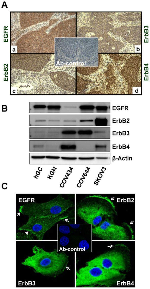
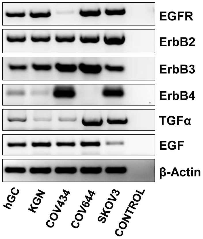
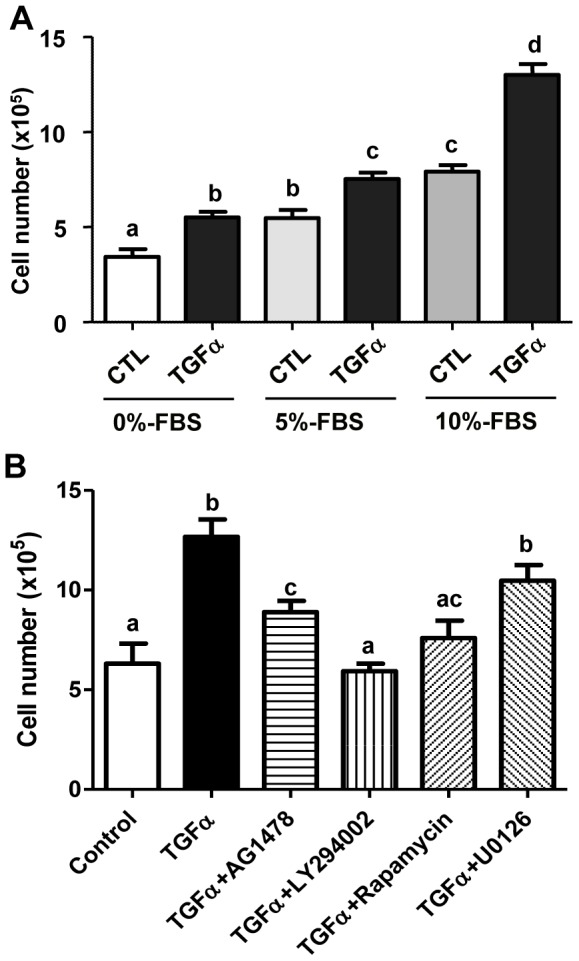
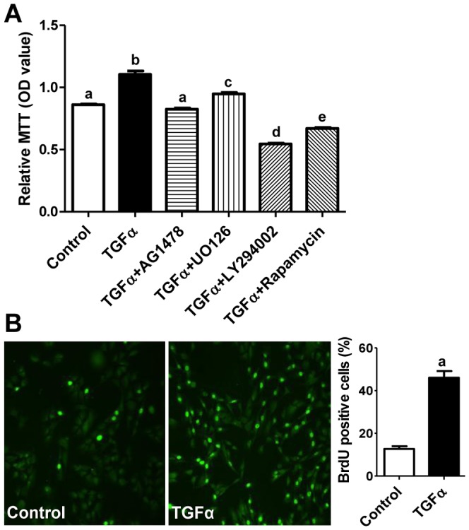
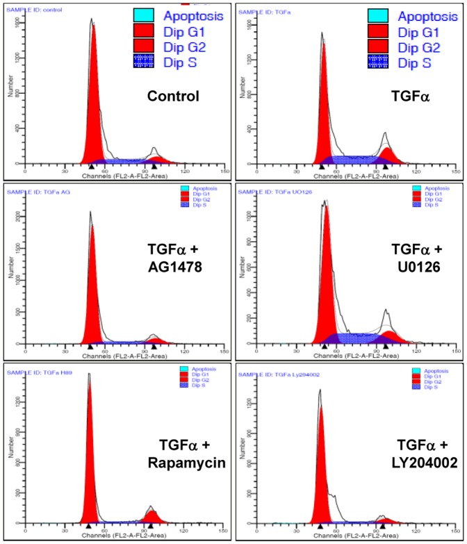
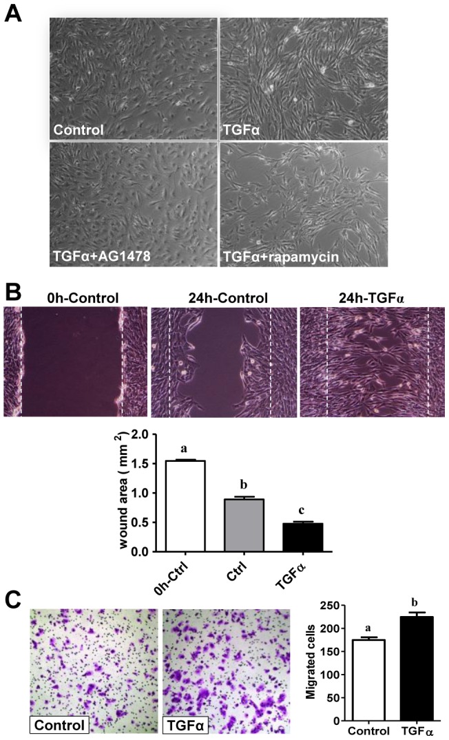
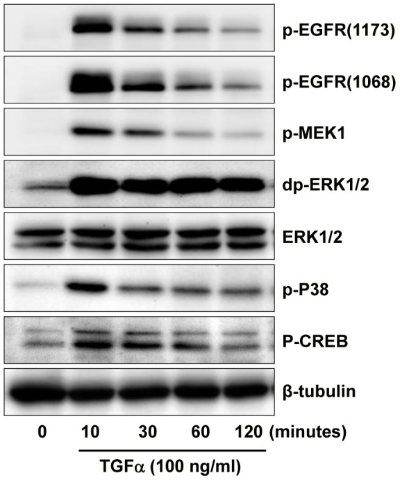
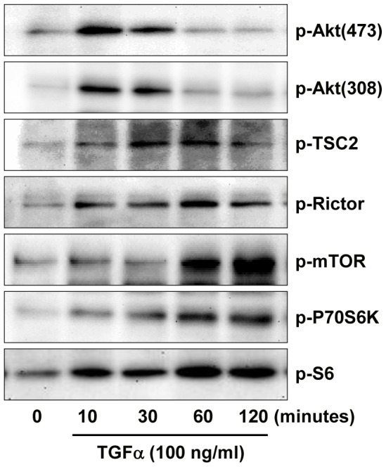
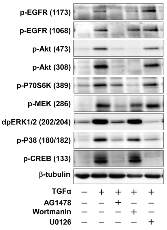
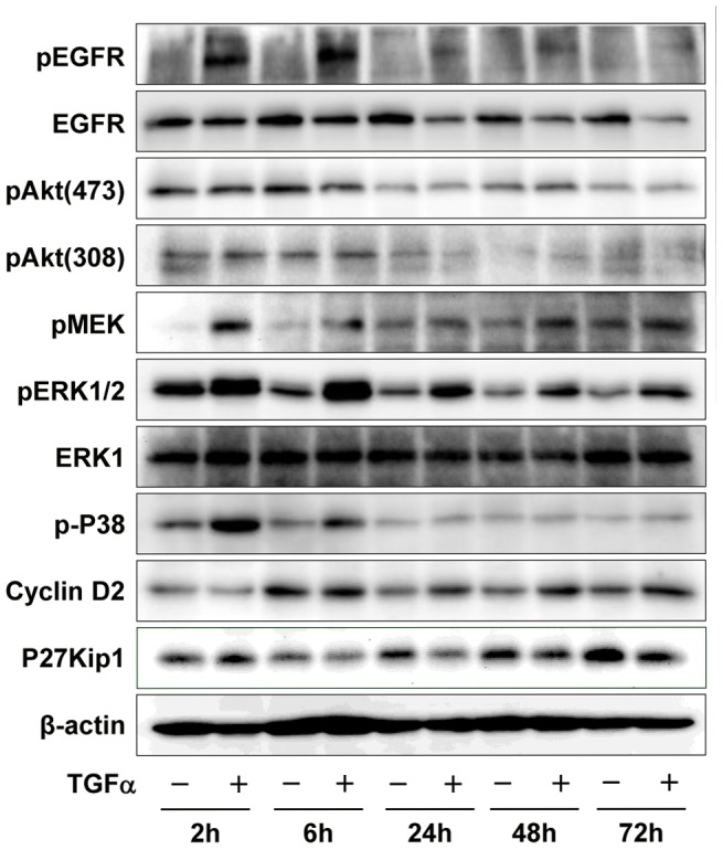
Granulosa cell tumors (GCTs) are the most common ovarian estrogen producing tumors, leading to symptoms of excessive estrogen such as endometrial hyperplasia and endometrial adenocarcinoma. These tumors have malignant potential and often recur. The etiology of GCT is unknown. TGFα is a potent mitogen for many different cells. However, its function in GCT initiation, progression and metastasis has not been determined. The present study aims to determine whether TGFα plays a role in the growth of GCT cells. KGN cells, which are derived from an invasive GCT and have many features of normal granulosa cells, were used as the cellular model. Immunohistochemistry, Western blot and RT-PCR results showed that the ErbB family of receptors is expressed in human GCT tissues and GCT cell lines. RT-PCR results also indicated that TGFα and EGF are expressed in the human granulosa cells and the GCT cell lines, suggesting that TGFα might regulate GCT cell function in an autocrine/paracrine manner. TGFα stimulated KGN cell DNA synthesis, cell proliferation, cell viability, cell cycle progression, and cell migration. TGFα rapidly activated EGFR/PI3K/Akt and mTOR pathways, as indicated by rapid phosphorylation of Akt, TSC2, Rictor, mTOR, P70S6K and S6 proteins following TGFα treatment. TGFα also rapidly activated the EGFR/MEK/ERK pathway, and P38 MAPK pathways, as indicated by the rapid phosphorylation of EGFR, MEK, ERK1/2, P38, and CREB after TGFα treatment. Whereas TGFα triggered a transient activation of Akt, it induced a sustained activation of ERK1/2 in KGN cells. Long-term treatment of KGN cells with TGFα resulted in a significant increase in cyclin D2 and a decrease in p27/Kip1, two critical regulators of granulosa cell proliferation and granulosa cell tumorigenesis. In conclusion, TGFα, via multiple signaling pathways, regulates KGN cell proliferation and migration and may play an important role in the growth and metastasis of GCTs.


Conflict of interest statement

Competing Interests: The authors have declared that no competing interests exist.

Figures

Figure 1
Figure 1. Expression of ErbB receptor family members in human ovarian granulosa cell tumors (GCTs).
A) Immunohistochemistry was used to detect the expression of EGFR (a), ErbB2 (b), ErbB3 (c) and ErbB4 (d) in paraffin-embedded human GCT tissues. Magnification, 400×. B) Western blot detection of expression of ErbB family receptor proteins in human ovarian granulosa cells and GCT cell lines. SKOV-3 and COV644 cells were used as controls for the detection system. C) Fluorescent immunohistochemistry localization of ErbB receptors in KGN cells. The images were captured with a confocal laser scanning microscope. Arrows indicate the membrane localization of ErbBs in KGN cells. Magnification, 630×; Ab-control: 2nd antibody only control, magnification, 200×.
Figure 2
Figure 2. Expression of TGFα, EGF and ErbB family receptor mRNA in KGN and COV434 GCT cell lines.
SKOV-3 (ErbB2 overexpressing cells) and COV644 cells (ErbB4 negative cells) were used as positive and negative controls. Primary cultures of normal human granulosa cells were used as a positive control for the detection of EGFR and TGFα. β-actin mRNA was used as an internal loading control.
Figure 3
Figure 3. Effect of TGFα on KGN cell proliferation.
A) KGN cells were treated without (CTL) or with TGFα (10 ng/ml) in DMEM supplied with increasing amounts of serum for 48 hours and cell numbers were counted. B) KGN cells were treated without (Control) or with TGFα and/or different kinase inhibitors in DMEM for 48 hours and cell numbers were counted. Bars represented means ± SEM, Bars with different letters are significantly different (P<0.05) from each other. CTL = control; AG1478 (100 nM): EGFR kinase inhibitor; U0126 (4 µM): MEK inhibitor; Rapamycin (20 nM): mTOR inhibitor; LY294002 (100 nM), PI3K inhibitor.
Figure 4
Figure 4. Effect of TGFα on KGN cell viability and DNA synthesis.
A) KGN cells were treated with or without TGFα for 48 hours and cell viability was detected with MTT assay. Bars represent means ± SEM. Bars with different letters are significantly different (P<0.05) from each other. B) Effect of TGFα on KGN cell DNA synthesis. KGN cells were treated with or without TGFα for 36 hours and DNA synthesis was detected by BrdU incorporation. A representative image for BrdU immunofluorescence is shown for both control and TGFα treated groups. Magnification, 200×. Bars represent means ± SEM. “a”, significantly different (P<0.05) from the control group.
Figure 5
Figure 5. Effect of TGFα and kinase inhibitors on KGN cell cycle progression.
KGN cells were treated without (Control) or with TGFα (10 ng/ml) and/or kinase inhibitors for 24 hours and cell cycle progression was determined by flow cytometry. AG1478 (100 nM): EGFR kinase inhibitor; U0126 (4 µM): MEK inhibitor; Rapamycin (20 nM): mTOR inhibitor; LY294002 (100 nM), PI3K inhibitor. Results are representative of three separate experiments.
Figure 6
Figure 6. Effects of TGFα on KGN cell morphology and migration.
A) Effect of TGFα on KGN cell morphology. TGFα treated cells become elongated and morphologically more fibroblast-like. Magnification, 200×. B) The would healing assay was performed to determine the effect of TGFα on KGN cell migration. The initial wound is highlighted by the white dashed line. The wound area was quantified with Microsuit™ FIVE software (Olympus American inc. Center Valley, PA). Bars represent mean areas ± SEM. Bars with different letters are significantly different (P<0.05) from each other. Magnification, 200×. C) Effects of TGFα on KGN cell migration analyzed with a Transwell migration assay system. Migrated cells were counted manually. Data are presented as means ± SEM. Bars with different letters are significantly (P<0.05) different from each other. Magnification, 100×.
Figure 7
Figure 7. Effect of TGFα on the activation of MAPK signaling pathways in cultured KGN cells.
KGN cells were incubated with or without 10 ng/ml of TGFα for 0, 10, 30, 60 or 120 minutes. Phosphorylation of proteins was detected with western blot. Results are representative of three separate experiments.
Figure 8
Figure 8. Effect of TGFα on the activation of PI3K/AKT and mTOR pathways in KGN cells.
KGN cells were incubated with or without 10 ng/ml of TGFα for 0, 10, 30, 60 or 120 minutes. Phosphorylated and non-phosphorylated proteins were detected with western blot. Results are representative of three separate experiments.
Figure 9
Figure 9. Effects of pathway specific kinase inhibitors on signaling pathways involved in the TGFα actions in KGN cells.
KGN cells were pretreated with or without pathway inhibitors for 3 hours before treatment with TGFα for 10 minutes. Phosphorylated and non-phosphorylated proteins were detected by Western blot. Results are representative of three separate experiments.
Figure 10
Figure 10. Long-term effects of TGFα on the activation of signaling pathways and expression of cell cycle regulators in KGN cells.
KGN cells were incubated with or without TGFα (10 ng/ml) for 2, 6, 24, 48 or 72 hours. Phosphorylated and non-phosphorylated proteins were detected by Western blotting. Results are representative of three separate experiments.

References

    1. Colombo N, Parma G, Zanagnolo V, Insinga A (2007) Management of ovarian stromal cell tumors. J Clin Oncol 25: 2944–2951. - PubMed
    1. Fuller PJ, Chu S (2004) Signalling pathways in the molecular pathogenesis of ovarian granulosa cell tumours. Trends Endocrinol Metab 15: 122–128. - PubMed
    1. Nishi Y, Yanase T, Mu Y, Oba K, Ichino I, et al. (2001) Establishment and characterization of a steroidogenic human granulosa-like tumor cell line, KGN, that expresses functional follicle-stimulating hormone receptor. Endocrinology 142: 437–445. - PubMed
    1. Schumer ST, Cannistra SA (2003) Granulosa cell tumor of the ovary. J Clin Oncol 21: 1180–1189. - PubMed
    1. Crew KD, Cohen MH, Smith DH, Tiersten AD, Feirt NM, et al. (2005) Long natural history of recurrent granulosa cell tumor of the ovary 23 years after initial diagnosis: a case report and review of the literature. Gynecol Oncol 96: 235–240. - PubMed

Publication types

MeSH terms

Substances