. 2012;7(11):e48875.
doi: 10.1371/journal.pone.0048875. Epub 2012 Nov 14.

Isolated Toll-like receptor transmembrane domains are capable of oligomerization

Affiliations

Isolated Toll-like receptor transmembrane domains are capable of oligomerization

James I Godfroy 3rd et al. PLoS One. 2012.

Abstract

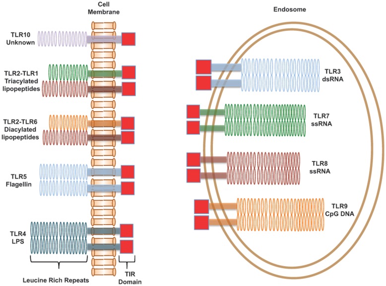
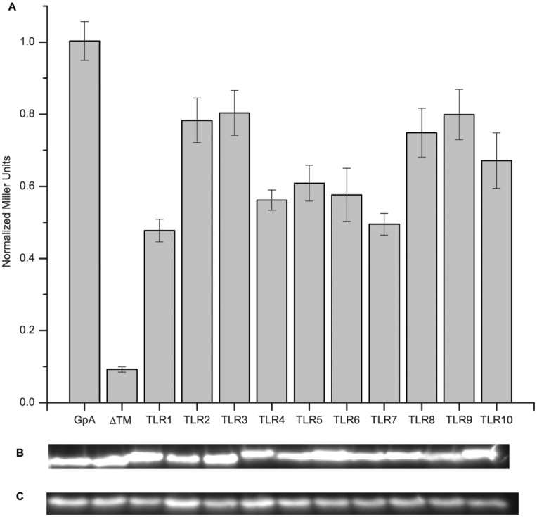
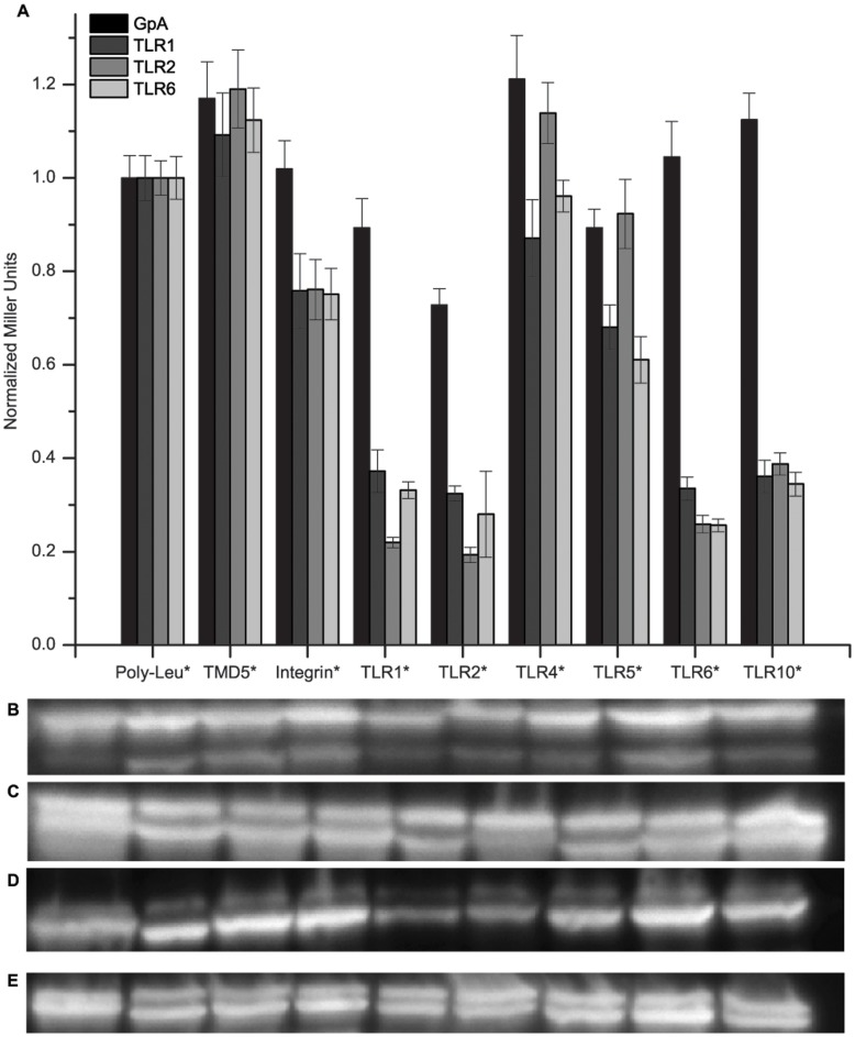
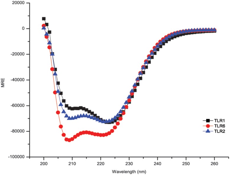
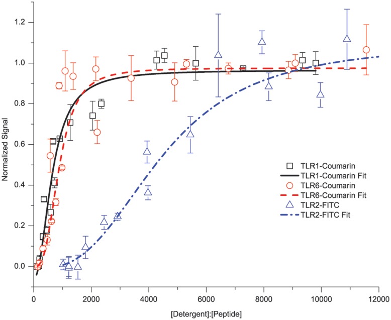
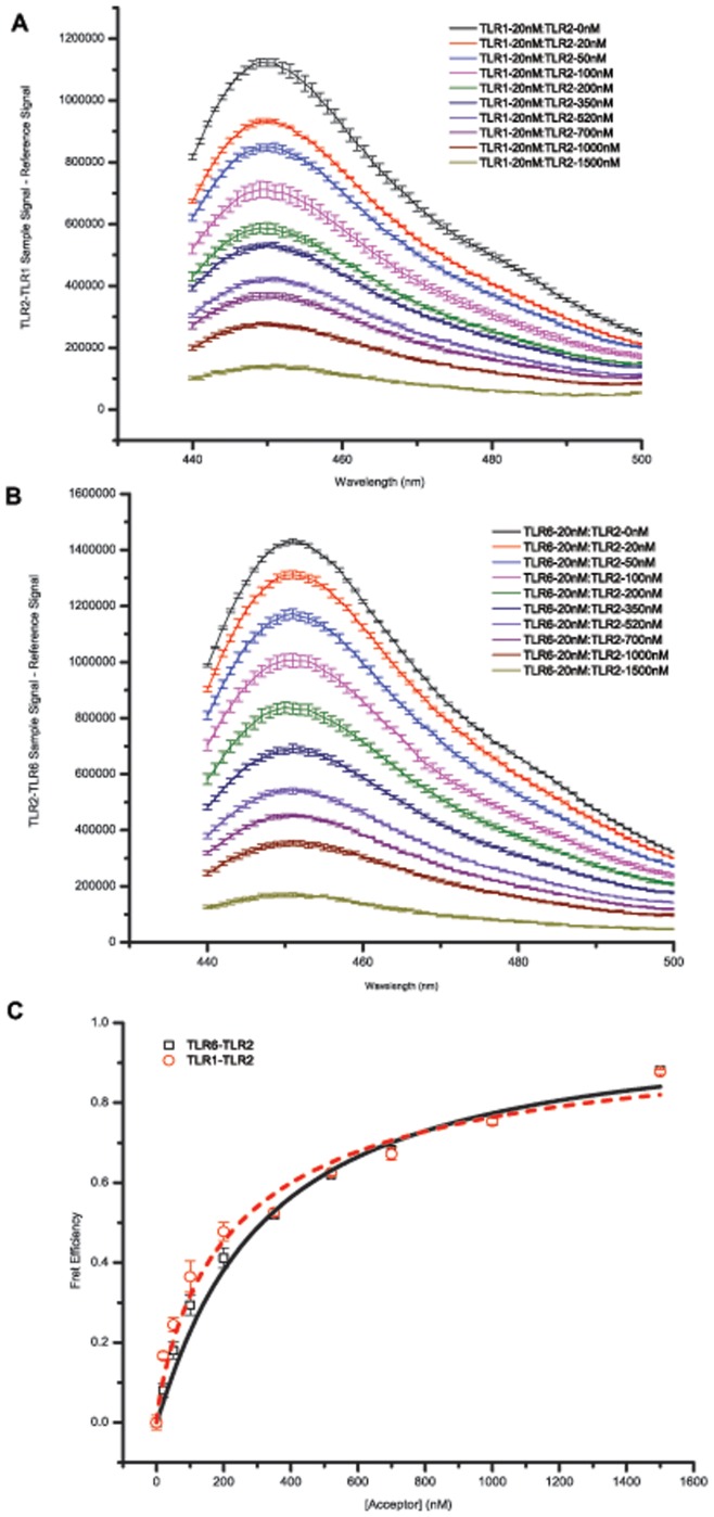
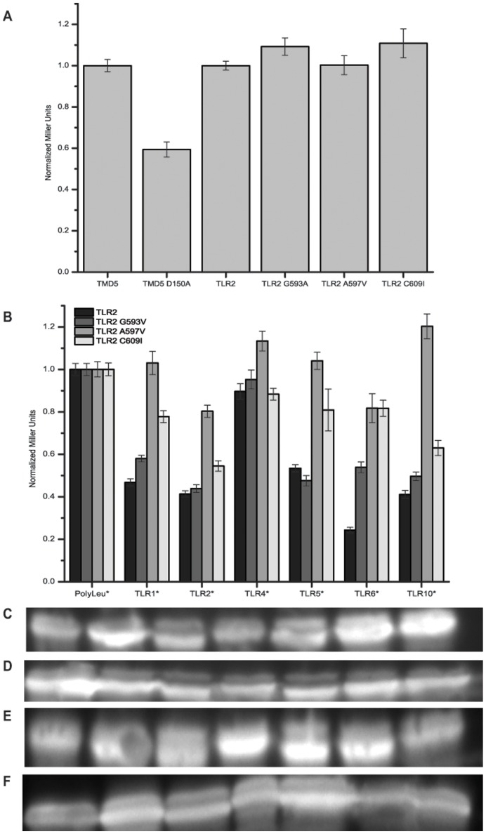
Toll-like receptors (TLRs) act as the first line of defense against bacterial and viral pathogens by initiating critical defense signals upon dimer activation. The contribution of the transmembrane domain in the dimerization and signaling process has heretofore been overlooked in favor of the extracellular and intracellular domains. As mounting evidence suggests that the transmembrane domain is a critical region in several protein families, we hypothesized that this was also the case for Toll-like receptors. Using a combined biochemical and biophysical approach, we investigated the ability of isolated Toll-like receptor transmembrane domains to interact independently of extracellular domain dimerization. Our results showed that the transmembrane domains had a preference for the native dimer partners in bacterial membranes for the entire receptor family. All TLR transmembrane domains showed strong homotypic interaction potential. The TLR2 transmembrane domain demonstrated strong heterotypic interactions in bacterial membranes with its known interaction partners, TLR1 and TLR6, as well as with a proposed interaction partner, TLR10, but not with TLR4, TLR5, or unrelated transmembrane receptors providing evidence for the specificity of TLR2 transmembrane domain interactions. Peptides for the transmembrane domains of TLR1, TLR2, and TLR6 were synthesized to further study this subfamily of receptors. These peptides validated the heterotypic interactions seen in bacterial membranes and demonstrated that the TLR2 transmembrane domain had moderately strong interactions with both TLR1 and TLR6. Combined, these results suggest a role for the transmembrane domain in Toll-like receptor oligomerization and as such, may be a novel target for further investigation of new therapeutic treatments of Toll-like receptor mediated diseases.


Conflict of interest statement

Competing Interests: The authors have declared that no competing interests exist.

Figures

Figure 1
Figure 1. Human Toll-like Receptors.
A schematic representation of the human Toll-like receptors. TLRs consist of three domains, an extracellular Leucine-rich repeat domain that recognizes the ligand, a single-pass transmembrane domain, and an intracellular TIR domain for signaling. Signaling is activated by the formation of either homodimers or heterodimers as depicted. TLRs are typically broken down into two classes, cell-surface receptors that recognize bacterial cell wall components, and endosomal receptors that recognize bacterial and viral nucleic acids.
Figure 2
Figure 2. Toll-like Receptor Transmembrane Domain Homotypic Interactions.
(A) The ToxR assay was used to study homotypic interactions of the TLR TMDs. A chimeric protein expressing the TMD of interest was monitored through β-galactosidase activity. In all cases, we saw that the TLRs have interaction potential solely from TMD-TMD interactions. Each TLR TMD interaction measurement analysis was performed on 3 technical replicates with >6 measurements for each replicate. Error bars depict the standard error of the mean (n – Table S1). Western blots staining for MBP were performed to monitor expression levels of the constructs – (B) chimeric maltose binding protein expression, (C) endogenous maltose binding protein expression. All samples were normalized to the GpA signal and expression levels. Significant differences were determined by use of the Tukey-Kramer test with all TLRs being significantly different than the negative control.
Figure 3
Figure 3. Cell- Surface Toll-like Recptor Heterotypic Interactions.
(A) A dominant-negative ToxR assay was used to study heterotypic interactions of the cell-surface TLRs. Two TLR TMDs were encoded in the FHK12 E.coli reporter strain, one with a functional ToxR domain (dominant phenotype) and one with a nonfunctional ToxR* domain (negative phenotype). Interaction between the two different TMDs leads to a reduction in signal from that seen for homotypic interactions. The TMDs for GpA, TLR1, TLR2, and TLR6 were used with the functional ToxR domain while TMDs for poly-Leucine, TMD5, integrin αIIb, TLR1, TLR2, TLR4, TLR5, TLR6, and TLR10 were used with the nonfunctional ToxR* domain. Interactions were most prominent for the TLRs known to have heterotypic interactions – TLR2-TLR1 and TLR2-TLR6. TLR10 also showed strong interactions with TLR2. We also saw interaction with the same TMD further validating the homotypic interactions. Moderate interaction was seen with other TMDs that could be attributed to non-specific interactions from similar TMD motifs as completely unrelated receptors showed similar levels of knockdown. Each dominant phenotype was done in 3 technical replicates with each negative phenotype and >6 measurements made for each replicate. Error bars depict the standard error of the mean (n- Tables S3, S5, S7, S9). Western blots staining for MBP were performed to monitor expression levels of the constructs with the upper band being functional ToxR chimeras and the lower band being nonfunctional ToxR* chimeras – (B) GpA, (C) TLR1, (D) TLR2, and (E) TLR6. Significant differences for the same dominant phenotype were determined by use of the Tukey-Kramer test.
Figure 4
Figure 4. Circular Dichroism Spectra of TLR1, TLR2, and TLR6 Synthetic TMD Peptides.
Far-UV spectra of the synthetic TMD peptides at concentrations ranging from 5–10 µM in the presence of 10 mM C14-betaine detergent micelles. Spectra were collected at 25°C with a step size of 1 nm and are the average of 9 scans. All peptides had helical content >99% (Table S11) as determined using CDNN .
Figure 5
Figure 5. TLR1, TLR2, and TLR6 Homotypic Interactions by Fluorescence Self-Quenching.
Synthetic TMD peptides were fluorescently tagged with fluorescein (TLR2) or coumarin (TLR1, TLR6). Homotypic interaction affinity was studied by changes of fluorescence intensity at different molar ratios of detergent to peptide. All samples were fixed at a peptide concentration of 1 µM, while the detergent concentration was varied. Increasing detergent competes off the TMD-TMD interactions and leads to an increase in fluorescence signal. TLR1 and TLR6 exhibit similar behavior characteristic of weak interactions as indicated by rapid release of quenched fluorescence with Kd of 645.63±49.08 and 883.57±86.92 respectively. TLR2 exhibits a different behavior indicative of a moderate interaction as it gradually releases quenched fluorescence with Kd of 4475.49±637.86. Each data point is a minimum of 3 measurements from 3 different sample preparations. Error bars are standard deviations of the measurements. Kd were determined using the Hill Equation.
Figure 6
Figure 6. TLR2-TLR1 and TLR2-TLR6 Heterotypic Interactions Measured by Förster Resonance Energy Transfer.
Synthetic TMD peptides were co-incubated in micelles with the FRET donor concentration fixed to be 20 nM and the FRET acceptor concentration varied from 0–1500 nM. The donor was excited at 415 nm and emission was monitored from 440–600 nm. Only donor emission is shown as the excitation-emission separation led to a high background signal seen in the acceptor channel. (A) TLR2-TLR1 donor channel. (B) TLR2-TLR6 donor channel. (C) FRET efficiency based on decrease in donor signal at increasing acceptor concentrations. Fitting these curves yields a TLR2-TLR1 interaction of Kd = 4332.0±410.7 (in molar fraction unit) and a TLR2-TLR6 interaction of Kd = 3490.4±190.1.
Figure 7
Figure 7. Toll-like Receptor 2 Interaction Interface Studied by Mutagenesis.
TLR2 residues were mutated to study effects on homotypic and heterotypic interactions using the ToxR assay. These mutations were at positions identified in the sequence as likely interaction locations. (A) Homotypic interactions of point mutations show no difference in interaction potential from wild-type TLR2 TMD. (B) Heterotypic interactions of point mutations show the potential importance of both the Ala597 and Cys609 positions for heterotypic interactions. Western blots staining for MBP were performed to monitor expression levels of the constructs with the upper band being functional ToxR chimeras and the lower band being nonfunctional ToxR* chimeras – (C) TLR2, (D) TLR2 G593V, (E) TLR2 A597V, and (F) TLR2 C609I.

References

    1. Medzhitov R (2001) Toll-like receptors and innate immunity. Nature Reviews Immunology 1: 135–145. - PubMed
    1. Kawai T, Akira S (2010) The role of pattern-recognition receptors in innate immunity: update on Toll-like receptors. Nature Immunology 11: 373–384. - PubMed
    1. Leonard JN, Ghirlando R, Askins J, Bell JK, Margulies DH, et al. (2007) The TLR3 signaling complex forms by cooperative receptor dimerization. PNAS 105: 258–263. - PMC - PubMed
    1. Liu L, Botos I, Wang Y, Leonard JN, Shiloach J, et al. (2008) Structural basis of Toll-like receptor 3 signaling with double-stranded RNA. Science 320: 379–381. - PMC - PubMed
    1. Park BS, Song DH, Kim HM, Choi B-S, Lee H, et al. (2009) The structural basis of lipopolysacchardie regonition by the TLR4-MD-2 complex. Nature 458: 1191–1195. - PubMed

Publication types

Substances