. 2012;7(11):e48884.
doi: 10.1371/journal.pone.0048884. Epub 2012 Nov 14.

Impaired skeletal muscle regeneration in the absence of fibrosis during hibernation in 13-lined ground squirrels

Affiliations

Impaired skeletal muscle regeneration in the absence of fibrosis during hibernation in 13-lined ground squirrels

Eva Andres-Mateos et al. PLoS One. 2012.

Abstract

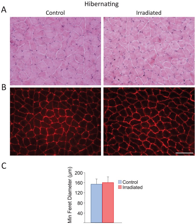
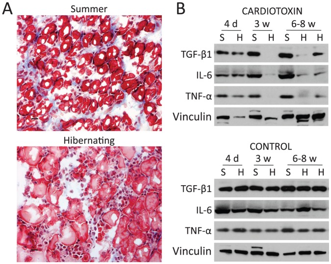
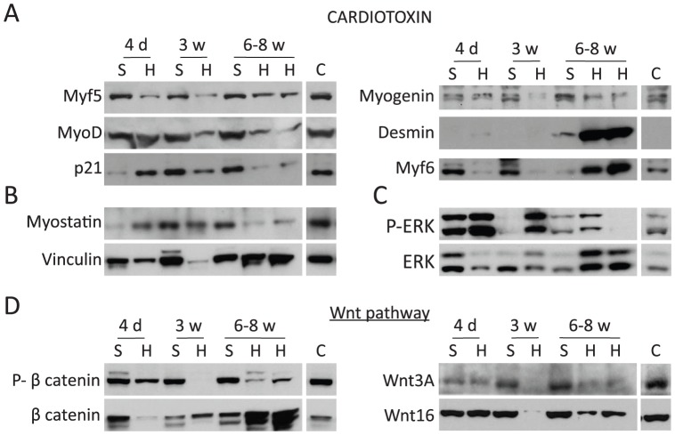
Skeletal muscle atrophy can occur as a consequence of immobilization and/or starvation in the majority of vertebrates studied. In contrast, hibernating mammals are protected against the loss of muscle mass despite long periods of inactivity and lack of food intake. Resident muscle-specific stem cells (satellite cells) are known to be activated by muscle injury and their activation contributes to the regeneration of muscle, but whether satellite cells play a role in hibernation is unknown. In the hibernating 13-lined ground squirrel we show that muscles ablated of satellite cells were still protected against atrophy, demonstrating that satellite cells are not involved in the maintenance of skeletal muscle during hibernation. Additionally, hibernating skeletal muscle showed extremely slow regeneration in response to injury, due to repression of satellite cell activation and myoblast differentiation caused by a fine-tuned interplay of p21, myostatin, MAPK, and Wnt signaling pathways. Interestingly, despite long periods of inflammation and lack of efficient regeneration, injured skeletal muscle from hibernating animals did not develop fibrosis and was capable of complete recovery when animals emerged naturally from hibernation. We propose that hibernating squirrels represent a new model system that permits evaluation of impaired skeletal muscle remodeling in the absence of formation of tissue fibrosis.


Conflict of interest statement

Competing Interests: Co-author Ronald Cohn is a PLOS ONE Editorial Board member. Co-author Tom G. Marr is employed by the commercial company Hiberna Corporation. This does not alter the authors' adherence to all the PLOS ONE policies on sharing data and materials. All the authors declare no potential competing interests.

Figures

Figure 1
Figure 1. Schematic diagram of myogenic and fibrosis regulatory pathways in mammals.
After skeletal muscle damage occurs, cytokines and growth factors are released from the injured blood vessels and from infiltrating inflammatory cells. As a result of these coordinated events, satellite cells undergo extensive proliferation upon activation. Activated satellite cells (also called MCPs or myoblasts) will differentiate into myotubes and fuse together with either damaged myofibers or form new myofibers, while some will undergo self-renewal to restore the satellite cell pool. If inflammatory cell infiltration and fibroblast activation persist, the aberrant tissue repair response will produce a non-functional mass of fibrotic tissue. Myogenic regulatory pathways and factors are indicated. In hibernating squirrels, activation of Wnt signaling pathway (Wnt) does not favor fibrosis formation as has been shown in other mammals (in red).
Figure 2
Figure 2. Satellite cells are not involved in maintenance of skeletal muscle during hibernation.
Irradiation was used to ablate satellite cells from one quadriceps muscle of hibernating squirrels. A: Hematoxylin-eosin (H&E) staining revealed no morphological differences between untreated control (left) and irradiated (right) quadriceps. B: Dystrophin immunolabeling outlined myofiber sarcolemmas enabling determination of percentage distribution of minimal Feret's diameter and revealed no effect of satellite cell ablation (scale bar 100 µm). C: Mean minimal Feret's diameter (µm) was not significantly different between control and irradiated quadriceps during hibernation (n = 3).
Figure 3
Figure 3. Skeletal muscle regeneration is impaired during hibernation.
H&E staining revealed the time course of degeneration, regeneration, and recovery in cardiotoxin-injected gastrocnemius muscles of summer (left) and hibernating (right) squirrels (n = 22; scale bars 50 µm). A–B: Uninjured control gastrocnemius from summer (left) and hibernating (right) squirrel illustrating normal myofiber morphology. C–D: 4 days after CTX injury, damage and inflammation was evident in both groups. E–F: 2 weeks after CTX injury, remodeling was underway in summer, but not hibernating, muscle. G–H: 3 weeks after CTX injury, recovery in hibernating muscle still lagged well behind that of summer muscle but displayed no fibrosis. I: 6 weeks after CTX injury, summer muscle was fully recovered. J: 4 weeks after arousal (following 6–10 weeks of hibernation after CTX injury), muscle achieved full recovery without fibrosis.
Figure 4
Figure 4. Regenerating fibers do not appear until 6 weeks after cardiotoxin injection in hibernating squirrels.
Developmental myosin immunofluorescence in summer (left) and torpid squirrels (right) sacrificed 3 and 6 weeks (w) after the injury. Summer (A) but not hibernating squirrels (B) have regenerating skeletal muscle fibers 3 weeks after cardiotoxin (CTX) injection. C: Muscle of summer squirrels was completely repaired by 6 w after CTX injection. D: First regenerating fibers in torpid animals appeared at 6 weeks after the injury (scale bar 100 µm).
Figure 5
Figure 5. Lack of fibrosis and suppression of inflammatory markers during hibernation.
A: Masson's trichrome staining showing collagen in blue, from summer and hibernating squirrels 6 weeks after cardiotoxin injury. B: Western blots of gastrocnemius muscle injected with cardiotoxin from summer active (S) and hibernating (H) squirrels using antibodies against fibrosis and inflammatory factors. d = days; w = weeks. Bars represent 20 µm.
Figure 6
Figure 6. Delayed differentiation of myocytes during regeneration in hibernating squirrels.
A–D: Western blots of gastrocnemius muscle injected with cardiotoxin from summer active (S) and hibernating (H) squirrels using antibodies against the proteins indicated. For comparison, a representative sample from the contralateral non-injected gastrocnemius is shown (C = Control). P = Phospho; d = days; w = weeks.

References

    1. Sandri M, Sandri C, Gilbert A, Skurk C, Calabria E, et al. (2004) Foxo transcription factors induce the atrophy-related ubiquitin ligase atrogin-1 and cause skeletal muscle atrophy. Cell 117: 399–412. - PMC - PubMed
    1. Debigare R, Cote CH, Maltais F (2001) Peripheral muscle wasting in chronic obstructive pulmonary disease. Clinical relevance and mechanisms. Am J Respir Crit Care Med 164: 1712–1717. - PubMed
    1. Degens H, Alway SE (2006) Control of muscle size during disuse, disease, and aging. Int J Sports Med 27: 94–99. - PubMed
    1. Jackman RW, Kandarian SC (2004) The molecular basis of skeletal muscle atrophy. Am J Physiol Cell Physiol 287: C834–843. - PubMed
    1. Andres-Mateos E, Brinkmeier H, Burks TN, Mejias R, Files DC, et al. (2012) Activation of Serum/Glucocorticoid-Induced Kinase (SGK) is Sufficient to Maintain Skeletal Muscle Homeostasis and Prevent Atrophy. EMBO Mol Med In press. - PMC - PubMed

Publication types