. 2012;7(11):e48934.
doi: 10.1371/journal.pone.0048934. Epub 2012 Nov 14.

Neurotensin and CRH interactions augment human mast cell activation

Affiliations

Neurotensin and CRH interactions augment human mast cell activation

Konstantinos-Dionysios Alysandratos et al. PLoS One. 2012.

Abstract

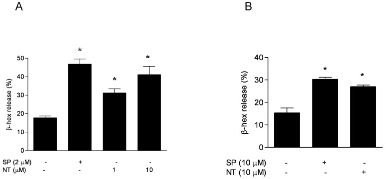
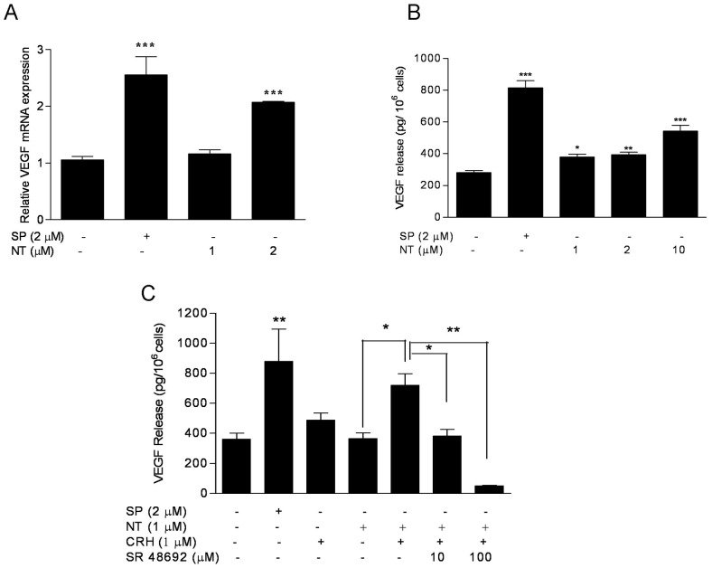
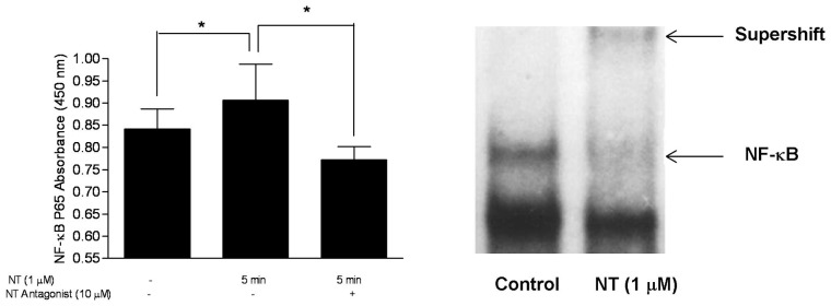
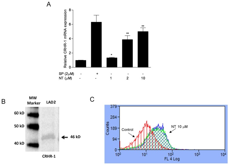
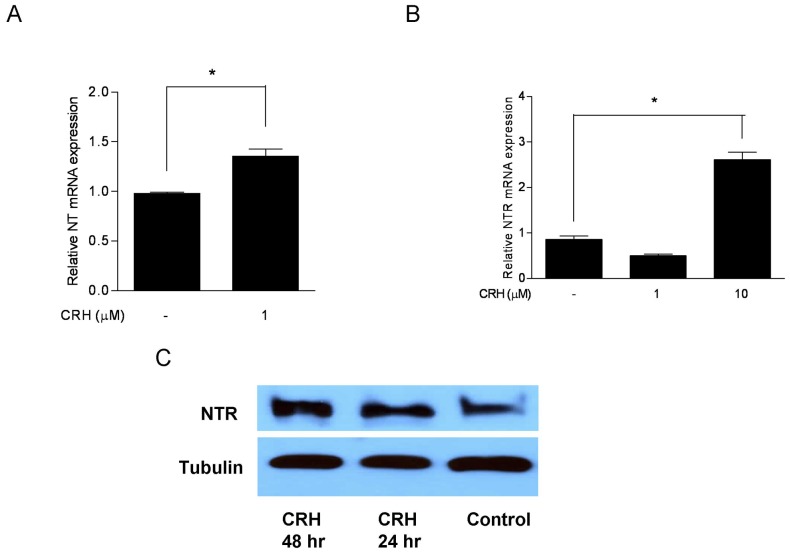
Stress affects immunity, but the mechanism is not known. Neurotensin (NT) and corticotropin-releasing hormone (CRH) are secreted under stress in various tissues, and have immunomodulatory actions. We had previously shown that NT augments the ability of CRH to increase mast cell-dependent skin vascular permeability in rodents. Here we show that NT triggered human mast cell degranulation and significantly augmented CRH-induced vascular endothelial growth factor (VEGF) release. Investigation of various signaling molecules indicated that only NF-κB activation was involved. These effects were blocked by pretreatment with the NTR antagonist SR48692. NT induced expression of CRH receptor-1 (CRHR-1), as shown by Western blot and FACS analysis. Interestingly, CRH also induced NTR gene and protein expression. These results indicate unique interactions among NT, CRH, and mast cells that may contribute to auto-immune and inflammatory diseases that worsen with stress.


Conflict of interest statement

Competing Interests: The authors have declared that no competing interests exist.

Figures

Figure 1
Figure 1. NT stimulates degranulation of human mast cells.
(A) LAD2 mast cells and (B) hCBMCs were stimulated with NT for 30 min at 37°C. SP was used as a “positive” control to stimulate mast cell degranulation. The release of β-hexosaminidase (β-hex) was significantly elevated as compared to the control (n = 3; *p<0.01).
Figure 2
Figure 2. NT increases VEGF gene expression and protein secretion in human mast cells, which is blocked by a NTR antagonist.
(A) VEGF mRNA expression was assessed following stimulation of LAD2 cells with NT (1, 2 µM) for 6 h; SP was used as a “positive” control to stimulate mast cell degranulation. (B) VEGF secretion from LAD2 cells was measured after stimulation with NT (1, 2, 10 µM) for 24 h; and (C) VEGF release from LAD2 cells treated for 48 hr with NT (1 µM) and/or followed with CRH (1 µM) for 24 h. The augmentation effect of NT and CRH is blocked by pre-treating with the NT antagonist SR 48692 (10 µM) for 30 min. For all experiments, n = 3; *p<0.05, **p<0.01, ***p<0.001 compared to control.
Figure 3
Figure 3. Effect of NT on NF-κB activation in human mast cells.
LAD2 mast cells were pretreated with/without the NTR antagonist SR 48692 (TOCRIS Bioscience, Ellisville, MO) (10 µM) for 30 min, then treated with NT (1 µM) for 5, 10, 20 min. (A) Different molecules were measured with PathScan® Sandwich ELISA Kit (Cell Signaling Technology, Inc. Danvers, MA) and the absorbance for NF-κB was determined spectrophotometrically at 450 nm. (B) NF-κB was determined by EMSA.
Figure 4
Figure 4. NT induces CRHR-1 expression in human mast cells.
(A) LAD2 cells CRHR-1 gene expression was assessed following incubation with different concentrations of NT for 6 h. SP was used as a “positive” control to stimulate mast cell degranulation. Relative mRNA expression was measured by quantitative qPCR, normalized to GAPDH, and expressed relatively to the untreated cells (control). (B) Detection of CRHR-1 in LAD2 cells by Western blot analysis following incubation with NT (10 µM) for 24 or 48 h is shown. For all experiments, n = 3; *p<0.05, **p<0.01, compared to control and (C) CRHR-1 detection by FACS analysis in LAD2 cells following treatment with NT (1, 10 µM) for 48 h; y-axis indicates “counts” of cells, while the x-axis indicates log fluorescence intensity. For all experiments, n = 3; *p<0.05, **p<0.01, compared to control.
Figure 5
Figure 5. CRH induces NT and NTR gene expression in human mast cells.
(A) NT and (B) NTR gene expression in LAD2 cells following incubation with the indicated concentrations of CRH for 6 h. Relative mRNA expression was measured by quantitative qPCR, normalized to GAPDH, and expressed relatively to the untreated cells (control). (C) Western blot analysis of NTR following incubation with CRH (10 µM) for 24 h. Tubulin was used as an internal control. For all the experiments, n = 3; *p<0.05, **p<0.01, compared to control.

References

    1. Carraway R, Leeman SE (1973) The isolation of a new hypotensive peptide, neurotensin, from bovine hypothalami. J Biol Chem 248: 6854–6861. - PubMed
    1. Tyler-McMahon BM, Boules M, Richelson E (2000) Neurotensin: peptide for the next millennium. Regul Pept 93: 125–136. - PubMed
    1. Mustain WC, Rychahou PG, Evers BM (2011) The role of neurotensin in physiologic and pathologic processes. Curr Opin Endocrinol Diabetes Obes 18: 75–82. - PubMed
    1. Singh LK, Pang X, Alexacos N, Letourneau R, Theoharides TC (1999) Acute immobilization stress triggers skin mast cell degranulation via corticotropin-releasing hormone, neurotensin and substance P: A link to neurogenic skin disorders. Brain Behav Immunity 13: 225–239. - PubMed
    1. Cochrane DE, Emigh C, Levine G, Carraway RE, Leeman SE (1982) Neurotensin alters cutaneous vascular permeability and stimulates histamine release from isolated skin. Ann NY Acad Sci 396–397.

Publication types

MeSH terms

Substances