. 2012;7(11):e49184.
doi: 10.1371/journal.pone.0049184. Epub 2012 Nov 14.

PLK1 interacts and phosphorylates Axin that is essential for proper centrosome formation

Affiliations

PLK1 interacts and phosphorylates Axin that is essential for proper centrosome formation

Ka Ruan et al. PLoS One. 2012.

Abstract

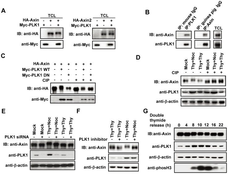
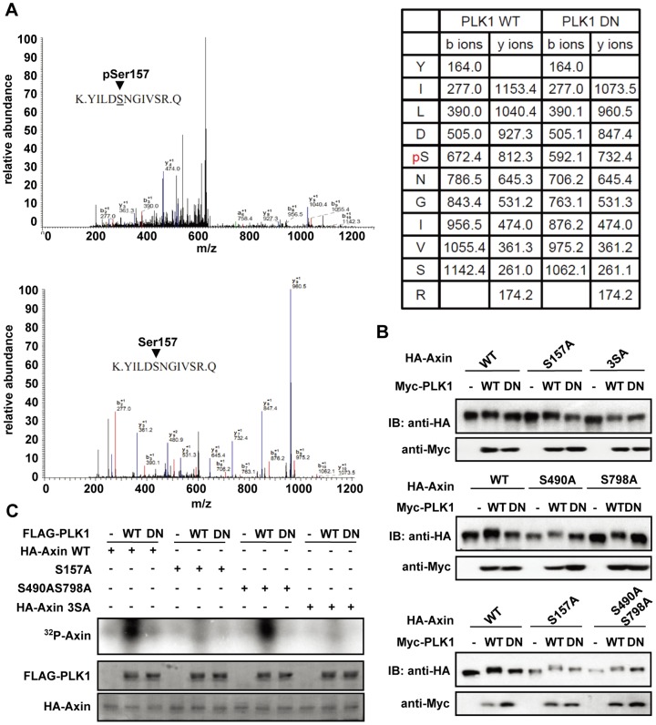
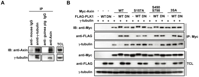
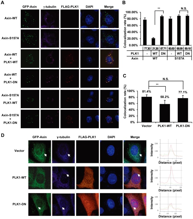
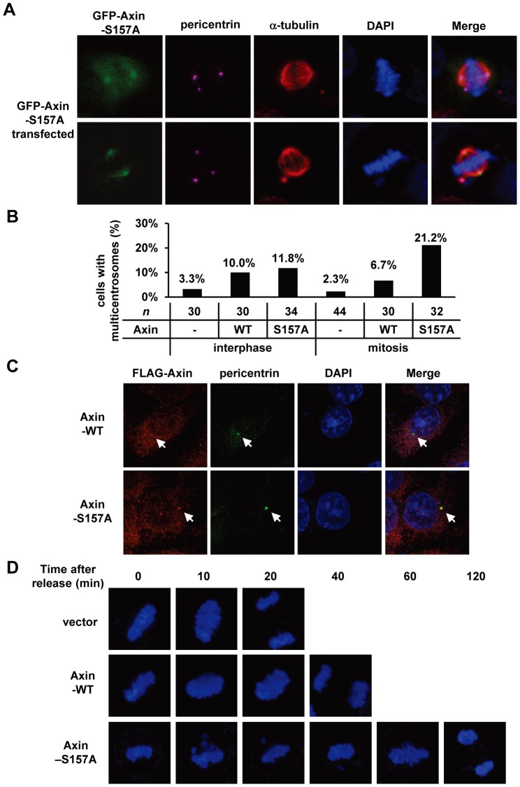
Abnormal amplification of centrosomes could lead to improper chromosome segregation and aneuploidy and is implicated in cancer development. Here, we demonstrate that Axin, a scaffolding protein in Wnt signaling, is phosphorylated by PLK1 during mitosis. Phosphorylation of Axin Ser-157 by PLK1 abolished Axin association with γ-tubulin, while substitution of Ser-157 with alanine exhibited sustained interaction with γ-tubulin. In addition, overexpression of Axin-S157A significantly increased the number of cells with multi-centrosomes. These results suggest that the phosphorylation status of Axin, mediated by PLK1, dynamically regulates its association with γ-tubulin and centrosome formation and segregation.


Conflict of interest statement

Competing Interests: The authors have declared that no competing interests exist.

Figures

Figure 1
Figure 1. Axin phosphorylation by PLK1.
(A) Total cell lysates (TCL) from cells expressing Axin or Axin2 with PLK1 or empty vector in HEK293T cells were analyzed by immunoblotting as indicated. (B) Axin interacts with PLK1 at its endogenous level. The HeLa cell lysates were immunoprecipitated with anti-Axin, mouse anti-PLK1 or control IgGs, respectively, followed by immunoblotting as indicated. (C) HA-Axin immunoprecipitates were subjected to CIP treatment and immunoblotting as indicated. (D) HeLa cells were mock treated or arrested in M-phase or early-S-phase by treatment with double-thymidine or thymidine-nocodazole. Axin immunoprecipitates were treated with CIP, followed by immunoblotting. (E, F) TCLs from cells expressing siRNA against PLK1 (E) or were treated with 50 nM of PLK1 inhibitor 10 h before harvest (F) were analyzed by immunoblotting as indicated. (G) PLK1 phosphorylation on Axin at G2/M transition. HeLa cells were synchronized by double-thymidine block and released in fresh medium for indicated times. TCLs were analyzed by western blot and probed with antibodies as indicated.
Figure 2
Figure 2. Identification of PLK1 phosphorylation sites on Axin.
(A) Mass spectrometry analysis using Axin immunoprecipitates shows that Ser-157 in Axin is one of the phosphorylation sites of PLK1. Upper panel: HA-Axin was immunoprecipitated from cells co-expressing PLK1-WT; lower panel: from cells co-expressing PLK1-DN. (B) Axin-3SA mutant no longer displays its mobility shift when co-expressed with PLK1. HA-tagged Axin-WT, Axin-S157A, Axin-3SA, Axin-S490A, and Axin-S798A, Axin-S490A/S798A mutants are co-expressed with Myc-PLK1-WT or Myc-PLK1-DN in HEK293T cells, respectively. At 24 h post-transfection, cell lysates were subjected to immunoblotting as indicated. (C) Phosphorylation of Axin by PLK1 in vitro. HEK293T cells were transfected with HA-Axin-WT or HA-Axin serine-to-alanine mutants, and cell extracts were immunoprecipitated with HA-antibody. The immunocomplexes were incubated with purified FLAG-PLK-WT or FLAG-PLK1-DN in the presence of [γ-32P] ATP for the kinase assay. Following the assay, samples were subjected to Coomassie Blue staining and autoradiography as indicated.
Figure 3
Figure 3. Axin phosphorylation by PLK1 regulates Axin-γ-tubulin association.
(A) Axin interacts with γ-tubulin at its endogenous level. (B) Axin-S157A and Axin-3SA show a sustained interaction with γ-tubulin. HEK293T cells were transfected as indicated. TCLs and Axin immunoprecipitates were subjected to immunoblotting as indicated.
Figure 4
Figure 4. Co-expression of PLK1 abolishes Axin-γ-tubulin co-localization.
(A) GFP-Axin-WT or GFP-Axin-S157A was co-transfected with empty vector, FLAG-PLK1-WT or FLAG-PLK1-DN into HeLa cells. Cells were fixed and immuno-stained as described in Materials and Methods. Axin was visualized by GFP, PLK1 by using anti-FLAG (red), γ-tubulin by using anti-γ-tubulin (pink), and nuclei by DAPI staining. (B) Bar chart showing the quantification data for the co-localization rates between γ-tubulin and GFP-Axin-WT or GFP-Axin-S157A at γ-tubulin foci in the background of FLAG-PLK1 or FLAG-PLK1-DN overexpression as shown in (A). n = 30 (“n” indicates the number of the cells quantified), N.S.: not significant; **P<0.01 (ANOVA followed by tukey). (C) The centrosome region was circled and analyzed using Volocity software. The co-localization rate was analyzed between 488 channel (Axin) and 642 channel (γ-tubulin). Statistical analysis was performed as in (B). (D) FLAG-PLK1-WT and FLAG-PLK1-DN were transfected respectively into HeLa cells. Goat anti-Axin, mouse anti-FLAG and rabbit anti-γ-tubulin were used for staining of endogenous Axin, PLK1 and γ-tubulin, respectively. The intensity of endogenous Axin and γ-tubulin staining along the γ-tubulin foci is shown by line profiles. A 2 µm line is drawn along the centrosome region and the Y axis shows the intensity of 488 channel (Axin) and 642 channel (γ-tubulin) along the distance of the line (X axis).
Figure 5
Figure 5. Altered centrosome number in Axin-S157A expressing mitotic cells.
(A) HeLa cells were transfected with GFP-Axin-S157A. Cells were treated with thymidine-nocodazole, then fixed and stained as described in Materials and Methods. GFP staining for Axin, anti-α-tubulin staining for microtubule (red), anti-pericentrin staining for centrosome (pink), and nuclear staining by DAPI were performed. (B) Bar chart representing the percentage of the interphase or mitotic HeLa cells with multiple centrosomes. Cells were transfected with GFP vector (control), GFP-Axin-WT or GFP-Axin-S157A mutant. “n” indicates the number of the mitotic cells quantified. The differences among interphase groups are not significant, P = 0.528; the differences among mitosis groups are significant (P = 0.014, Vectora; Axin-WTa,b; Axin-S157Ab, Chi-Square Tests). (C) Centrosome numbers are not altered in Axin-WT or Axin-S157A expressing interphase cells. HeLa cells were transfected with FLAG-Axin-WT or FLAG-Axin-S157A. Axin was visualized by using anti-FLAG (red), pericentrin by anti-pericentrin (green), and nuclei by DAPI staining. (D) Axin-S157A overexpression delays the chromosome segregation. The cells were synchronized at metaphase and released in fresh medium followed by staining with DAPI.

References

    1. Swami M (2010) Chromosomal instability: Coping with extra copies. Nat Rev Cancer 10: 737. - PubMed
    1. Holland AJ, Cleveland DW (2009) Boveri revisited: chromosomal instability, aneuploidy and tumorigenesis. Nat Rev Mol Cell Biol 10: 478–487. - PMC - PubMed
    1. Doxsey S (2002) Duplicating dangerously: linking centrosome duplication and aneuploidy. Mol Cell 10: 439–440. - PubMed
    1. Wong C, Stearns T (2003) Centrosome number is controlled by a centrosome-intrinsic block to reduplication. Nat Cell Biol 5: 539–544. - PubMed
    1. Lingle WL, Barrett SL, Negron VC, D'Assoro AB, Boeneman K, et al. (2002) Centrosome amplification drives chromosomal instability in breast tumor development. Proc Natl Acad Sci U S A 99: 1978–1983. - PMC - PubMed

Publication types