. 2012;7(11):e49221.
doi: 10.1371/journal.pone.0049221. Epub 2012 Nov 14.

Human breast cancer cells are redirected to mammary epithelial cells upon interaction with the regenerating mammary gland microenvironment in-vivo

Affiliations

Human breast cancer cells are redirected to mammary epithelial cells upon interaction with the regenerating mammary gland microenvironment in-vivo

Karen M Bussard et al. PLoS One. 2012.

Abstract

Breast cancer is the second leading cause of cancer deaths in the United States. At present, the etiology of breast cancer is unknown; however the possibility of a distinct cell of origin, i.e. a cancer stem cell, is a heavily investigated area of research. Influencing signals from the tissue niche are known to affect stem cells. Literature has shown that cancer cells lose their tumorigenic potential and display 'normal' behavior when placed into 'normal' ontogenic environments. Therefore, it may be the case that the tissue microenvironment is able to generate signals to redirect cancer cell fate. Previously, we showed that pluripotent human embryonal carcinoma cells could be redirected by the regenerating mammary gland microenvironment to contribute epithelial progeny for 'normal' gland development in-vivo. Here, we show that that human metastatic, non-metastatic, and metastasis-suppressed breast cancer cells proliferate and contribute to normal mammary gland development in-vivo without tumor formation. Immunochemistry for human-specific mitochondria, keratin 8 and 14, as well as human-specific milk proteins (alpha-lactalbumin, impregnated transplant hosts) confirmed the presence of human cell progeny. Features consistent with normal mammary gland development as seen in intact hosts (duct, lumen formation, development of secretory acini) were recapitulated in both primary and secondary outgrowths from chimeric implants. These results suggest the dominance of the tissue microenvironment over cancer cell fate. This work demonstrates that cultured human breast cancer cells (metastatic and non-metastatic) respond developmentally to signals generated by the mouse mammary gland microenvironment during gland regeneration in-vivo.


Conflict of interest statement

Competing Interests: The authors have declared that no competing interests exist.

Figures

Figure 1
Figure 1. Human breast cancer cells differentiate in the mouse mammary gland.
Whole mounts of chimeric mammary gland outgrowths (A–D) were formed from the implantation of fragments from first generation chimeric mammary gland outgrowths (Figure S1A–D). Twelve weeks post implantation, these secondary chimeric mammary gland outgrowths were harvested, fixed in Carnoy's fixative, and stained overnight with Carmine Alum. A) Second generation whole mount of chimeric mammary gland outgrowth formed from implantation of a fragment from an original MDA-MB-468 chimera (1 K MDA-MB-468 human non-metastatic breast cancer cells plus 50 K primary mouse mammary epithelial cells); B) Second generation whole mount of chimeric mammary gland outgrowth formed from implantation of a fragment from an original MDA-MB-231-GFP chimera (10 K MDA-MB-231-GFP human metastatic breast cancer cells plus 50 K primary mouse mammary epithelial cells); C) Second generation whole mount of chimeric mammary gland outgrowth formed from implantation of a fragment from an original MDA-MB-231BRMS-GFP chimera (1 K MDA-MB-231BRMS-GFP metastasis-suppressed breast cancer cells plus 50 K primary mouse mammary epithelial cells); D) Second generation whole mount of chimeric mammary gland outgrowth formed from implantation of a fragment from an original hTERT-HME1 chimera (10 K hTERT-HME1 human mammary epithelial cells plus 50 K mouse mammary epithelial cells). Scale bars 1000 µm.
Figure 2
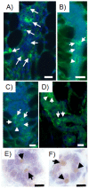
Figure 2. Human breast cancer cells are present in chimeric mammary outgrowths.
Human breast cancer cells incorporated into the mammary gland and express human-specific mitochondria as indicated by arrows (A–D) (green; Alexa Fluor 488) and green fluorescent protein as demonstrated by immunoperoxidase (E–F) (brown, DAB). Human specific mitochondria is expressed in chimeric mammary gland outgrowths produced with A) MDA-MB-468 plus mammary epithelial cells transplant fragment; B) MDA-MB-231-GFP plus mammary epithelial cells transplant fragment; C) MDA-MB-231BRMS-GFP plus mammary epithelial cells transplant fragment; D) hTERT-HME1 plus mammary epithelial cells transplant fragment. Green fluorescent protein (GFP) as demonstrated by immunoperoxidase staining is expressed in chimeric mammary gland outgrowths generated using E) MDA-MB-231-GFP plus mammary epithelial cells transplant fragment; F) MDA-MB-231BRMS-GFP plus mammary epithelial cells transplant fragment. All outgrowths are second generation chimera transplants. Scale bars 40 µm.
Figure 3
Figure 3. Human breast cancer cells contribute to the formation of luminal epithelial cells and secrete human milk proteins in the chimeric mammary gland.
Human breast cancer cells incorporated into the mammary gland express human-specific luminal epithelial cell marker keratin 8 as indicated by the white arrows (green; Alexa Fluor 488) and secrete milk proteins in impregnated hosts (arrows, human alpha-lactalbumin, green, FITC; mouse casein, red, rhodamine). Human-specific luminal epithelial cell marker keratin 8 (arrows) is expressed in chimeric mammary gland outgrowths produced with A) MDA-MB-468 plus mammary epithelial cells transplant fragment, B) MDA-MB-231-GFP plus mammary epithelial cells transplant fragment; C) MDA-MB-231BRMS-GFP plus mammary epithelial cells transplant fragment; D) hTERT-HME1 human epithelial cells plus mammary epithelial cells transplant fragment. Anti-human alpha-lactalbumin (arrows) is expressed in chimeric mammary gland outgrowths generated using E) MDA-MB-231BRMS-GFP plus mammary epithelial cells transplant fragment; F) intact host. Anti-mouse caseins (arrows) is expressed in chimeric mammary gland outgrowths generated using G) MDA-MB-231-GFP plus mammary epithelial cells transplant fragment; H) intact host. All outgrowths are second generation chimeras (human breast cancer cells plus mouse mammary epithelial cells). Intact glands are from lactating hosts. Scale bars 10 µm.
Figure 4
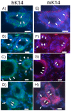
Figure 4. Human keratin 14 and mouse keratin 14 are expressed in basal cells of consecutive sections of the same second generation chimeric duct.
Human breast cancer cells contribute to the formation of basal cellular structures via the expression of the myoepithelial cell marker keratin 14 in the mouse mammary gland. Human-specific myoepithelial cell marker keratin 14 (arrows) is expressed in chimeric mammary gland outgrowths produced with A, E) MDA-MB-468 plus mammary epithelial cell transplant fragment; B, F) MDA-MB-231-GFP plus mammary epithelial cell transplant fragment; C,G) MDA-MB-231BRMS-GFP plus mammary epithelial cell transplant fragment; D,H) hTERT-HME1 plus mammary epithelial cell transplant fragment. A–D) Human-specific keratin 14; E–H) mouse-specific keratin 14. All outgrowths are second generation chimeras (human breast cancer/human mammary epithelial cells plus mouse mammary epithelial cells). Scale bars 10 µm.
Figure 5
Figure 5. Neither human or mouse specific antibodies were cross-reactive.
Human-specific keratin 8 reacts with A) human tissue, but not B) mouse tissue. Human-specific keratin 14 reacts with C) human tissue, but not D) mouse tissue. Mouse-specific keratin 14 reacts with E) mouse tissue but not F) human tissue. Human-specific mitochondria reacts with G) human tissue, but not H) mouse tissue. Normal human breast tissue was obtained from a female undergoing reduction mammoplasty with no evidence of breast disease. Normal mouse mammary tissue was obtained from the untreated, intact abdominal mammary gland of a 15-week-old female athymic nude mouse. All insets, magnification of representative areas with merge of Alexa Fluor 488 (green), FITC (green), or rhodamine (red) plus DAPI (blue). Scale bars 40 µm.
Figure 6
Figure 6. Human breast cancer cells form tumorspheres, which can be propagated after dissociation.
Breast cancer cells pre-implantation were cultured under non-adherent conditions in order to elicit the formation of tumorspheres via the tumor-initiating cells in each population. Upon dissociation by enzymatic digestion, tumorspheres were propagated up to three passages. A) MDA-MB-468; B) MDA-MB-231-GFP; C) MDA-MB-231BRMS-GFP; D) hTERT-HME1 cells. All representative images. Scale bars 100–500 µm.
Figure 7
Figure 7. A population of human breast cancer cells expresses markers for human keratin 8, human keratin 14, and human mitochondria in chimeric mammary outgrowths.
MDA-MB-231-GFP human breast cancer cells were magnetically sorted for CD44, then separated into CD44-enriched and CD44-depleted populations. Ten thousand of either CD44-enriched or CD44-depleted breast cancer cells were mixed with fifty thousand mouse mammary epithelial cells and inoculated into the epithelium-divested fat pads of three-week-old female athymic nude mice. Twelve weeks later, fat pad outgrowths were harvested and sections made for immunochemistry. Sections were stained for the human-specific luminal epithelial cell marker human keratin 8, the human-specific myoepithelial cell marker human keratin 14, the mouse-specific myoepithelial cell marker mouse keratin 14, and the human-specific marker human mitochondria. A,E) Human keratin 8 (green, Alexa Fluor 488); B, F) human keratin 14 (green, Alexa Fluor 488); C, G) mouse keratin 14 (red, rhodamine); D, H) human mitochondria (red, rhodamine). A–D) CD44-enriched; E–H) CD44-depleted. All outgrowths are first generation chimeras (human breast cancer cells plus mouse mammary epithelial cells). All are representative images. Scale bars 10 µm.

References

    1. Siegel R, Naishadham D, Jemal A (2012) Cancer statistics, 2012. CA Cancer J Clin 62: 10–29. - PubMed
    1. Lorusso G, Ruegg C (2008) The tumor microenvironment and its contribution to tumor evolution toward metastasis. Histochem Cell Biol 130: 1091–1103. - PubMed
    1. Mintz B, Illmensee (1975) Normal genetically mosaic mice produced from malignant teratocarcinoma cells. Proc Natl Acad Sci U S A 72: 3585–3589. - PMC - PubMed
    1. Illmensee K, Mintz B (1976) Totipotency and normal differentiation of single teratocarcinoma cells cloned by injection into blastocysts. Proc Natl Acad Sci U S A 73: 549–553. - PMC - PubMed
    1. Felsher DW (2003) Cancer revoked: Oncogenes as therapeutic targets. Nat Rev Cancer 3: 375–380. - PubMed

Publication types