

Ribosomal frameshifting used in influenza A virus expression occurs within the sequence UCC_UUU_CGU and is in the +1 direction
- PMID: 23155484
- PMCID: PMC3498833
- DOI: 10.1098/rsob.120109
Ribosomal frameshifting used in influenza A virus expression occurs within the sequence UCC_UUU_CGU and is in the +1 direction
Abstract
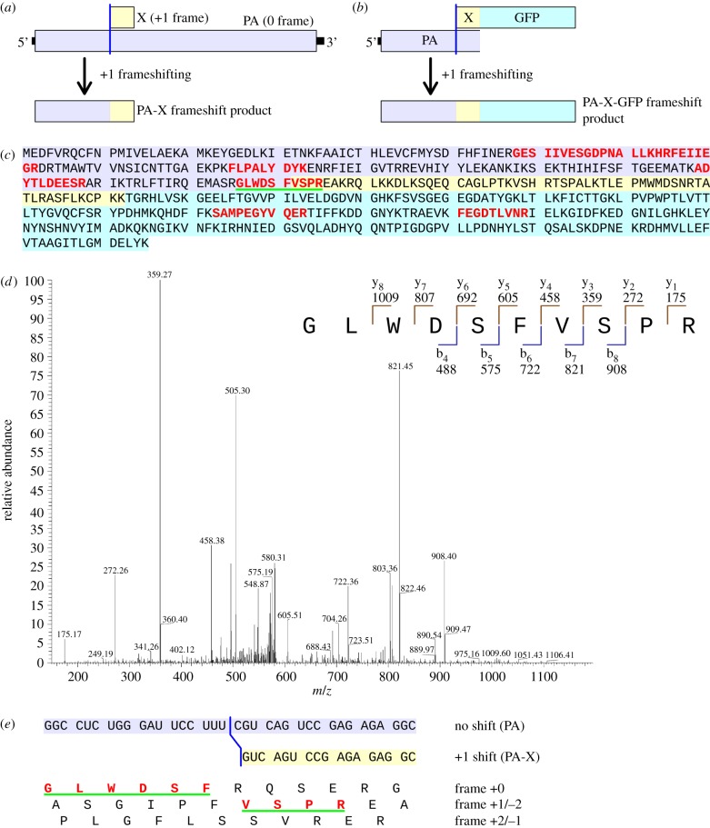
Programmed ribosomal frameshifting is used in the expression of many virus genes and some cellular genes. In eukaryotic systems, the most well-characterized mechanism involves -1 tandem tRNA slippage on an X_XXY_YYZ motif. By contrast, the mechanisms involved in programmed +1 (or -2) slippage are more varied and often poorly characterized. Recently, a novel gene, PA-X, was discovered in influenza A virus and found to be expressed via a shift to the +1 reading frame. Here, we identify, by mass spectrometric analysis, both the site (UCC_UUU_CGU) and direction (+1) of the frameshifting that is involved in PA-X expression. Related sites are identified in other virus genes that have previously been proposed to be expressed via +1 frameshifting. As these viruses infect insects (chronic bee paralysis virus), plants (fijiviruses and amalgamaviruses) and vertebrates (influenza A virus), such motifs may form a new class of +1 frameshift-inducing sequences that are active in diverse eukaryotes.
Keywords: PA-X; genetic recoding; influenza virus; mass spectrometry; ribosomal frameshifting; translation.
Figures

References
-
- Craigen WJ, Caskey CT. 1986. Expression of peptide chain release factor 2 requires high efficiency frameshift. Nature 322, 273–275 10.1038/322273a0 (doi:10.1038/322273a0) - DOI - PubMed
-
- Bekaert M, Atkins JF, Baranov PV. 2006. ARFA: a program for annotating bacterial release factor genes, including prediction of programmed ribosomal frameshifting. Bioinformatics 22, 2463–2465 10.1093/bioinformatics/btl430 (doi:10.1093/bioinformatics/btl430) - DOI - PubMed
-
- Ivanov IP, Atkins JF. 2007. Ribosomal frameshifting in decoding antizyme mRNAs from yeast and protists to humans: close to 300 cases reveal remarkable diversity despite underlying conservation. Nucleic Acids Res. 35, 1842–1858 10.1093/nar/gkm035 (doi:10.1093/nar/gkm035) - DOI - PMC - PubMed
-
- Kurian L, Palanimurugan R, Gödderz D, Dohmen RJ. 2011. Polyamine sensing by nascent ornithine decarboxylase antizyme stimulates decoding of its mRNA. Nature 477, 490–494 10.1038/nature10393 (doi:10.1038/nature10393) - DOI - PubMed
-
- Ivanov IP, Gesteland RF, Matsufuji S, Atkins JF. 1998. Programmed frameshifting in the synthesis of mammalian antizyme is +1 in mammals, predominantly +1 in fission yeast, but –2 in budding yeast. RNA 4, 1230–1238 10.1017/S1355838298980864 (doi:10.1017/S1355838298980864) - DOI - PMC - PubMed
Publication types
MeSH terms
Substances
Grants and funding
LinkOut - more resources
Full Text Sources
Research Materials