Review
. 2012 Oct;4(20):2457-69.
doi: 10.4155/bio.12.246.

Defining putative glycan cancer biomarkers by MS

Affiliations
Review

Defining putative glycan cancer biomarkers by MS

Yehia Mechref et al. Bioanalysis. 2012 Oct.

Abstract

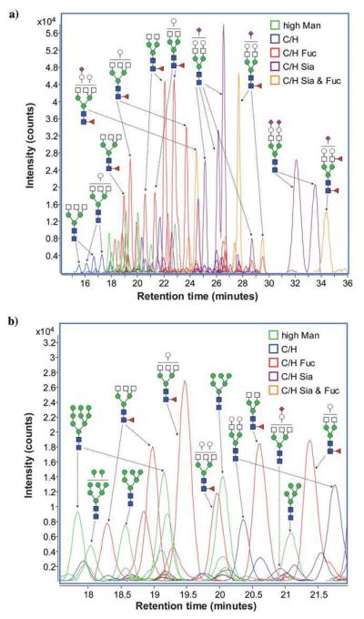
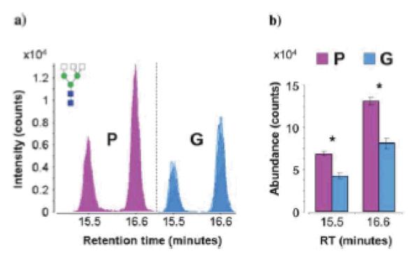
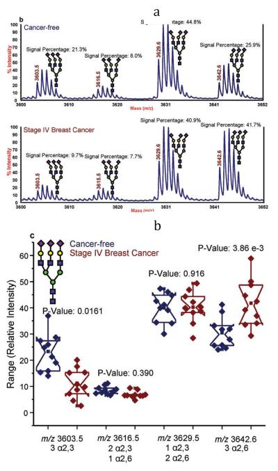
For decades, the association between aberrant glycosylation and many types of cancers has been shown. However, defining the changes of glycan structures has not been demonstrated until recently. This has been facilitated by the major advances in MS and separation science, which allows the detailed characterization of glycan changes associated with cancer. MS glycomics methods have been successfully employed to compare the glycomic profiles of different human specimens collected from disease-free individuals and patients with cancer. Additionally, comparing the glycomic profiles of glycoproteins purified from specimen collected from disease-free individuals and patients with cancer has also been performed. These types of glycan analyses employing MS or LC-MS allow the characterization of native, labeled and permethylated glycans. This review discusses the different glycomic and glycoproteomic methods employed for defining glycans as cancer biomarkers of different organs, including breast, colon, esophagus, liver, lung, ovarian, pancreas and prostate.


Figures

Figure 1
Figure 1
Representative extracted-ion chromatograms for two statistically different N-linked glycans between cancer-free samples and Stage IV breast cancer samples derived from human blood serum glycoproteins. (a) The extracted-ion chromatogram for an N-linked biantennarymonosialylated glycan showing a decrease in relative abundance in the late-stage breast cancer sample and (b) the extractedion-chromatogram for an N-linked fucosylated triantennary-trisialylated glycan, showing a significant increase in this N-linked glycan's relative abundance in the pathological sample. Symbols: red triangle, fucose; blue square, HexNAc; green circle, mannose; yellow circle, galactose. Magenta diamond, sialic acid. Reproduced with permission from [36].
Figure 2
Figure 2
Area under the receiver operating characteristics (ROC) curve (AuROC) for three glycans comparing a blinded validation set of hepatocellular carcinoma (HCC) cases (n = 47) and CLD controls (n = 27). Symbols: as in Figure 1. Reproduced with permission from [39].
Figure 3
Figure 3
N-linked carbohydrates were separated by microhydrophilic interaction LC and online detected by ESI-TOF MS. The combined mass spectra of retention time 27–28 min and retention time 28–29 min are shown in (a) and (b). All of the peaks presented here are doubly charged. The spectrum for cancer sample is shown above the normal sample. The zoomed spectrum depicts the differences between cancer and control. Symbols: black triangle, fucose; black star, N-acetyl neuraminic acid (sialic acid); black square, HexNAc; white circle, mannose; white diamond, galactose. Reproduced with permission from [47].
Figure 4
Figure 4
Average drift time profiles of (A) [S1H5N4+3Na]3+ and (B) [S1F1H5N4+3Na]3+ glycans showing both conformational and intensity differences with respect to disease state. Note that in the case of S1H5N4, the disease states exhibit lower overall intensities than the healthy state, while S1F1H5N4 shows higher overall drift time intensities in the case of diseased states than in the healthy state. This might be due to increased fucosylation of glycans with cancer and cirrhosis. Additionally, for both ions, careful inspection of drift time profiles show variances in intensities of finer features between the three groups. Symbols: as in Figure 3. Reproduced with permission from [51].
Figure 5
Figure 5
(a) Extracted compound chromatograms (ECCs) of glycans found in a representative serum sample. (b) Magnified view of a short segment of the glycan elution profile, showing the high sensitivity and resolution achieved by nano-LC separation. Colors denote different glycan classes. Symbols: red triangle, fucose; blue square and white square, HexNAc; green circle, mannose; white circle, galactose. magenta diamond, sialic acid. Reproduced with permission from [53].
Figure 6
Figure 6
(a) Overlaid chromatograms of the isomers of complex triantennary glycan composition Hex3-HexNAc5. Overlaps are represented by varying degrees of translucency. (b) Bar graph representation of average abundances and standard error for the isomers of Hex3-HexNAc5. Asterisks denote statistically significant differences between patient groups. Group P represents patients with poor prognoses based on elevated PSA levels post-radical retropubic prostatectomy (N=4), while Group G represents patients with good prognoses based on undetectable PSA levels post-radical retropubic prostatectomy (N=4). Symbos: as in Figure 5. Reproduced with permission from [53].
Figure 7
Figure 7
(a) Representative MALDI-TOF-MS spectra of a cancer-free patient (top) and a breast cancer patient (bottom); and (b) notched-box plots for the different isomers associated with the triantennary-trisialylated glycan structures derived from the blood sera of the 10 cancer-free patients and 10 breast cancer patients. Symbols: as in Figure 1. In this figure, sialic acid directed toward the left are linked α2,3 while those directed toward the right are linked α2,6. Reproduced with permission from [54].

References

    1. Helenius A, Aebi M. Intracellular functions of N-linked glycans. Science. 2001;291(5512):2364–2369. - PubMed
    1. Rudd PM, Woods RJ, Wormald MR, et al. The effects of variable glycosylation on the functional activities of ribonuclease, plasminogen and tissue plasminogen activator. Biochem. Biophys. Acta. 1995;1248(1):1–10. - PubMed
    1. Rudd PM, Wormald MR, Stanfield RL, et al. Roles for glycosylation of cell surface receptors involved in cellular immune recognition. J. Mol. Biol. 1999;293(2):351–366. - PubMed
    1. Varki A. Biological roles of oligosaccharides: all of the theories are correct. Glycobiology. 1993;3(2):97–130. - PMC - PubMed
    1. Dwek RA. Glycobiology: Toward understanding the function of sugars. Chem. Rev. 1996;96(2):683–720. - PubMed

Publication types