Review
. 2013:75:69-93.
doi: 10.1146/annurev-physiol-030212-183737. Epub 2012 Nov 13.

Functions of microRNAs in cardiovascular biology and disease

Affiliations
Review

Functions of microRNAs in cardiovascular biology and disease

Akiko Hata. Annu Rev Physiol. 2013.

Abstract

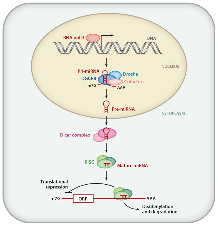
In 1993, lin-4 was discovered as a critical modulator of temporal development in Caenorhabditis elegans and, most notably, as the first in the class of small, single-stranded noncoding RNAs now defined as microRNAs (miRNAs). Another eight years elapsed before miRNA expression was detected in mammalian cells. Since then, explosive advancements in the field of miRNA biology have elucidated the basic mechanism of miRNA biogenesis, regulation, and gene-regulatory function. The discovery of this new class of small RNAs has augmented the complexity of gene-regulatory programs as well as the understanding of developmental and pathological processes in the cardiovascular system. Indeed, the contributions of miRNAs in cardiovascular development and function have been widely explored, revealing the extensive role of these small regulatory RNAs in cardiovascular physiology.


Figures

Figure 1
Figure 1
Schematic representation of miRNA biogenesis and mechanisms of miRNA action. RNA polymerase II (RNA pol II) mediates transcription of miRNA genes. The primary miRNA transcripts (pri-miRNAs) can range from a few hundred nucleotides (nt) to a few kilobases long. In the nucleus, the pri-miRNAs are recognized by ribonuclease III (RNase III) endonuclease Drosha (light blue) together with an essential partner, DiGeorge syndrome critical region gene 8 (DGCR8; also known as Pasha) (dark blue). The Drosha complex also contains other cofactors ( pink) that modulate the catalytic activity of Drosha, such as RNA helicases (p68 and p72), transcription factors [Smad, p53, estrogen receptor α(ERα)], and RNA-binding proteins (KSRP and hnRNP A1). The cleavage product of pri-miRNA, precursor miRNA (pre-miRNA), is ~70 nt long and configures a stem-loop structure. Exportin-5 exports pre-miRNAs from the nucleus to the cytoplasm. In the cytoplasm, another RNase III enzyme, Dicer, cleaves pre-miRNA to generate double-stranded mature miRNA that is ~21 nt long. The mature miRNA duplex is then incorporated into the RNA-induced silencing complex (RISC). In mammals, the RISC is composed of Argonaute proteins 1–4 (Ago1–4) and several cofactors, such as PACT and TARBP1/2. MiRNA loaded onto the RISC undergoes strand separation, interaction with typically the 3′ untranslated regions of mRNA targets through Watson-Crick base pairing between bases 2 and 7 at the 5′ end of the miRNA known as the seed sequence, and repression of mRNA translation through sequestering translational machinery from the miRNAs or mRNA destabilization through the recruitment of poly(A) nuclease, followed by deadenylation. ORF, m7G, and AAA denote open reading frame, 5′ capping, and 3′ poly(A) tail, respectively. Adapted from Reference .
Figure 2
Figure 2
Posttranscriptional mechanisms of regulation of the miRNA biogenesis pathway. MiRNA biosynthesis requires two steps of processing: (1) processing from primary miRNA transcripts (pri-miRNA) to precursor miRNA (pre-miRNA) by the ribonuclease III (RNase III) Drosha in the nucleus and (2) processing from pre-miRNA to a mature miRNA duplex by the RNase III Dicer in the cytoplasm. Both steps can be regulated by various RNA-binding proteins and DNA-binding proteins as summarized in panels ad. (a) Heterogeneous nuclear ribonucleoprotein A1 (hnRNP A1) ( green) recognizes the terminal loop of pri-miRNAs (red ) and promotes the structural remodeling of the stem region of pri-miRNAs, which generates a favorable Drosha (light blue)-binding site and enhances processing. The nuclear factor 90/nuclear factor 40 (NF90/NF40) complex ( purple) associates with the stem region of pri-miRNAs, which precludes association with the Drosha-processing complex and inhibits processing. (b) Similar to hnRNP A1 in panel a, KH-type splicing regulatory protein (KSRP) ( gray) binds to the terminal loop of pri-miRNAs and promotes both Drosha (light blue) and Dicer ( yellow) processing. (c) Binding of Lin28 (orange) to the terminal loop of pri-miRNAs prevents the association of both Drosha (light blue) and Dicer ( yellow). Additionally, Lin28 acts as a scaffold to promote the association of terminal RNA uridyltransferase 4 (TUT4; also known as Zcchc11) (light green) with the pre-miRNA. TUT4 promotes the 3′-uridinylation of pre-miRNA, which is then rapidly degraded. (d ) The RNA helicases p68 and p72 (teal ) play a critical role in the posttranscriptional regulation of numerous miRNAs in response to cellular signals, including transforming growth factor β(TGF-β) stimulation, p53-mediated DNA damage response, and estrogen stimulation. The downstream mediators of TGF-βstimulation and DNA damage, Smads and p53 ( pink), promote miRNA processing. Conversely, when bound to E2 ( pink), ERαreduces the processing of a subset of miRNAs. Adapted from Reference .
Figure 3
Figure 3
Schematic representation of mouse heart developmental stages and miRNAs that play a role in the regulation of heart development. Anterior is toward the top. Mouse cardiogenesis begins when two populations of cells termed the first heart field (FHF) (red ) and the second heart field (SHF) (blue) in the mesoderm commit to a cardiogenic fate at embryonic day 7.5 (E7.5). These cardiac cells, localized to the cardiac crescent, migrate ventromedially to form the linear primitive heart tube at E8.0. Shortly after E8.0, rhythmic contractions start. Subsequently, looping morphogenesis, chamber specification, cardiac valve formation, and neural crest cell migration and contribution give rise to the four-chambered heart by E10.5. By E12.5, the four chambers of the heart (RV and LV) are well delineated, and septation (shown as a broken line) of the outflow tract (OFT) is observed. By the late postnatal stages, the OFT is completely septated, and both ventricular septation and atrial septation are complete in preparation for postnatal life. Representative miRNAs and functional targets at the different stages of development are indicated. In the early stages of cardiac development, miR-1 and miR-133 cooperatively promote mesoderm differentiation in embryonic stem (ES) cells and repress ectodermal and endodermal cell fate. At a later stage, miR-1 and miR-133 exhibit opposing effect in cardiomyocyte differentiation. miR-27 is involved in early cardiac development by modulating MEF2c expression. The miR-17~92 cluster promotes myogenic differentiation in the secondary heart field. Deletion of the miR-17~92 cluster causes ventricular-septum defects in mice. The miR-15 family of miRNAs inhibits postnatal proliferation of cardiac myocytes. miR-208a plays a role in cardiomyocyte-specific gene expression, the development of electrophysiological properties of the heart, and hypertrophic growth upon cardiac stress. Abbreviations: Ao, aorta; LA, left atrium; LV, left ventricle; PT, pulmonary tract; RA, right atrium; RV, right ventricle; Dll1, Delta-like-1; Irx5, Iroquois homeobox 5; SRF, serum response factor; Tbx1, T-box 1; Isl1, Islet 1; Thrap1, thyroid hormone receptor–associated protein 1; Chek1, checkpoint kinase 1; PDK4, pyruvate dehydrogenase kinase 4; MHC, myosin heavy chain.
Figure 4
Figure 4
Schematic representation of approaches to manipulate miRNA activity. The activity of a specific miRNA can be manipulated to modulate the expression level of all miRNA target genes (steps 13) or of a specific miRNA target gene (step 4). (1) Under physiological conditions, endogenous miRNA (red ) associates with a partially complementary sequence known as the seed sequence ( gray box) that is often located in the 3′UTR of target gene transcripts. This association results in downregulation of the target. (2) To augment the expression and activity of a specific miRNA, an miRNA mimic ( purple), which is composed of a chemically modified and stabilized ribonucleotide duplex, can be delivered. This structure mimics endogenous miRNA action and further downregulates target gene expression. (3) In contrast, to inhibit the activity of specific endogenous miRNAs, antisense oligonucleotides that are complementary to an endogenous miRNA (anti-miRNA; brown) can be delivered. These antisense oligonucleotides prevent the endogenous miRNA from binding to the seed sequence in the target transcripts, thus derepressing the expression of all target genes. (4) Alternatively, overexpression of exogenous mRNA (orange) that contains multiple copies of the seed sequence of a specific miRNA (an miRNA sponge; orange box) can be used to prevent endogenous miRNA from targeting target gene transcripts. (5) To protect a particular target transcript from silencing by endogenous miRNA (target protection), an antisense oligonucleotide designed to associate with the miRNA target sequence ( green) can be delivered. ORF and m7G denote open reading frame and 5′ capping, respectively. Adapted from Reference .
Figure 5
Figure 5
Role of miRNAs in healthy and diseased vasculature. Various miRNAs, i.e., miR-24, miR-126, miR-150, miR-155, miR-221/222, miR-223, and miR-451, are critical for hematopoiesis. Blood vessels are composed of three layers: the outermost adventitia ( pink), vascular smooth muscle cells (VSMCs; red ), and endothelial cells (ECs; orange). Upon vascular injury or upon pathological stimuli, a healthy blood vessel (left) undergoes vascular remodeling, which is characterized by aberrant proliferation and migration of ECs and/or VSMCs. In some cases an aneurysm or an atherosclerotic lesion forms. Many miRNAs are expressed in a cell type–specific manner. For example, miR-143/145 are specifically expressed in VSMCs and play a role in VSMC development and function. miR-10, miR-17~92, miR-126, and miR-128 are enriched in ECs and play a role in angiogenesis. Aberrant expression of some of these miRNAs is linked to pathological changes in the vasculature, such as vascular remodeling (i.e., miR-21, miR-26, miR-145, miR-146a, and miR-221/222), aortic aneurysm (i.e., miR-21, miR-26, and miR-29), and atherosclerosis (i.e., miR-92a, miR-126, and miR-146). Therefore, these miRNAs are potential therapeutic targets for vascular disorders.
Figure 6
Figure 6
The role of miRNAs in the normal and diseased heart. Upon injury, such as vascular injury, pressure overload, hypoxia, and pharmacological stimuli, a normal heart (left) undergoes pathological remodeling and develops into a hypertrophic heart with fibrosis (right). miRNAs that are expressed in cardiac tissue and contribute to normal function or pathological remodeling along with their corresponding functions are indicated. miR-1 and miR-133 are involved in the development of a normal heart by regulating proliferation, differentiation, and cardiac conduction. Both miR-1 and miR-133 downregulate cell cycle regulators and thereby block proliferation. However, miR-1 and miR-133 exhibit opposing effects in cardiomyocyte differentiation. The miR-17~92 cluster promotes myogenic differentiation in the secondary heart field. miR-208a also contributes to the regulation of the conduction system. After cardiac injury, various miRNAs contribute to pathological remodeling of the heart and the progression to heart failure. miR-29 and miR-21 block and promote cardiac fibrosis, respectively. miR-21 and miR-29 are expressed in cardiac fibroblasts. Whereas miR-29 blocks fibrosis by inhibiting the expression of extracellular matrix (ECM) components, miR-21 promotes fibrosis by stimulating mitogen-activated protein kinase (MAPK) signaling. miR-208 controls this myosin isoform switching, cardiac hypertrophy, and fibrosis. miR-23a is induced in cardiac myocytes by different hypertrophic stimuli and promotes hypertrophic responses. Ischemic injury leads to the downregulation of miR-24, miR-29, and miR-199 in cardiomyocytes; such downregulation promotes inhibition of apoptosis and fibrosis. Downregulation of miR-320 upon ischemic injury contributes to hypertrophic responses as well. RV and LV denote right ventricle and left ventricle, respectively.
Figure 7
Figure 7
Schematic diagram of the mechanisms of release of cellular miRNAs. In the cytoplasm, both precursor miRNAs (pre-miRNAs) and mature miRNAs can be incorporated into small vesicles termed exosomes (orange), which are released from the cell when multivesicular bodies (MVB) are fused to the plasma membrane. Mature miRNAs and pre-miRNAs can also be released by microvesicles. Some miRNAs associate with high-density lipoproteins (HDL; purple) or bind to RNA-binding proteins, such as nucleophosmin 1 (NPM1; teal ) and Argonaute 2 (Ago2; yellow), a component of the RNA-induced silencing complex (RISC) and released through an unknown mechanism ( green arrow). It was initially speculated that dead cells passively release miRNAs. Recent studies suggest that some miRNAs may be actively released into circulation through an unknown mechanism, potentially through a channel. How miRNAs and pre-miRNAs are sorted out to multiple mechanisms of release is unclear. Whether mature miRNA and pre-miRNA are segregated in different compartments is also unclear. RNA Pol II denotes RNA polymerase II.

References

    1. Wightman B, Ha I, Ruvkun G. Posttranscriptional regulation of the heterochronic gene lin-14 by lin-4 mediates temporal pattern formation in C. elegans. Cell. 1993;75:855–62. - PubMed
    1. Lee RC, Feinbaum RL, Ambros V. The C. elegans heterochronic gene lin-4 encodes small RNAs with antisense complementarity to lin-14. Cell. 1993;75:843–54. - PubMed
    1. Lagos-Quintana M, Rauhut R, Lendeckel W, Tuschl T. Identification of novel genes coding for small expressed RNAs. Science. 2001;294:853–58. - PubMed
    1. Bruneau BG. The developmental genetics of congenital heart disease. Nature. 2008;451:943–48. - PubMed
    1. Cordes KR, Srivastava D. MicroRNA regulation of cardiovascular development. Circ Res. 2009;104:724–32. - PMC - PubMed

Publication types

MeSH terms