. 2013 Jan:52:140-51.
doi: 10.1016/j.mcn.2012.11.008. Epub 2012 Nov 15.

The role of autophagy in Nmnat-mediated protection against hypoxia-induced dendrite degeneration

Affiliations

The role of autophagy in Nmnat-mediated protection against hypoxia-induced dendrite degeneration

Yuhui Wen et al. Mol Cell Neurosci. 2013 Jan.

Abstract

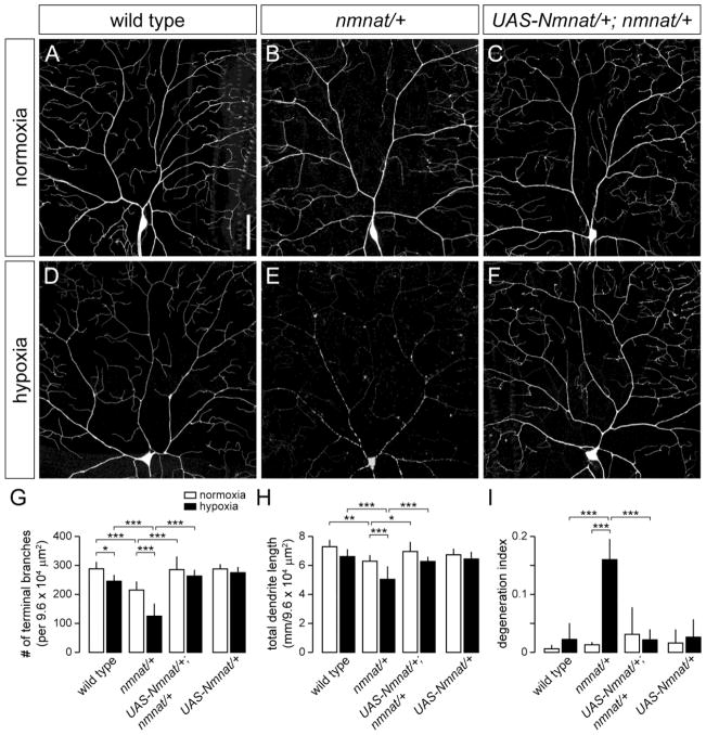
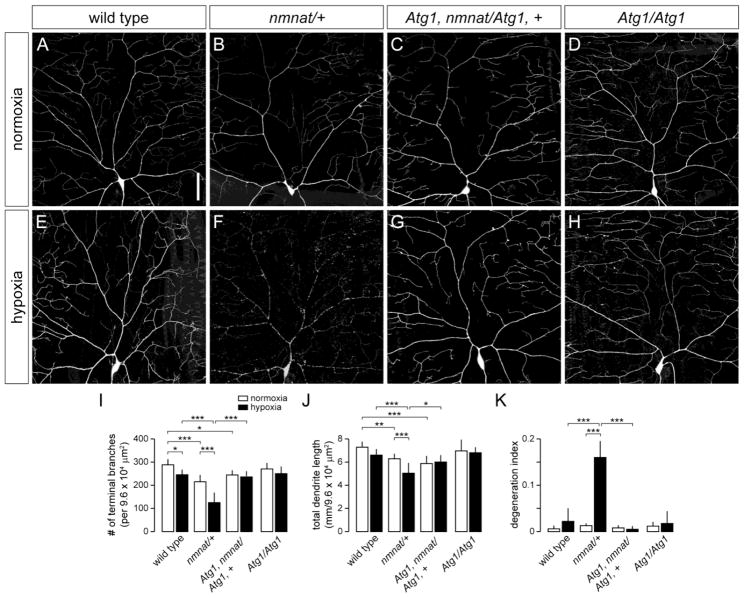
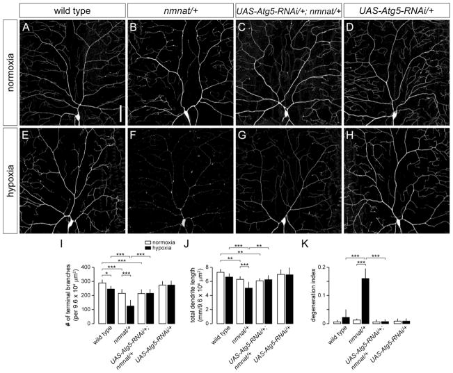
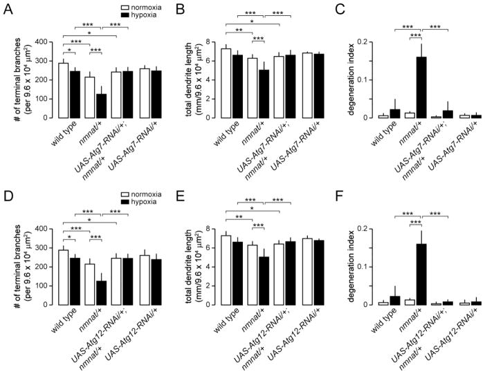
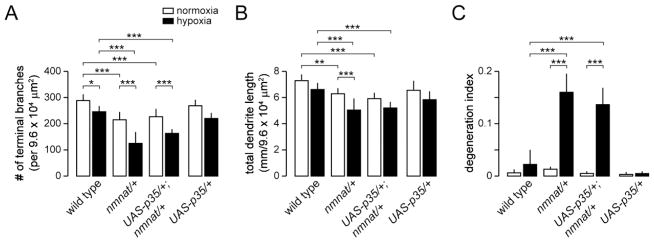
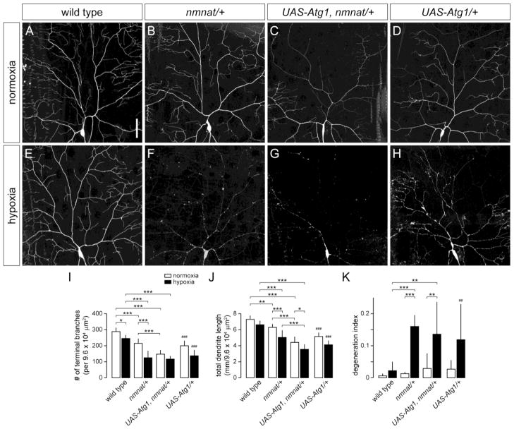
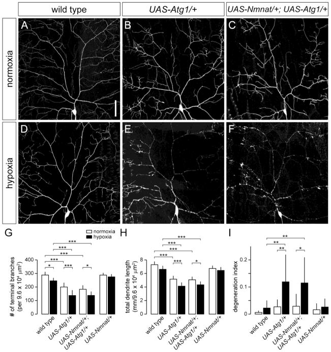
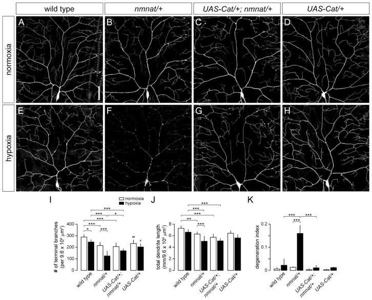
The selective degeneration of dendrites precedes neuronal cell death in hypoxia-ischemia (HI) and is a neuropathological hallmark of stroke. While it is clear that a number of different molecular pathways likely contribute to neuronal cell death in HI, the mechanisms that govern HI-induced dendrite degeneration are largely unknown. Here, we show that the NAD synthase nicotinamide mononucleotide adenylyltransferase (Nmnat) functions endogenously to protect Drosophila class IV dendritic arborization (da) sensory neurons against hypoxia-induced dendritic damage. Whereas dendrites of wild-type class IV neurons are largely resistant to morphological changes during prolonged periods of hypoxia (<1.0% O(2)), class IV neurons of nmnat heterozygous mutants exhibit significant dendrite loss and extensive fragmentation of the dendritic arbor under the same hypoxic conditions. Although basal levels of autophagy are required for neuronal survival, we demonstrate that autophagy is dispensable for maintaining the dendritic integrity of class IV neurons. However, we find that genetically blocking autophagy can suppress hypoxia-induced dendrite degeneration of nmnat heterozygous mutants in a cell-autonomous manner, suggestive of a self-destructive role for autophagy in this context. We further show that inducing autophagy by overexpression of the autophagy-specific kinase Atg1 is sufficient to cause dendrite degeneration of class IV neurons under hypoxia and that overexpression of Nmnat fails to protect class IV dendrites from the effects of Atg1 overexpression. Our studies reveal an essential neuroprotective role for endogenous Nmnat in hypoxia and demonstrate that Nmnat functions upstream of autophagy to mitigate the damage incurred by dendrites in neurons under hypoxic stress.


Figures

Figure 1
Figure 1. Nmnat protects against dendritic damage in hypoxia-exposed larvae
(A,B) Dendrite morphology of dorsal class IV ddaC neuron in a wild-type (A) and nmnatΔ4792 heterozygous mutant (nmnat/+) (B) third instar larva (110-114 hrs AEL) after exposure to 14 hrs of normoxia. (C) Selective overexpression of wild-type Nmnat in class IV neurons rescues the dendrite regression phenotype of nmnat heterozygous mutants under normoxia. (D,E) Dendrite morphology of ddaC neuron in a wild-type (D) and nmnat heterozygous mutant (E) third instar larva after exposure to 14 hrs of hypoxia (< 1.0% O2). Class IV neurons of hypoxia-exposed nmnat heterozygotes show extensive breaks along the dendritic arbor. (F) Selective overexpression of wild-type Nmnat in class IV neurons rescues the dendrite degeneration phenotype of nmnat heterozygous mutants under hypoxia. (G–I) Quantification of total number of terminal dendritic branches (G) and total dendrite length (H) per 9.6 x104 μm2 field (mean ± SD), and degeneration index (DI) (I) for ddaC neurons in wild type, nmnat heterozygous mutant (nmnat/+), nmnat heterozygous mutant background overexpressing Nmnat (UAS-Nmnat/+; nmnat/+), and wild-type background overexpressing Nmnat (UAS-Nmnat/+) under normoxia and hypoxia (* p < 0.05, ** p < 0.01, *** p < 0.001). Statistical comparison of UAS-Nmnat/+ with other groups not shown for simplification purposes. Dorsal is up and anterior is to the left in this and all subsequent figures. Scale bar, 50 μm.
Figure 2
Figure 2. Loss of Atg1 suppresses hypoxia-induced dendrite degeneration in nmnat mutants
(A,B) Dendrite morphology of class IV ddaC neuron in a wild-type (A) and nmnat heterozygous mutant (nmnat/+) (B) third instar larva after exposure to 14 hrs of normoxia. (C) Loss of Atg1 fails to suppress the dendrite regression phenotype of class IV neurons in nmnat heterozygous mutants under normoxia. (D) Class IV dendritic arborization patterns are largely unaltered in Atg1 homozygous mutants under normoxia. (E,F) Dendrite morphology of ddaC neuron in a wild-type (E) and nmnat heterozygous mutant (F) third instar larva after exposure to 14 hrs of hypoxia. (G) Loss of Atg1 suppresses the dendrite regression and degeneration phenotypes of class IV neurons in nmnat heterozygous mutants under hypoxia. (H) Class IV dendritic arborization patterns are largely unaltered in Atg1 homozygous mutants under hypoxia. (I–K) Quantification of total number of terminal dendritic branches (I), total dendrite length (J), and degeneration index (DI) (K) for ddaC neurons in wild type, nmnat heterozygous mutant (nmnat/+), Atg1 homozygous mutant with loss of one copy of nmnat (Atg1, nmnat/Atg1, +), and Atg1 homozygous mutant (Atg1/Atg1) under normoxia and hypoxia (* p < 0.05, ** p < 0.01, *** p < 0.001). Statistical comparison of Atg1/Atg1 with other groups not shown for simplification purposes. Scale bar, 50 μm.
Figure 3
Figure 3. Knockdown of Atg5 suppresses hypoxia-induced dendrite degeneration in nmnat mutants
(A,B) Dendrite morphology of class IV ddaC neuron in a wild-type (A) and nmnat heterozygous mutant (nmnat/+) (B) third instar larva after exposure to 14 hrs of normoxia. (C) Cell-specific knockdown of Atg5 fails to suppress the dendrite regression phenotype of class IV neurons in nmnat heterozygous mutants under normoxia. (D) Knockdown of Atg5 has no effect on the development or maintenance of class IV neurons under normoxia. (E,F) Dendrite morphology of ddaC neuron in a wild-type (E) and nmnat heterozygous mutant (F) third instar larva after exposure to 14 hrs of hypoxia. (G) Cell-specific knockdown of Atg5 suppresses the dendrite regression and degeneration phenotypes of class IV neurons in nmnat heterozygous mutants under hypoxia. (H) Knockdown of Atg5 has no effect on the development or maintenance of class IV neurons under hypoxia. (I–K) Quantification of total number of terminal dendritic branches (I), total dendrite length (J), and degeneration index (DI) (K) for ddaC neurons in wild type, nmnat heterozygous mutant (nmnat/+), nmnat heterozygous mutant background expressing Atg5-RNAi (UAS-Atg5-RNAi/+; nmnat/+), and wild-type background expressing Atg5-RNAi (UAS-Atg5-RNAi/+) under normoxia and hypoxia (* p < 0.05, ** p < 0.01, *** p < 0.001). Statistical comparison of UAS-Atg5-RNAi/+ with other groups not shown for simplification purposes. Scale bar, 50 μm.
Figure 4
Figure 4. Knockdown of Atg7 and Atg12 suppresses hypoxia-induced dendrite degeneration in nmnat mutants
(A–B) Quantification of total number of terminal dendritic branches (A), total dendrite length (B), and degeneration index (DI) (C) for ddaC neurons in wild type, nmnat heterozygous mutant (nmnat/+), nmnat heterozygous mutant background expressing Atg7-RNAi (UAS-Atg7-RNAi/+; nmnat/+), and wild-type background expressing Atg7-RNAi (UAS-Atg7-RNAi/+) under normoxia and hypoxia (* p < 0.05, ** p < 0.01, *** p < 0.001). Statistical comparison of UAS-Atg7-RNAi/+ with other groups not shown for simplification purposes. (D-F) Quantification of total number of terminal dendritic branches (D), total dendrite length (E), and degeneration index (DI) (F) for ddaC neurons in wild type, nmnat heterozygous mutant (nmnat/+), nmnat heterozygous mutant background expressing Atg12-RNAi (UAS-Atg12-RNAi/+; nmnat/+), and wild-type background expressing Atg12-RNAi (UAS-Atg12-RNAi/+) under normoxia and hypoxia (* p < 0.05, ** p < 0.01, *** p < 0.001). Statistical comparison of UAS-Atg12-RNAi/+ with other groups not shown for simplification purposes.
Figure 5
Figure 5. Hypoxia-induced dendrite degeneration in nmnat mutants is independent of the apoptotic machinery
(A–B) Quantification of total number of terminal dendritic branches (A), total dendrite length (B), and degeneration index (DI) (C) for ddaC neurons in wild type, nmnat heterozygous mutant (nmnat/+), nmnat heterozygous mutant background expressing p35 (UAS-p35/+; nmnat/+), and wild-type background expressing p35 (UAS-p35/+) under normoxia and hypoxia (* p < 0.05, ** p < 0.01, *** p < 0.001). Statistical comparison of UAS-p35/+ with other groups not shown for simplification purposes.
Figure 6
Figure 6. Overexpression of Atg1 induces dendrite degeneration under hypoxia
(A,B) Dendrite morphology of class IV ddaC neuron in a wild-type (A) and nmnat heterozygous mutant (nmnat/+) (B) third instar larva after exposure to 14 hrs of normoxia. (C) Overexpression of Atg1 enhances the dendrite regression phenotype of nmnat heterozygous mutants under normoxia. (D) Class IV neurons overexpressing Atg1 show reduced dendritic branching. (E,F) Dendrite morphology of ddaC neuron in a wild-type (E) and nmnat heterozygous mutant (F) third instar larva after exposure to 14 hrs of hypoxia. (G) Overexpression of Atg1 enhances dendritic regression, but not dendrite degeneration, phenotype of class IV neurons in nmnat heterozygous mutants under hypoxia. (H) Class IV neurons overexpressing Atg1 show reduced dendritic branching and extensive breaks along the arbors. (I–K) Quantification of total number of terminal dendritic branches (I), total dendrite length (J), and degeneration index (DI) (K) for ddaC neurons in wild type, nmnat heterozygous mutant (nmnat/+), nmnat heterozygous mutant background overexpressing Atg1 (UAS-Atg1, nmnat/+), and wild-type background overexpressing Atg1 (UAS-Atg1/+) under normoxia and hypoxia (* p < 0.05, ** p < 0.01, *** p < 0.001). Statistical comparison of UAS-Atg1/+ shown only with wild-type controls (## p < 0.01, ### p < 0.001). Scale bar, 50 μm.
Figure 7
Figure 7. Overexpression of Nmnat fails to suppress dendritic phenotypes associated with Atg1 overexpression
(A,B) Dendrite morphology of class IV ddaC neuron in wild-type (A) or overexpressing Atg1 (B) under normoxia. (C) Overexpression of Nmnat fails to suppress the dendritic branching defect of class IV neurons overexpressing Atg1. (D,E) Dendrite morphology of class IV ddaC neuron in wild-type (D) or overexpressing Atg1 (E) under hypoxia. (F) Overexpression of Nmnat fails to suppress the dendritic branching and degeneration phenotype of class IV neurons overexpressing Atg1. (G–I) Quantification of total number of terminal dendritic branches (G), total dendrite length (H), and degeneration index (DI) (I) for ddaC neurons in wild type, wild-type background overexpressing Atg1 (UAS-Atg1), wild-type background overexpressing Nmnat and Atg1 (UAS-Nmnat/+; UAS-Atg1/+), and wild-type background overexpressing Nmnat (UAS-Nmnat/+) under normoxia and hypoxia (* p < 0.05, ** p < 0.01, *** p < 0.001). Statistical comparison of UAS-Nmnat/+ with other groups not shown for simplification purposes. Scale bar, 50 μm.
Figure 8
Figure 8. Overexpression of Catalase suppresses the dendrite fragmentation phenotype of hypoxia-exposed nmnat mutants
(A,B) Dendrite morphology of class IV ddaC neuron in a wild-type (A) and nmnat heterozygous mutant (nmnat/+) (B) third instar larva after exposure to 14 hrs of normoxia. (C) Overexpression of Catalase does not suppress the dendrite regression phenotype of class IV neurons in nmnat heterozygous mutants under normoxia. (D) Dendrite morphology of class IV ddaC neuron overexpressing Catalase under normoxia. (E,F) Dendrite morphology of ddaC neuron in a wild-type (E) and nmnat heterozygous mutant (F) third instar larva after exposure to 14 hrs of hypoxia. (G) Overexpression of Catalase suppresses the dendrite fragmentation phenotype of class IV neurons in nmnat heterozygous mutants under hypoxia. (H) Dendrite morphology of class IV ddaC neuron overexpressing Catalase under hypoxia. (I–K) Quantification of total number of terminal dendritic branches (I), total dendrite length (J), and degeneration index (DI) (K) for ddaC neurons in wild type, nmnat heterozygous mutant (nmnat/+), nmnat heterozygous mutant background expressing Catalase (UAS-Cat/+; nmnat/+), and wild-type background expressing Catalase (UAS-Cat/+) under normoxia and hypoxia (* p < 0.05, ** p < 0.01, *** p < 0.001). Statistical comparison of UAS-Cat/+ shown only with wild-type controls (# p < 0.05, ## p < 0.01). Scale bar, 50 μm.

Similar articles

Cited by

References

    1. Abramov AY, Scorziello A, Duchen MR. Three distinct mechanisms generate oxygen free radicals in neurons and contribute to cell death during anoxia and reoxygenation. J Neurosci. 2007;27:1129–1138. - PMC - PubMed
    1. Adhami F, Liao G, Morozov YM, Schloemer A, Schmithorst VJ, Lorenz JN, Dunn RS, Vorhees CV, Wills-Karp M, Degen JL, Davis RJ, Mizushima N, Rakic P, Dardzinski BJ, Holland SK, Sharp FR, Kuan CY. Cerebral ischemia-hypoxia induces intravascular coagulation and autophagy. Am J Pathol. 2006;169:566–583. - PMC - PubMed
    1. Alers S, Loffler AS, Wesselborg S, Stork B. The incredible ULKs. Cell Commun Signal. 2012;10:7. - PMC - PubMed
    1. Ali YO, McCormack R, Darr A, Zhai RG. Nicotinamide mononucleotide adenylyltransferase is a stress response protein regulated by the heat shock factor/hypoxia-inducible factor 1alpha pathway. J Biol Chem. 2011;286:19089–19099. - PMC - PubMed
    1. Ali YO, Ruan K, Zhai RG. NMNAT suppresses Tau-induced neurodegeneration by promoting clearance of hyperphosphorylated Tau oligomers in a Drosophila model of tauopathy. Hum Mol Genet. 2012;21:237–250. - PMC - PubMed

Publication types

Substances

LinkOut - more resources