. 2012 Dec 7;57(23):8061-78.
doi: 10.1088/0031-9155/57/23/8061. Epub 2012 Nov 16.

Ultrasonic atomization of tissue and its role in tissue fractionation by high intensity focused ultrasound

Affiliations

Ultrasonic atomization of tissue and its role in tissue fractionation by high intensity focused ultrasound

Julianna C Simon et al. Phys Med Biol. .

Abstract

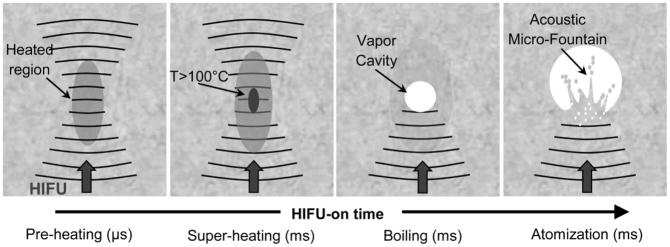
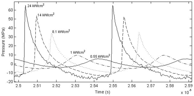
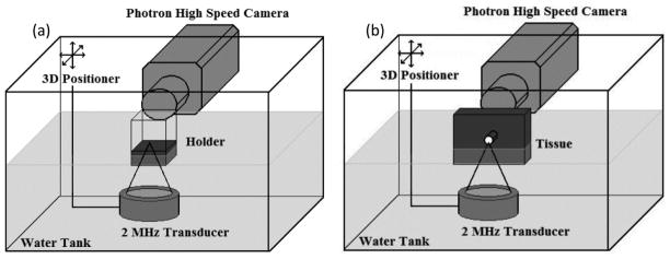
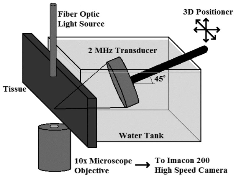
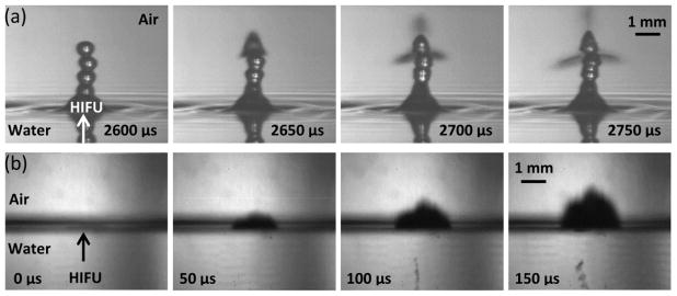
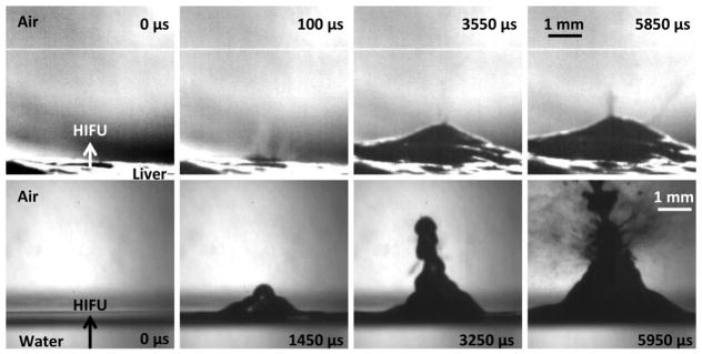
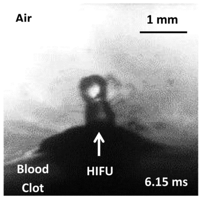
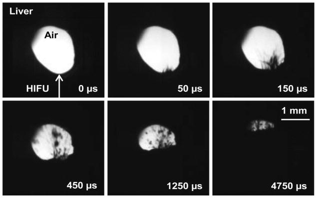
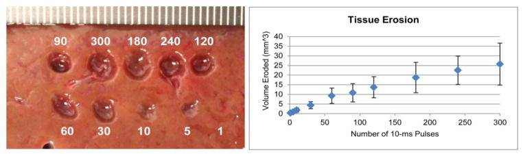
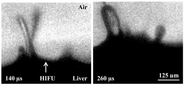
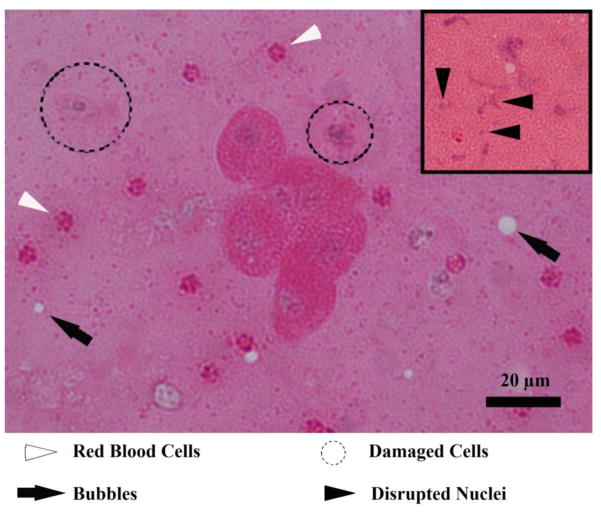
Atomization and fountain formation is a well-known phenomenon that occurs when a focused ultrasound wave in liquid encounters an air interface. High intensity focused ultrasound (HIFU) has been shown to fractionate a tissue into submicron-sized fragments in a process termed boiling histotripsy, wherein the focused ultrasound wave superheats the tissue at the focus, producing a millimetre-sized boiling or vapour bubble in several milliseconds. Yet the question of how this millimetre-sized boiling bubble creates submicron-sized tissue fragments remains. The hypothesis of this work is that the tissue can behave as a liquid such that it atomizes and forms a fountain within the vapour bubble produced in boiling histotripsy. We describe an experiment, in which a 2 MHz HIFU transducer (maximum in situ intensity of 24 000 W cm(-2)) was aligned with an air-tissue interface meant to simulate the boiling bubble. Atomization and fountain formation was observed with high-speed photography and resulted in tissue erosion. Histological examination of the atomized tissue showed whole and fragmented cells and nuclei. Air-liquid interfaces were also filmed. Our conclusion was that HIFU can fountain and atomize tissue. Although this process does not entirely mimic what was observed in liquids, it does explain many aspects of tissue fractionation in boiling histotripsy.


Figures

Figure 1
Figure 1
Proposed mechanism of tissue fractionation by boiling histotripsy.
Figure 2
Figure 2
Waveforms with calculated linear intensities corresponding to experimental conditions for the 2.165 MHz transducer.
Figure 3
Figure 3
Macro experimental setups. (a) Shows the planar interface for liquids or tissues in a holder while (b) shows the curved, bubble-like interface for tissue.
Figure 4
Figure 4
Micro experimental setup.
Figure 5
Figure 5
Air-water interface filmed with (a) the camera lens in air such that the reflections we see in the bottom of the frames are due to an optical reflection of the same jet and (b) the camera lens positioned so that half the objective is facing air and the other half facing water to simultaneously observe effects in water and air. (a) Shows a sequence of images taken at very low intensities (180 W/cm2) with the camera positioned at a slightly downward angle to catch the surface of the water. Atomization does not occur until the drop chain emerges. At some point, the top drop becomes unstable and atomization jets are released. (b) A sequence of images showing the air to water interface at high acoustic intensities of 24,000 W/cm2. Cavitation occurs before atomization or jetting and is faintly visible just to the right of the HIFU arrowhead in the first frame, which is taken 20 μs after the acoustic wave arrives at the water surface. As the ultrasonic pulse continues, the cavitation bubbles just below the water surface are joined by a cavitation bubble cloud further below the surface, possibly due to the standing wave pattern that develops upon the reflection of the acoustic wave. In both cases, the total HIFU-on time was 10 ms. This figure is available in movie form in supplement 1.
Figure 6
Figure 6
A direct comparison of liver (upper) and water (lower) at intensities slightly above their respective atomization thresholds (8100 W/cm2 derated for liver and 550 W/cm2 for water). In both cases, the first frame occurs 20 μs after the ultrasonic wave arrives at the interface. The second shows the initial spray of atomization in liver (upper) and the mound forming in water (lower) with no atomization. The third frame shows the small spray of atomization from the mound in liver and the first case of atomization for water; whereas the final frame shows atomization at its most significant. The timing is fairly similar between liver and water; the only difference is that liver has that first initial spray of atomization before the mound forms and enhances atomization, while at this intensity water forms the mound before atomization occurs. In both cases, the total HIFU-on time was 10 ms. This figure is available as a movie in supplement 2.
Figure 7
Figure 7
A drop-chain fountain emerging from the surface of a blood clot at 1000 W/cm2, derated, an intensity just above the atomization threshold in blood clots. In this case, the total HIFU-on time was 10 ms.
Figure 8
Figure 8
Cylindrical, bubble-like tunnel through bovine liver at linear intensity of 14,000 W/cm2, derated. At this intensity, there is a spurt of atomization that becomes more pronounced as the mound forms in the tissue. After around 5 ms, the hole becomes occluded with the spray. In this case, the total HIFU-on time was 10 ms. This movie is available in supplement 4.
Figure 9
Figure 9
Left: Tissue erosion observed on the surface of bovine liver at the maximum acoustic intensities of 14,000 W/cm2, derated, after a varying number of 10-ms pulses all at 1 Hz PRF. Right: A plot of the overall tissue erosion plotted as volume eroded per number of 10-ms pulses.
Figure 10
Figure 10
Magnified jets emitted from the surface of bovine liver. The jet(s) on the left side of each frame begin as thin streams and thicken over time. The jet on the right side of each frame shows how jets often start as the emission of a fog before developing into more significant jets. Furthermore, the right frame shows more jets forming between the established jets. The total HIFU-on time was 10-ms.
Figure 11
Figure 11
H&E stain of the collected fountain projectiles. In the centre of the image, a cell cluster consisting of six whole cells is present. In addition, there are red blood cells (white arrowheads), damaged or dying cells (dotted circles), and vapour bubbles (black arrows). The insert shows smeared and fragmented nuclei (black arrowheads).

References

    1. Antonevich J. Ultrasonic atomization of liquids. IRE Prof Group Ultrason Eng. 1959;6:6–15.
    1. Bailey M, Khokhlova V, Sapozhnikov O, Kargl S, Crum L. Physical mechanisms of the therapeutic effect of ultrasound (A review) Acoust Phys. 2003;49:369–88.
    1. Barreras F, Amaveda H, Lozano A. Transient high-frequency ultrasonic water atomization. Exp Fluids. 2002;33:405–13.
    1. Bassett J, Bright A. Observations concerning the mechanism of atomisation in an ultrasonic fountain. J Aerosol Sci. 1976;7:47–51.
    1. Bessonova O, Khokhlova V, Canney M, Bailey M, Crum L. A derating method for therapeutic applications of high intensity focused ultrasound. Acoust Phys. 2010;56:376–85. - PMC - PubMed

Publication types