. 2012 Dec 1;11(23):4462-73.
doi: 10.4161/cc.22794. Epub 2012 Nov 16.

Estrogen receptor α is a novel target of the Von Hippel-Lindau protein and is responsible for the proliferation of VHL-deficient cells under hypoxic conditions

Affiliations

Estrogen receptor α is a novel target of the Von Hippel-Lindau protein and is responsible for the proliferation of VHL-deficient cells under hypoxic conditions

Youn-Sang Jung et al. Cell Cycle. .

Abstract

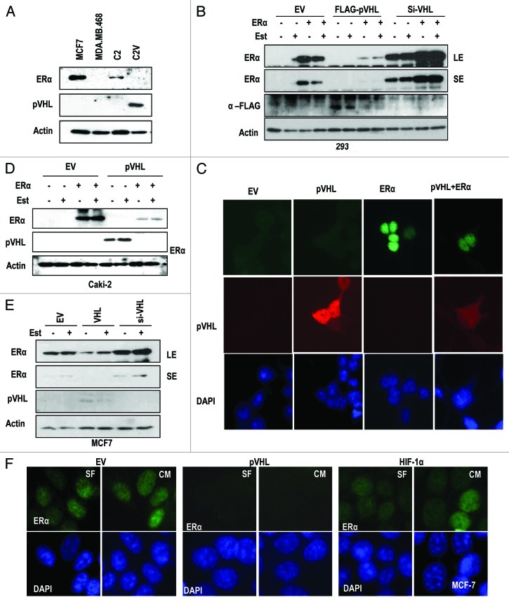
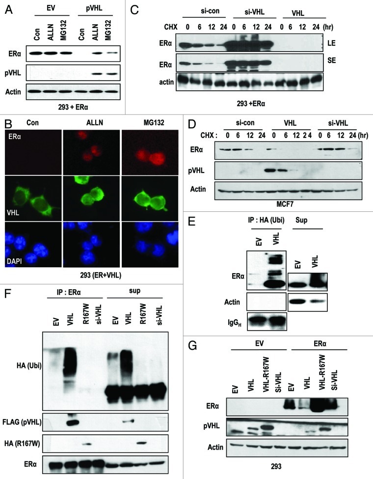
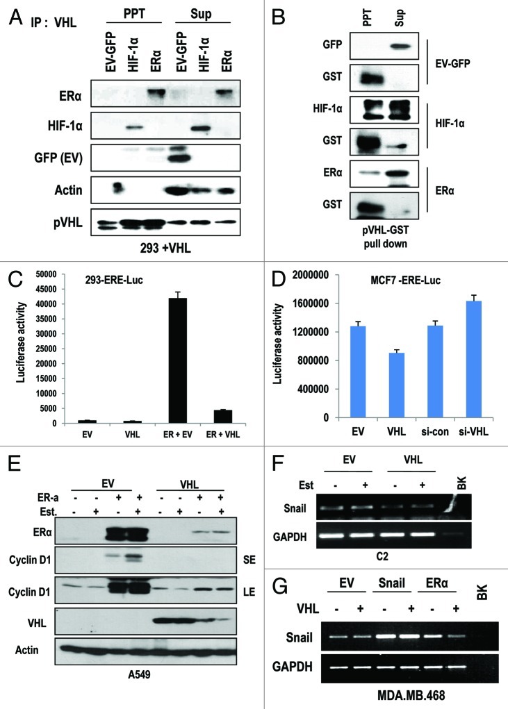
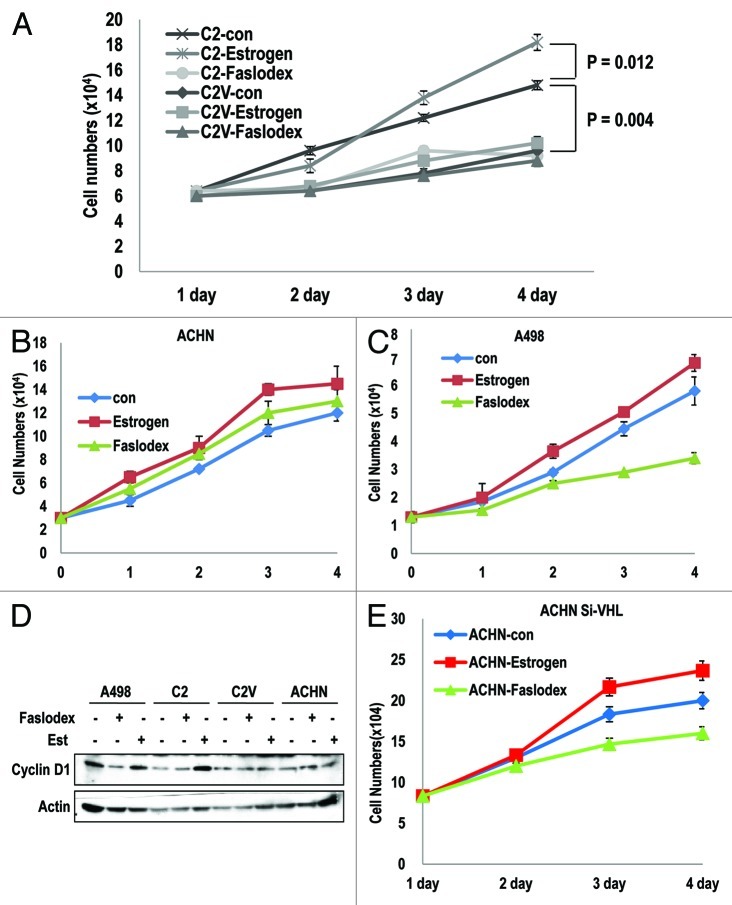
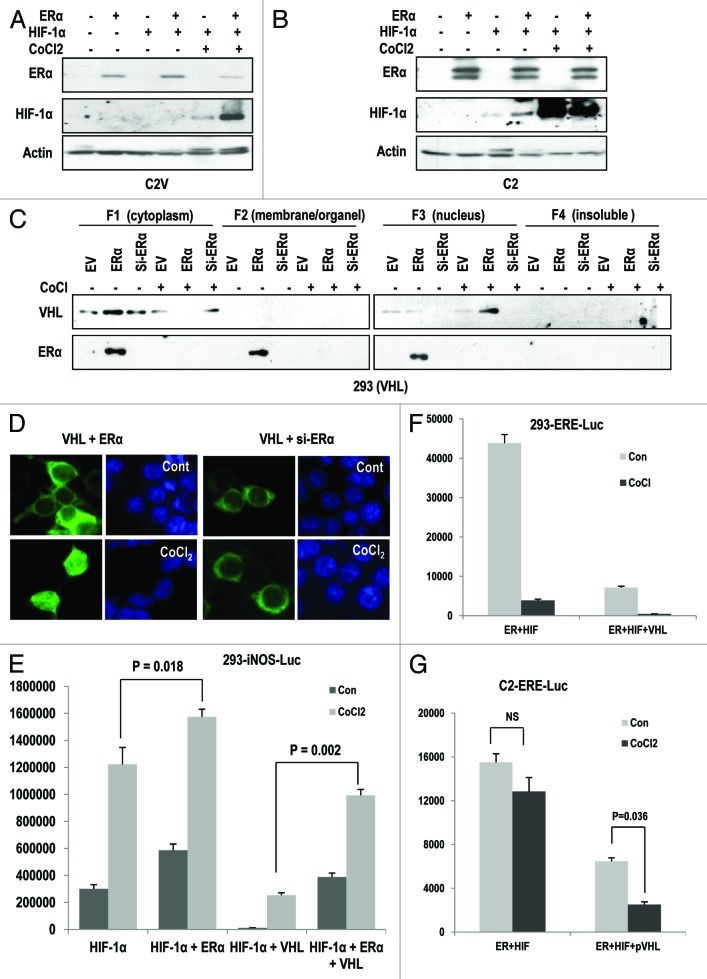
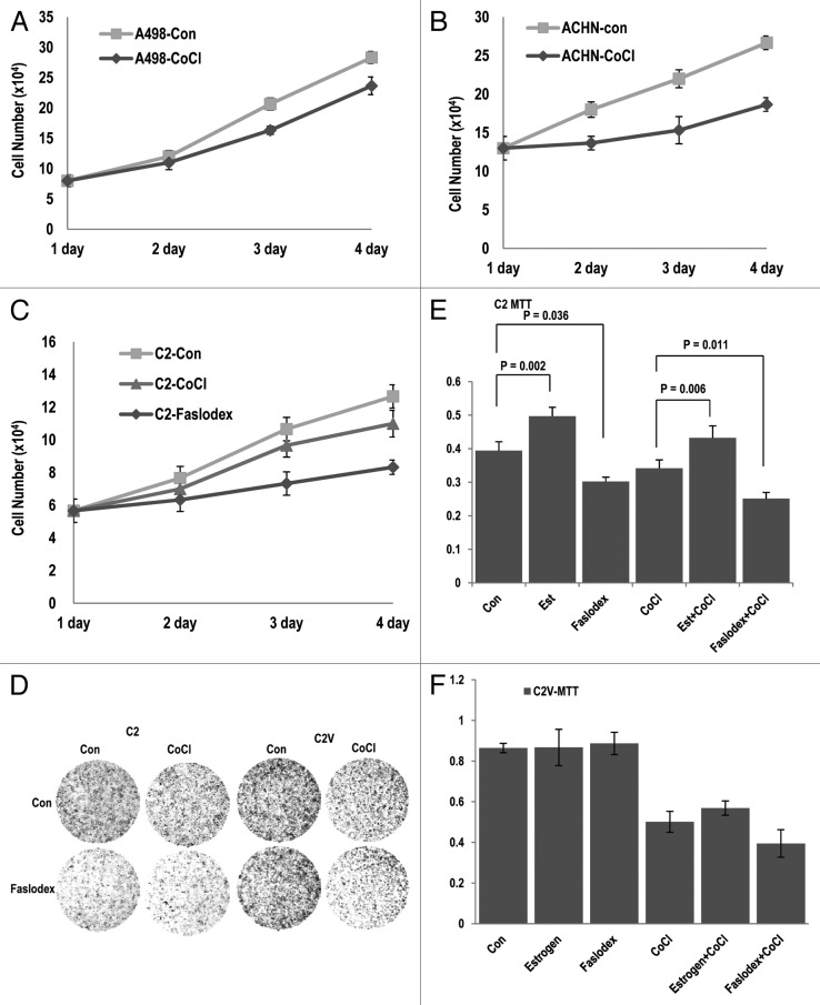
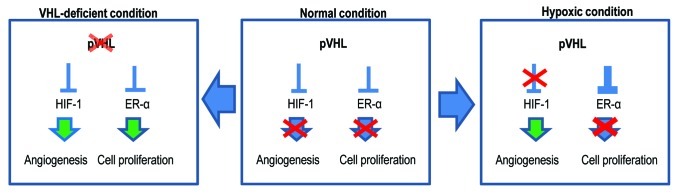
The Von Hippel-Lindau gene (VHL) is frequently deleted or mutated in human renal cell carcinoma (RCC) at the early stage. According to the well-established theory, pVHL acts as a tumor suppressor through its E3 ligase activity, which targets hypoxia-inducing factor-1α (HIF-1α). However, the elevated expression of HIF-1α did not promote cell proliferation, indicating that there would be another target, which could promote cell proliferation at the early cancer stage of RCC. In this study, we show that estrogen receptor-α (ER-α) is a novel proteasomal degradation target of the pVHL E3 ligase. Indeed, the overexpression of VHL suppresses exo- and endogenous ER-α expression, whereas si-pVHL can increase ER-α expression. The negative regulation of pVHL on ER-α expression is achieved by its E3 ligase activity. Thus, pVHL can promote the ER-α ubiquitinylation. In addition, we revealed that ER-α and HIF-1α are competitive substrates of pVHL. Thus, under normal conditions, ER-α overexpression can increase the transcription factor activity of HIF-1α. Under the hypoxic condition, where HIF-1α is not a suitable target of pVHL, ER-α is more rapidly degraded by pVHL. However, in VHL-deficient cells, the expression of ER-α and HIF-1α is retained, so that the hypoxic condition did not suppress cell proliferation obviously compared with cells that are expressing pVHL. Thus, blocking of ER-α using its inhibitor could suppress the proliferation of VHL-deficient cells as effectively as hypoxia-induced growth suppression. Considering our results, blocking of ER-α signaling in VHL-deficient cancer cells would be beneficial for cancer suppression. Indeed, we showed the anti-proliferative effect of Faslodex in VHL-deficient cells.


Figures

None
Figure 1. pVHL suppresses ER-α. (A) Expression of ER-α in VHL-deficient C2 cells. MCF7 and MDA-MB.468 (MDA) cells were used as positive and negative controls of ER-α expression. Actin was used as a loading control. (B) pVHL suppresses ER-α. After transfection with the indicated vectors or si-RNAs for 24 h, 293 cells were treated with estrogen (Est; 600 μg/ml) for 6 h. The expression of ER-α was determined by an ER-α-specific Ab, and the expression of exogenous pVHL was determined by a FLAG-Ab. EV indicates an empty vector transfection. LE and SE indicate a long exposure and a short exposure, respectively. (C) The reduction of ER-α and pVHL. Two hundred and ninety-three cells were transfected with VHL and/or ER-α for 24 h. After fixation with Me-OH, 293 cells were stained with anti-ER-α (green), anti-pVHL (red) and DAPI (blue). (D) pVHL suppresses ER-α in pVHL-mutant Caki-2 cells. Caki-2 cells were transfected with the indicated vectors for 24 h and were treated with estrogen for 6 h. (E) pVHL suppresses endogenous ER-α. MCF7 cells were transfected with the indicated vectors or si-RNAs for 24 h and were treated with estrogen for 6 h. LE and SE indicate a long exposure and a short exposure, respectively. (F) The reduction of endogenous ER-α by VHL transfection. MCF-7 cells were transfected with VHL or HIF-1α for 24 h. After fixation with Me-OH, MCF7 cells were stained with anti-ER-α (green) and DAPI (blue). To check for any effects that could have been caused by the serum, cells were incubated in serum-free medium (SF) or complete medium (CM) for 6 h before harvest.
None
Figure 2. pVHL promotes ER-α degradation through an E3 ligase. (A) Blocking of proteasomal degradation induces ER-α expression. Two hundred and ninety-three cells were transfected with the indicated vectors or si-RNAs for 24 h and were treated with ALLN and MG132 (50 μg/ml) for 6 h. SE and LE indicate short exposure and long exposure. (B) Proteasome inhibitors block pVHL-induced reduction in nuclear ER-α. 293 cells were co-transfected with VHL and ER-α for 24 h and incubated with ALLN or MG132 for 6 h. After fixation with Me-OH, 293 cells were stained with anti-ER-α (red), anti-pVHL (green) and DAPI (blue). (C) si-VHL extends the half-life of ER-α. Two hundred and ninety-three cells were co-transfected with ER-α and si-VHL or pVHL for 24 h. To block de novo synthesis, cycloheximide (CHX; 5 μg/ml) was added for the indicated times. Although VHL transfection completely eliminated ER-α, si-VHL extended the ER-α half-life. LE and SE indicate a long exposure and a short exposure, respectively. (D) pVHL can reduce the half-life of ER-α. MCF7 cells were transfected with the indicated vectors or si-RNAs for 24 h. After washing with serum-free medium, de novo synthesis was blocked by cycloheximide treatment for indicated times, and the expression of ER-α was measured. (E and F) ER-α is ubiquitinylated through the E3 ligase activity of pVHL. Immunoprecipitation was performed using HA or ER-α antibodies, and the co-precipitated proteins were analyzed using the indicated antibodies. Whole-cell extracts were obtained from 293 cells transfected with the indicated vectors. (G) ER-α can be suppressed by wild-type but not mutant pVHL. Two hundred and ninety-three cells were transfected with the indicated vectors or si-RNAs for 24 h.
None
Figure 3. pVHL suppressed ER-α through a direct interaction. (A) Direct interaction between ER-α and pVHL. Immunoprecipitation was performed using a pVHL antibody, and the co-precipitated proteins were analyzed using the indicated antibodies. Whole-cell extracts were obtained from 293 cells transfected with the indicated vectors. (B) GST pull-down assay. Agarose-conjugated GST-pVHL was incubated with whole-cell extracts from 293 cells transfected with the indicated vectors in RIPA buffer for 4 h. PPT indicates proteins that co-precipitated with bead-conjugated pVHL, whereas Sup indicates the supernatant. (C) pVHL suppresses transcription activity of ER-α. Two hundred and ninety-three cells were transfected with ERE-Luc and pVHL or ER-α. The increase in ERE-Luc activity was clearly reduced by VHL transfection. (D) pVHL regulates endogenous ER-α transcription activity. MCF-7 cells were transfected with ERE-Luc and VHL or si-VHL for 24 h. ER-α transcription activity was determined by a luciferase activity. (E) pVHL can suppress cyclin D1 expression. After transfection with the indicated vectors for 24 h, estrogen was added for 6 h in A549 cells. Immunoblot analysis was performed using the indicated antibodies. LE and SE indicate a long exposure and a short exposure, respectively. (F and G) pVHL can suppress Snail expression. After transfection with indicated vectors for 24 h. RT-PCR was performed to determine the expression of Snail using matched specific primers.
None
Figure 4. ER-α blocker suppresses cell proliferation in VHL-deficient cells. (A–C) Cell growth was estimated. Each cell line was treated with estrogen or Faslodex (5 μg/ml). Cells were maintained for maximum of 3 d, and cell numbers were calculated daily. (D) VHL-intact cell lines did not respond to estrogen or Faslodex. Each cell line was treated with estrogen or Faslodex for 6 h. After treatment, an immunoblot analysis was performed using the indicated antibodies. (E) The sensitivity to estrogen of Faslodex can be recovered by adding si-VHL to ACHN cells. Cell growth was estimated. After transfection with si-VHL, ACHN cells were treated with estrogen or Faslodex for 6 h. Cells were maintained for 4 d, and cell numbers were calculated daily.
None
Figure 5. Hypoxia promotes pVHL-mediated ER-α degradation. (A and B) Hypoxia reduces the expression of ER-α in a pVHL-dependent manner. After transfection with the indicated vectors for 24 h, CoCl2 (400 μM) was added for 12 h in each cell line. Immunoblot analysis was performed using the indicated antibodies. (C) pVHL translocates from the cytoplasm to the nucleus under the hypoxic condition. Two hundred and ninety-three cells were transfected with the indicated vectors and were treated with CoCl2 for 12 h. After treatment, cells were harvested to isolate the cytoplasm, membrane/organelle, nucleus and insoluble fractions and these samples were analyzed by immunoblot. (D) Knockdown of ER-α blocks the nuclear translocation of pVHL. Two hundred and ninety-three cells were transfected with the indicated vectors or si-RNAs for 24 h and were treated with CoCl2 for 12 h in 293 cells. After fixation with Me-OH, cells were stained with anti-pVHL (green) and DAPI (blue). (E) ER-α promotes HIF-1α transcription activity. Two hundred and ninety-three cells were co-transfected with an iNOS-Luc reporter containing a HIF-1α response element and the indicated vectors for 24 h. In addition, cells were incubated with CoCl2 for 12 h to induce the hypoxic conditions. (F) Hypoxia-induced suppression of ER-α activity is achieved by pVHL. Two hundred and ninety-three cells transfected with ERE-Luc were incubated with CoCl2 for 12 h. A clear reduction in ERE-Luc activity in response to hypoxia was detected. (G) The resistance of hypoxia-induced ERE-Luc suppression in VHL-deficient cells. Under similar conditions to those described above, a reduction in ERE-Luc activity in response to CoCl2 in C2 cells was not observed. Instead, pVHL transfection could restore sensitivity to hypoxia.
None
Figure 6. Blocking of ER-α suppresses cell proliferation in VHL-deficient tumors. (A and B) Hypoxia-induced growth arrest is dependent on pVHL status. The growth of cells that lacked VHL (A498; A) was not suppressed by hypoxia. In contrast, ACHN (VHL-positive) cells were sensitive to hypoxia-induced growth suppression. Each cell line was treated with CoCl2. Cells were maintained for 4 d, and cell numbers were calculated daily. (C) The inhibition of ER-α is more effective than hypoxia in cell growth suppression in VHL-deficient cells. C2 cells were incubated in CoCl2 or Faslodex-treated medium for 4 d. (D) Comparison of the growth of C2 and C2V cells. Differentially from C2 cells, where hypoxia did not notably suppress cell growth, C2V cells showed the sensitivity to hypoxia-induced growth suppression. In contrast, the growth of C2 cells was reduced by Faslodex. Cells were incubated with CoCl2 or Faslodex-containing medium for 3 d and fixed with PFA. Cells were visualized by staining with trypan blue. (E and F) pVHL is a critical factor for the determination of hypoxia- or Faslodex-induced growth suppression. Compared with C2V cells, in which Est or Faslodex did not alter cell viability, C2 cells were affected by Faslodex and Est. The opposite effect was observed with CoCl2 treatment. Cells were incubated with the indicated chemicals for 4 d. Cell viability was determined by a MTT assay.
None
Figure 7. Diagram for the working mechanism. Under normal conditions (center), pVHL suppresses HIF-1α and ER-α through its E3 ligase activity. However, in the hypoxic condition (right panel), pVHL sequesters ER-α more strongly so that cells cannot proliferate. Instead, HIF-1α can be activated and induce angiogenesis. Because the hypoxic condition is generated by tissue damage, this mechanism may contribute to blocking an overgrowth of damaged cells. However, when pVHL is deleted or mutated (left panel), ER-1α-induced cell proliferation can be apparent. In addition, nutrients are then supplied by HIF-1α-mediated angiogenesis. These conditions would increase the probability of the occurrence of cancer.

Similar articles

Cited by

References

    1. Kaelin WG., Jr. Molecular basis of the VHL hereditary cancer syndrome. Nat Rev Cancer. 2002;2:673–82. doi: 10.1038/nrc885. - DOI - PubMed
    1. Kaelin WG., Jr. Von Hippel-Lindau disease. Annu Rev Pathol. 2007;2:145–73. doi: 10.1146/annurev.pathol.2.010506.092049. - DOI - PubMed
    1. Schofield CJ, Ratcliffe PJ. Oxygen sensing by HIF hydroxylases. Nat Rev Mol Cell Biol. 2004;5:343–54. doi: 10.1038/nrm1366. - DOI - PubMed
    1. Kim WY, Kaelin WG. Role of VHL gene mutation in human cancer. J Clin Oncol. 2004;22:4991–5004. doi: 10.1200/JCO.2004.05.061. - DOI - PubMed
    1. Semenza GL, Wang GL. A nuclear factor induced by hypoxia via de novo protein synthesis binds to the human erythropoietin gene enhancer at a site required for transcriptional activation. Mol Cell Biol. 1992;12:5447–54. - PMC - PubMed

Publication types

MeSH terms

Substances

LinkOut - more resources