. 2012 Nov;3(11):1386-400.
doi: 10.18632/oncotarget.709.

Tumor suppressive microRNA-218 inhibits cancer cell migration and invasion through targeting laminin-332 in head and neck squamous cell carcinoma

Affiliations

Tumor suppressive microRNA-218 inhibits cancer cell migration and invasion through targeting laminin-332 in head and neck squamous cell carcinoma

Takashi Kinoshita et al. Oncotarget. 2012 Nov.

Abstract

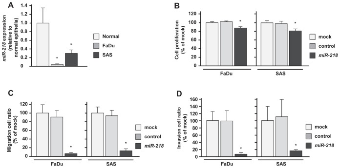
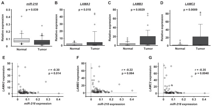
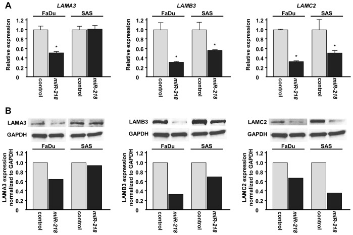
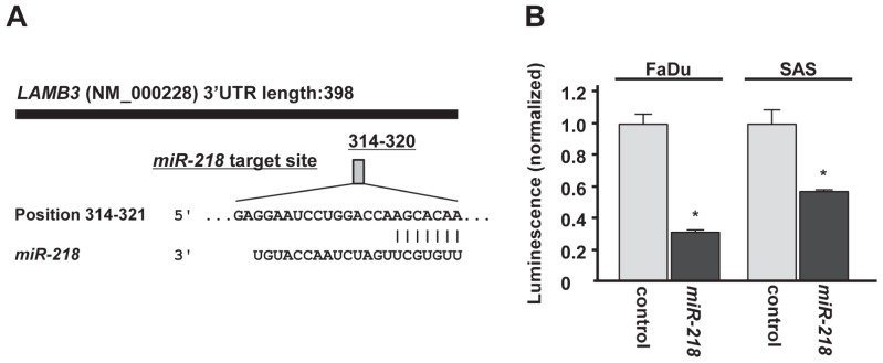
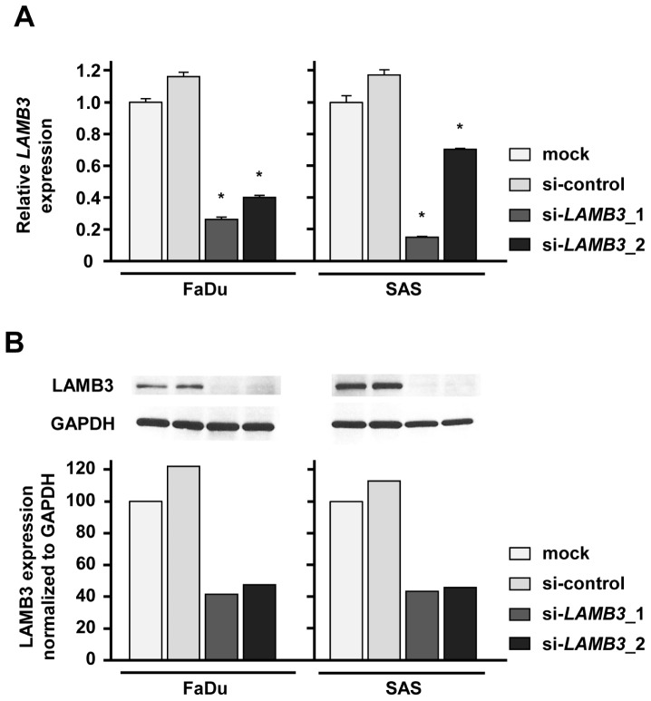
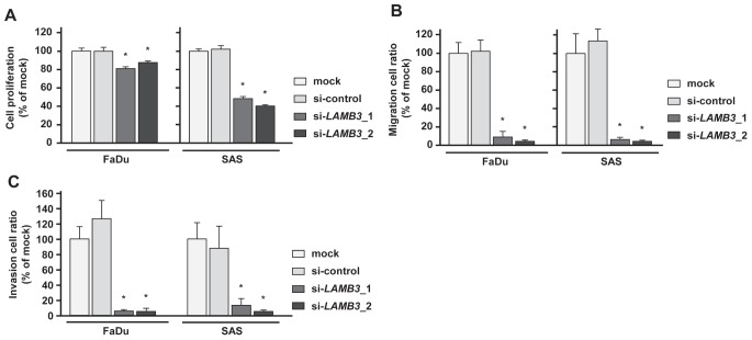
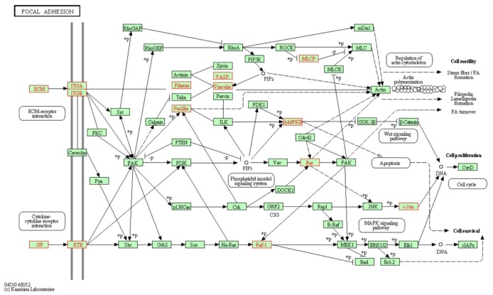
Recent our microRNA (miRNA) expression signature revealed that expression of microRNA-218 (miR-218) was reduced in cancer tissues, suggesting a candidate of tumor suppressor in head and neck squamous cell carcinoma (HNSCC). The aim of this study was to investigate the functional significance of miR-218 and its mediated moleculer pathways in HNSCC. Restoration of miR-218 in cancer cells led to significant inhibition of cell migration and invasion activities in HNSCC cell lines (FaDu and SAS). Genome-wide gene expression analysis of miR-218 transfectants and in silico database analysis showed that focal adhesion pathway was a promising candidate of miR-218 target pathways. The laminins are an important and biologically active part of the basal lamina, the function of that are various such as influencing cell differentiation, migration and adhesion as well as proliferation and cell survival. Interestingly, all components of laminin-332 (LAMA3, LAMB3 and LAMC2) are listed on the candidate genes in focal adhesion pathway. Furthermore, we focused on LAMB3 which has a miR-218 target site and gene expression studies and luciferase reporter assays showed that LAMB3 was directly regulated by miR-218. Silencing study of LAMB3 demonstrated significant inhibition of cell migration and invasion. In clinical specimens with HNSCC, the expression levels of laminin-332 were significantly upregulated in cancer tissues compared to adjacent non-cancerous tissues. Our analysis data showed that tumor suppressive miR-218 contributes to cancer cell migration and invasion through regulating focal adhesion pathway, especially laminin-332. Tumor suppressive miRNA-mediated novel cancer pathways provide new insights into the potential mechanisms of HNSCC oncogenesis.


Figures

Figure 1
Figure 1. Effects of miR-218 transfection on HNSCC cell lines, FaDu and SAS
(A)Real-time RT-PCR showed that expression levels of miR-218 was significantly lower in both cell lines, FaDu and SAS than in normal epithelial specimens. (B)Suppression of FaDu and SAS cell proliferation after transfection (72h) with miR-218 (10nM) was determined by XTT assay. (C) Suppression of FaDu and SAS cell migration activity after transfection (72h) with miR-218 (10nM) was determined by migration assay. (D) Suppression of FaDu and SAS cell invasion activity after transfection (72h) with miR-218 (10nM) was determined by Matrigel invasion assay. *p < 0.0167.
Figure 2
Figure 2. The expression levels of miR-218 and laminin-332 in HNSCC clinical specimens
The expression levels of miR-218 (A), LAMA3 (B), LAMB3 (C) and LAMC2 (D) in tumor tissues and adjacent normal epithelium tissues of 35 HNSCC patients were determined by qRT-PCR. RNU48 and GUSB were used as internal controls. The correlated expression of miR-218 and laminin-332 was determined in HNSCC specimens. The correlation coefficient indicates that miR-218 expression was highly correlated with that of laminin-332, LAMA3 (E), LAMB3 (F) and LAMC2 (G).
Figure 3
Figure 3. Laminin-332 expression was suppressed by miR-218 transfection at both the mRNA and protein levels in HNSCC cell lines
(A) mRNA expression of LAMA3, LAMB3 and LAMC2 as revealed by qRT-PCR 48 h after transfection with 10 nM of miR-218. GUSB was used as internal controls. *p< 0.05. (B) Protein expression of LAMA3, LAMB3 and LAMC2 as revealed by western blot analysis 48 h after transfection with 10 nM of miR-218. GAPDH was used as loading controls. The expression ratio of LAMA3, LAMB3 and LAMC2 were evaluated using ImageJ software.
Figure 4
Figure 4. miR-218 directly regulates LAMB3 by luciferase reporter assay
(A)Putative miR-218 binding site in the 3'UTR of LAMB3 mRNA was identified with the TargetScan database. (B) Luciferase reporter assay was performed using the vector encoding partial sequences of 3'UTR which contained the putative miR-218 target site. The vector (10 ng) and miR-218 or miR-control (10 nM) were cotransfected into FaDu and SAS cell lines. Renila luciferase activity was measured after 24h transfection. The results were normalized by firefly luciferase values. *p < 0.05.
Figure 5
Figure 5. LAMB3 expression was suppressed by si-LAMB3 transfection at both the mRNA and protein levels in HNSCC cell lines
(A) mRNA expression of LAMB3 as revealed by real-time qRT-PCR 48 h after transfection with 10 nM of si-LAMB3. *p < 0.0083. (B) Protein expression of LAMB3 as revealed by western blot analysis 48 h after transfection with 10 nM of si-LAMB3. GAPDH was used as loading controls. The expression ratio of LAMB3 was evaluated using ImageJ software.
Figure 6
Figure 6. Effects of LAMB3 knockdown by si-LAMB3 transfection on HNSCC cell lines, FaDu and SAS
(A) Cell proliferation activities as revealed by XTT assay in HNSCC cell lines, FaDu and SAS. *p < 0.0083 (B) Cell migration activities (migration assay) of HNSCC cells. *p < 0.0083 (C) Cell invasion activities (Matrigel invasion assay) of HNSCC cells. *p < 0.0083
Figure 7
Figure 7. Putative LAMB3 regulated genes in “focal adhesion pathway”
Putative LAMB3 regulated genes in “focal adhesion pathway” from Kyoto Encyclopedia of Genes and Genomes (KEGG). The putative LAMB3 regulated genes (highlighted in red) as defined by KEGG pathway and determined through GENECODIS analysis.

References

    1. Parkin DM, Bray F, Ferlay J, Pisani P. Global cancer statistics, 2002. CA Cancer J Clin. 2005;55:74–108. - PubMed
    1. Leemans CR, Braakhuis BJ, Brakenhoff RH. The molecular biology of head and neck cancer. Nat Rev Cancer. 2011;11:9–22. - PubMed
    1. Mattick JS. RNA regulation: a new genetics? Nat Rev Genet. 2004;5:316–323. - PubMed
    1. Bartel DP. MicroRNAs: genomics, biogenesis, mechanism, and function. Cell. 2004;116:281–297. - PubMed
    1. Tran N, O'Brien CJ, Clark J, Rose B. Potential role of micro-RNAs in head and neck tumorigenesis. Head Neck. 2010;32:1099–1111. - PubMed

Publication types

MeSH terms