

A genome-wide association study identifies susceptibility loci for nonsyndromic sagittal craniosynostosis near BMP2 and within BBS9
- PMID: 23160099
- PMCID: PMC3736322
- DOI: 10.1038/ng.2463
A genome-wide association study identifies susceptibility loci for nonsyndromic sagittal craniosynostosis near BMP2 and within BBS9
Abstract
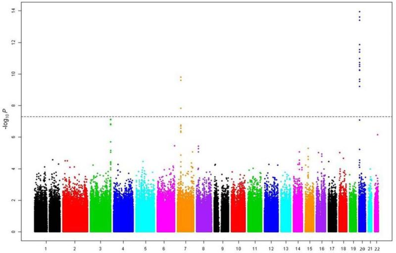
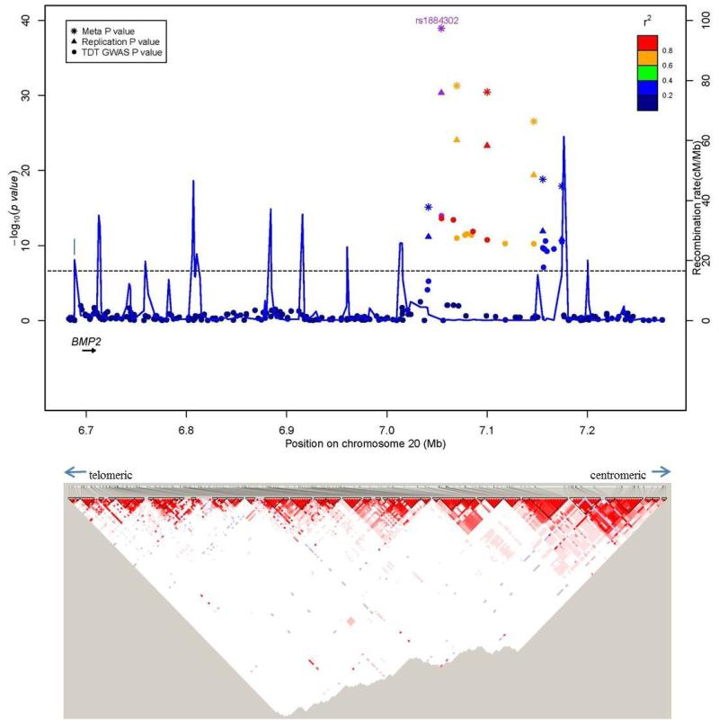
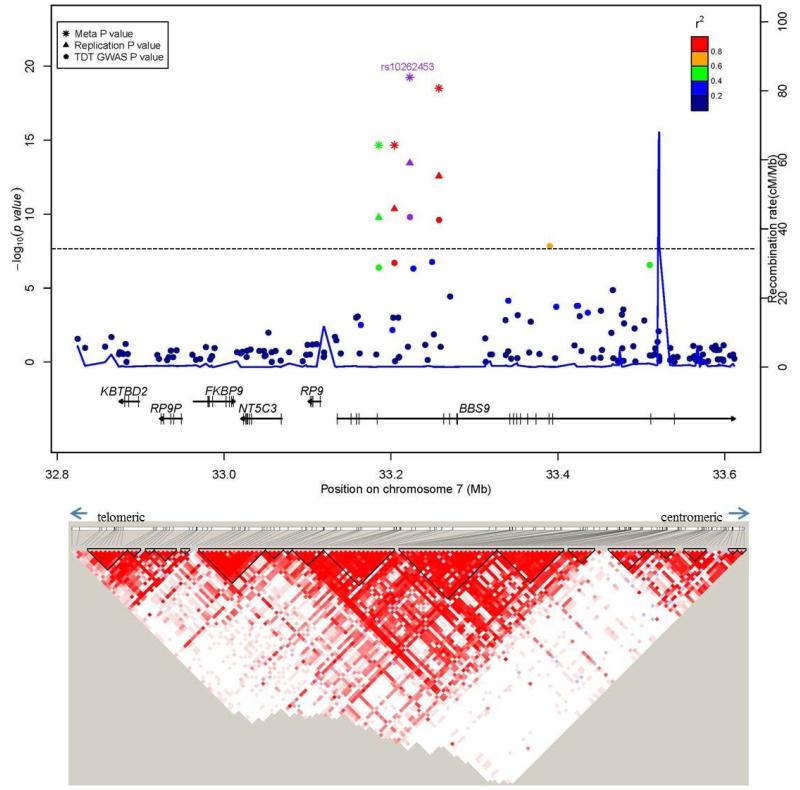
Sagittal craniosynostosis is the most common form of craniosynostosis, affecting approximately one in 5,000 newborns. We conducted, to our knowledge, the first genome-wide association study for nonsyndromic sagittal craniosynostosis (sNSC) using 130 non-Hispanic case-parent trios of European ancestry (NHW). We found robust associations in a 120-kb region downstream of BMP2 flanked by rs1884302 (P = 1.13 × 10(-14), odds ratio (OR) = 4.58) and rs6140226 (P = 3.40 × 10(-11), OR = 0.24) and within a 167-kb region of BBS9 between rs10262453 (P = 1.61 × 10(-10), OR = 0.19) and rs17724206 (P = 1.50 × 10(-8), OR = 0.22). We replicated the associations to both loci (rs1884302, P = 4.39 × 10(-31) and rs10262453, P = 3.50 × 10(-14)) in an independent NHW population of 172 unrelated probands with sNSC and 548 controls. Both BMP2 and BBS9 are genes with roles in skeletal development that warrant functional studies to further understand the etiology of sNSC.
Figures
References
-
- Cohen MM. Craniosynostosis: Diagnosis, Evaluation, and Management. Oxford University Press; New York: 2000.
-
- Johnson D, et al. A novel mutation, Ala315Ser, in FGFR2: a gene-environment interaction leading to craniosynostosis? Eur J Hum Genet. 2000;8(8):571–577. - PubMed
-
- Seto ML, et al. Isolated sagittal and coronal craniosynostosis associated with TWIST box mutations. Am J Med Genet A. 2007;143(7):678–86. - PubMed
-
- Weber I, et al. Molecular analyisis of 74 patients with craniosynostosis. Eur J Hum Genet. 2001;9(Sup.1):P0409, 179.
-
- Merrill AE, et al. Cell mixing at a neural crest-mesoderm boundary and deficient ephrin-Eph signaling in the pathogenesis of craniosynostosis. Hum Mol Genet. 2006;15(8):1319–28. - PubMed
Publication types
MeSH terms
Substances
Grants and funding
- 093329/WT_/Wellcome Trust/United Kingdom
- N01-DK-7-3431/DK/NIDDK NIH HHS/United States
- U01 DD000492/DD/NCBDD CDC HHS/United States
- R37 DE012711/DE/NIDCR NIH HHS/United States
- M01-RR00052/RR/NCRR NIH HHS/United States
- R01 DE016886/DE/NIDCR NIH HHS/United States
- K23 DE000462/DE/NIDCR NIH HHS/United States
- R21DE022419/DE/NIDCR NIH HHS/United States
- 3R01 DE018500-02S1/DE/NIDCR NIH HHS/United States
- R01 DE022988/DE/NIDCR NIH HHS/United States
- R01 DE018500/DE/NIDCR NIH HHS/United States
- HHSN268200782096C/HG/NHGRI NIH HHS/United States
- K12-HD05954/HD/NICHD NIH HHS/United States
- 5 R01 DD000350/DD/NCBDD CDC HHS/United States
- M01 RR000052/RR/NCRR NIH HHS/United States
- UL1 TR000067/TR/NCATS NIH HHS/United States
- ImNIH/Intramural NIH HHS/United States
- K23 DE00462/DE/NIDCR NIH HHS/United States
- 5U01DD000492/DD/NCBDD CDC HHS/United States
- UL1TR000067/TR/NCATS NIH HHS/United States
- R01 DE018227/DE/NIDCR NIH HHS/United States
- R01 DD000350/DD/NCBDD CDC HHS/United States
- R21 DE022419/DE/NIDCR NIH HHS/United States
- R03 DE016342/DE/NIDCR NIH HHS/United States
- 3R37DE012711-13S1/DE/NIDCR NIH HHS/United States
LinkOut - more resources
Full Text Sources
Medical
Molecular Biology Databases
