. 2012 Dec 12;31(24):4511-23.
doi: 10.1038/emboj.2012.298. Epub 2012 Nov 16.

Steroid-induced microRNA let-7 acts as a spatio-temporal code for neuronal cell fate in the developing Drosophila brain

Affiliations

Steroid-induced microRNA let-7 acts as a spatio-temporal code for neuronal cell fate in the developing Drosophila brain

Mariya M Kucherenko et al. EMBO J. .

Abstract

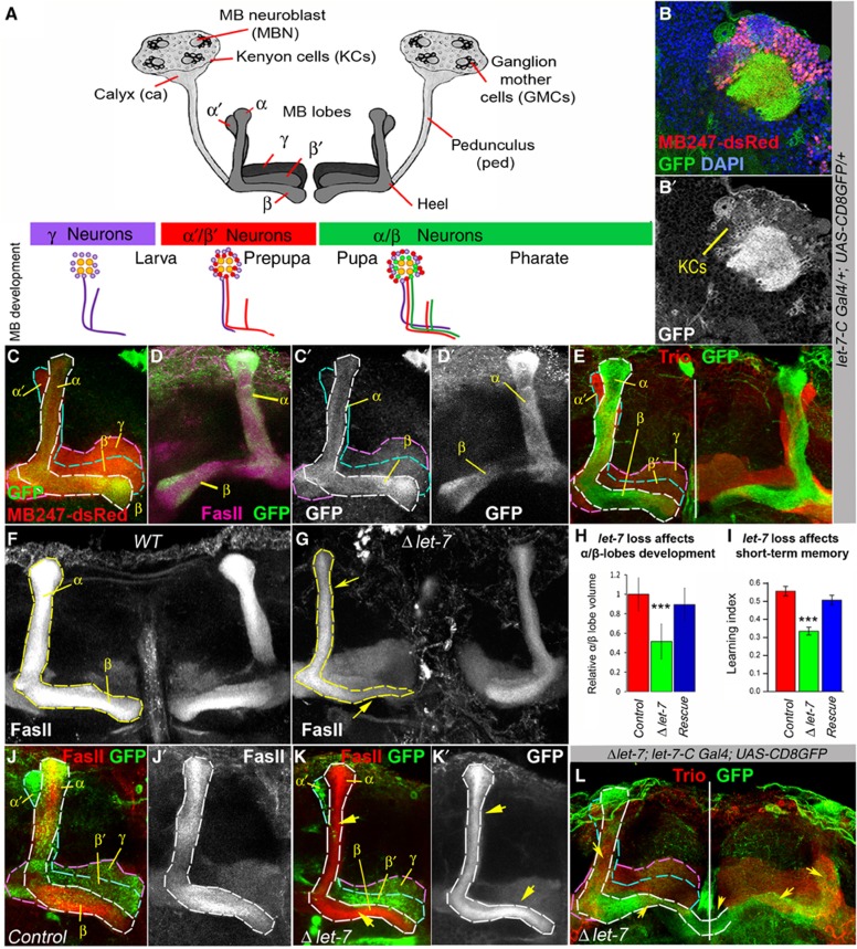
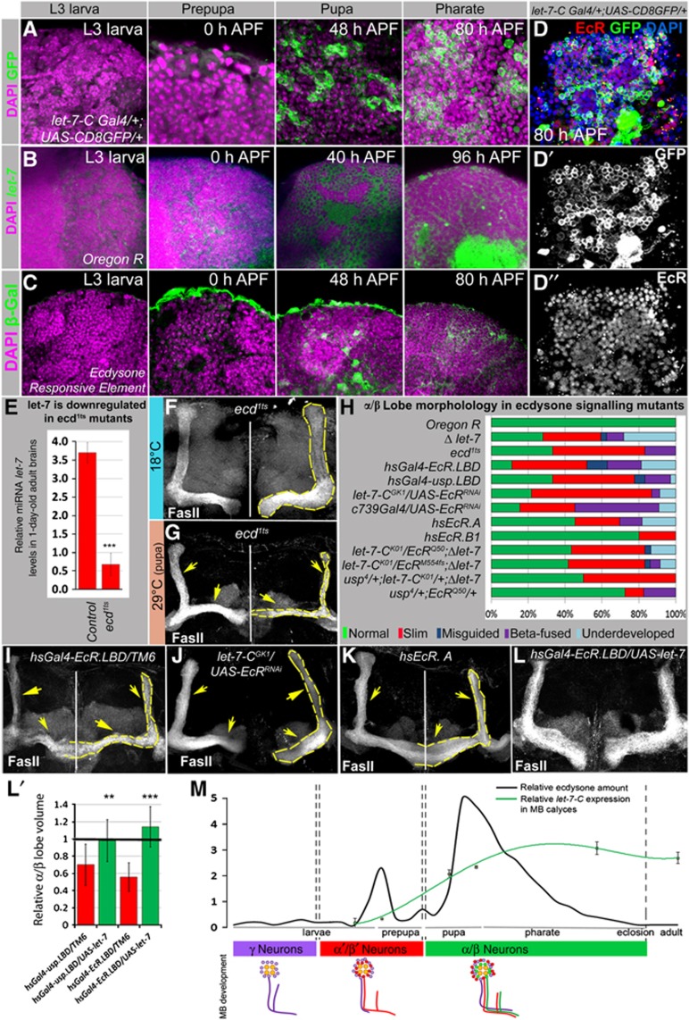
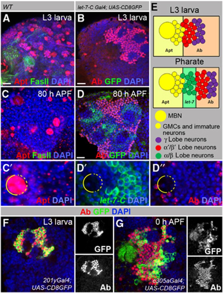
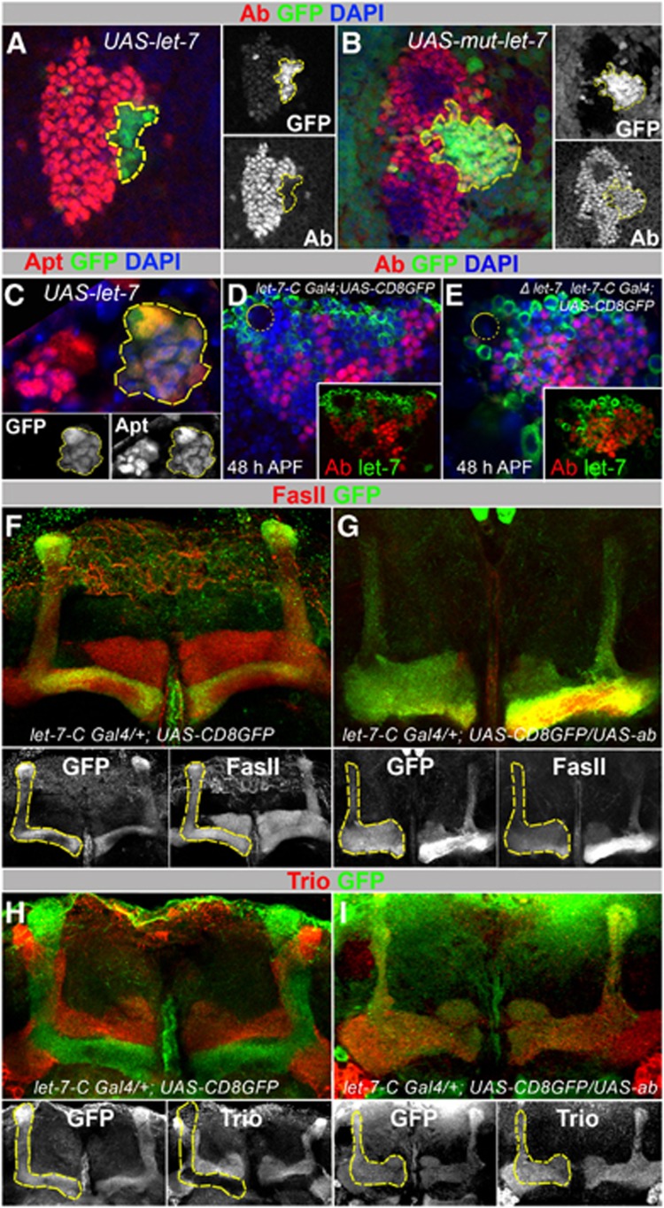
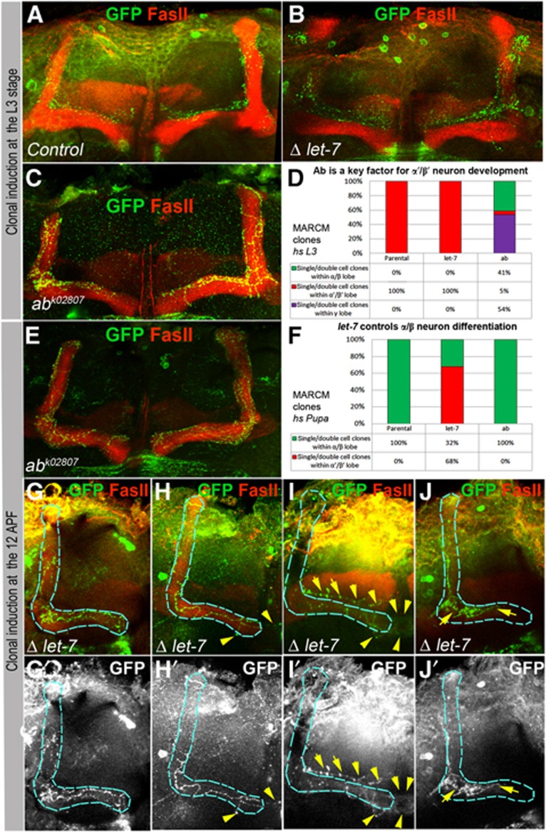
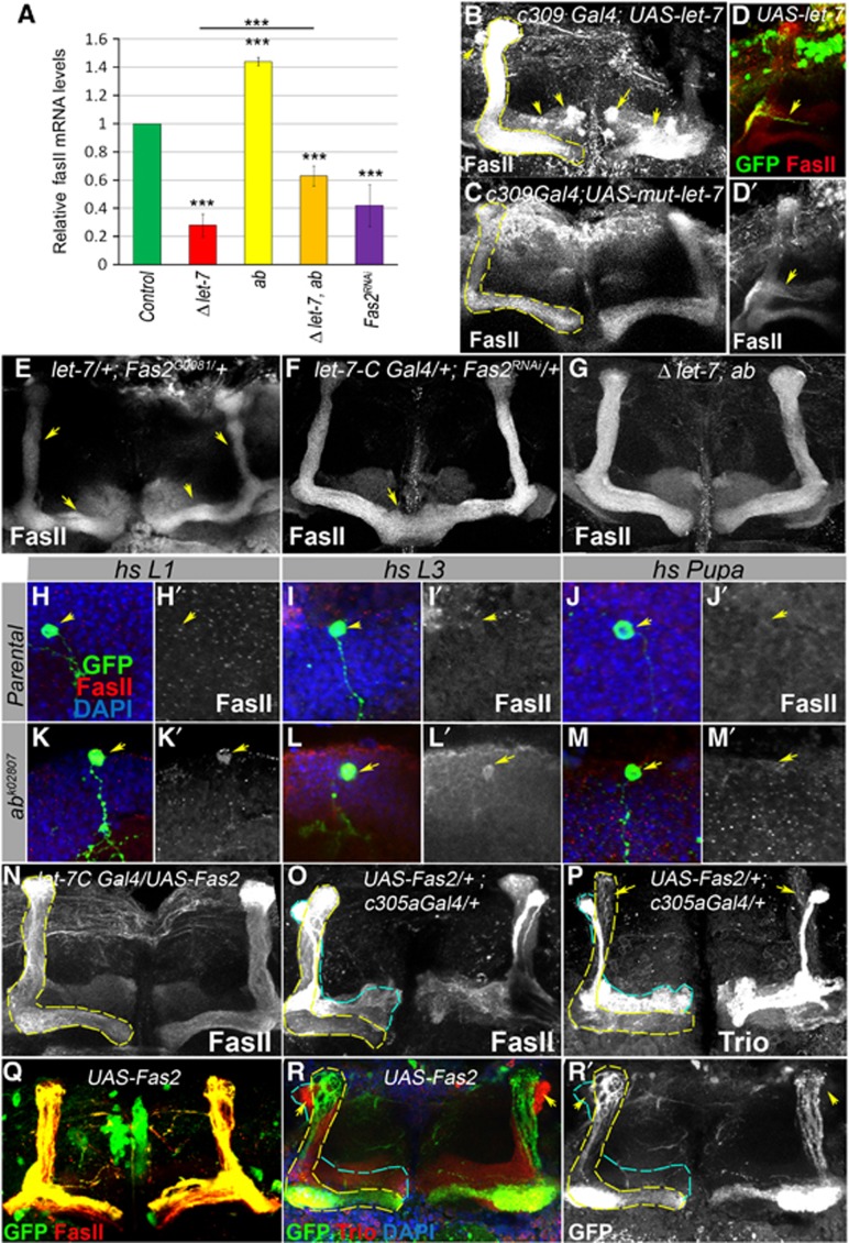
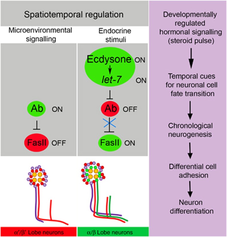
Mammalian neuronal stem cells produce multiple neuron types in the course of an individual's development. Similarly, neuronal progenitors in the Drosophila brain generate different types of closely related neurons that are born at specific time points during development. We found that in the post-embryonic Drosophila brain, steroid hormones act as temporal cues that specify the cell fate of mushroom body (MB) neuroblast progeny. Chronological regulation of neurogenesis is subsequently mediated by the microRNA (miRNA) let-7, absence of which causes learning impairment due to morphological MB defects. The miRNA let-7 is required to regulate the timing of α'/β' to α/β neuronal identity transition by targeting the transcription factor Abrupt. At a cellular level, the ecdysone-let-7-Ab signalling pathway controls the expression levels of the cell adhesion molecule Fasciclin II in developing neurons that ultimately influences their differentiation. Our data propose a novel role for miRNAs as transducers between chronologically regulated developmental signalling and physical cell adhesion.


Figures

Figure 1
Figure 1
let-7 miRNA is expressed in α/β MB lobes and its loss results in morphological and behavioural defects. (A) Schematic view of the Drosophila mushroom bodies (MBs), which are paired structures originating from four identical MB neuroblasts (MBNs). Within MB cell body cluster, proliferating MBNs give rise to ganglion mother cells (GMCs) that divide one more time to generate intrinsic MB neurons or Kenyon cells (KCs). There are three main types of KCs that each form distinct lobes (γ, α′/β′ and α/β), which connect to the calyx via the pedunculus (ped). The scheme below depicts subsequent birth of γ, α′/β′ and α/β neurons during different stages of Drosophila development. (B) In MBs, let-7-C is detected in a subset of KCs and in the calyx (ca). (C) In the 3- to 6-day-old adult Drosophila brain, high let-7-C expression levels (visualized here by GFP expression, green) are detected in α/β and very weak levels in γ MB lobes; dsRed marks all MB lobes (γ, α′/β′ and α/β). (D) let-7-C expression pattern also coincides with Fas II, which is a marker for α/β lobes. (E) let-7-C expression is not detected in α′/β′ lobes marked with Trio. (F, G) Loss of the miRNA let-7 results in abnormally developed α/β MB lobes in comparison to OregonR control (F). Note the ‘slim-α/β-lobe’ phenotype (G). (H) The volume of α/β lobes is significantly decreased in let-7-deficient brains, which can be rescued via exogenous expression of one copy of let-7 (n=17 MB lobes for Control, n=18 for Δlet-7 and n=8 for Rescue; Supplementary Table 1). (I) Absence of let-7 causes short-term memory impediment, which can be rescued via re-introducing a copy of let-7 (n=16 experiments; Supplementary Table 2); ***P⩽0.001. (JK) Induction of MARCM clones at the first instar larval stage shows underdeveloped let-7 clonal (GFP positive) α/β lobes as a result of let-7 depletion in MB neurons. Compare outlined α/β lobes in (J) (Control, hsFlp UAS CD8GFP; tubGal80 FRT 40A/FRT 40A; tubGal4/+) and (K) (Δlet-7, hsFlp UAS CD8GFP; tubGal80 FRT 40A/let-7 miR-125 FRT 40A; tubGal4/P{W8, let-7-CΔlet-7}). (L) In let-7 mutants, α/β neurons migrate into α′/β′ lobe as can be seen by co-localization of Trio (red) with let-7-C>GFP (green). Yellow arrows show morphological defects in α/β lobes, white vertical line marks midline in (E) and (L), α/β MB lobes are visualized by the presence of let-7-C Gal4; UAS-CD8GFP (CE), γ, α/β marker Fas II (D, F, G, JK), and the absence of γ, α′/β′ marker Trio (E, L).
Figure 2
Figure 2
A developmentally regulated ecdysone pulse induces let-7-C expression and affects MB α/β lobe development. (A) let-7-C expression (let-7-CGK1; UAS-CD8GFP:UAS-nLacZ), (B) let-7 LNA in situ hybridization and (C) active ecdysone signalling (EcRE.lacZ) in MB cell body clusters of wild-type animals at different stages of development visualized by anti-GFP, fluorescent in situ hybridization, and anti-β-Gal staining, respectively. Strong let-7-C and let-7 miRNA expression is detected at the pupal stage, which correlates with the ecdysone-signalling peak. Note that let-7-C and let-7 miRNA expression after being induced continues through pharate and adult stages (as seen in Figure 1B–E), while the activity of ecdysone signalling starts at prepupa, peaks at pupa and diminishes at later stages (Supplementary Figure S3). (DD″) Ecdysone receptor expression pattern shows co-expression of EcR and let-7 in MB cell body clusters. (E) The bar graph represents decreased let-7 expression levels in the newly eclosed adult brains of ecd1ts mutants kept at the restrictive temperature during metamorphosis (n=3 experiments; Supplementary Table 3). (F, G) Abnormally developed α/β lobes were observed in ecd1ts mutants kept at 29°C at the pupal stage (G, yellow arrows), compared to control ecd1ts animals kept at 18°C (F). (HK) Graph showing the frequency of α/β morphology defects in inducible ecdysone signalling mutants stimulated at the pupal stage (hsGal4-EcRLBD, hsGal4-uspLBD, hs EcR.A, hs EcR.B1; I, K). Note that overexpression of EcR.B1 isoform that has been shown to be specific for γ lobe remodelling (Lee et al, 2000) had less severe effects than overexpression of EcR.A isoform. Similar α/β phenotypes were observed due to cell autonomous downregulation of EcR in α/β lobes (let-7GK1; EcRRNAi and c739Gal4; EcRRNAi), as well as a result of transheterozygous genetic interactions between miRNA let-7 and ecdysone pathway mutants (let-7-CK01/EcRQ50;P{W8, let-7-CΔlet-7}/+, let-7-CK01/EcRM554fs;P{W8, let-7-CΔlet-7}/+, let-7-CK01/usp4;P{W8, let-7-CΔlet-7}/+, usp4/+;EcRQ50/+ (H, J), see also Supplementary Table 5). (LL′) This defect can be rescued by overexpression of let-7 (hsGal4-EcR-L.B.D./UAS-let-7, hsGal4-usp-L.B.D./UAS-let-7, compare (I) and (L), see Supplementary Table 4). (M) Schematic drawing illustrating developmentally regulated ecdysone pulses (Riddiford, 1993), chronology of different MB lobes formation (Lee et al, 1999), and let-7 complex expression in MB cell body clusters during development. let-7-C expression was analysed via measuring relative GFP intensity in the MB cell body clusters of different stages let-7-CGK1; UAS-CD8GFP animals using ImageJ: L3 larva 0.18±0.19; 0 h APF 1.00±0.13; 48 h APF 2.08±0.16; 58 h APF 2.35±0.05; 80 h APF 3.09±0.24; 1- to 3-day old adults 2.7±0.22; at least three brains for each stage were analysed. Note the correlation between the ecdysone activity, initiation of let-7-C expression in MB neurons, and a transition of their differentiation into α/β type neurons. **P⩽0.01, ***P⩽0.001.
Figure 3
Figure 3
Putative let-7 targets Apt and Ab are expressed in MB cell body clusters. (A, C) In wild-type animals (OregonR), Apt is expressed in the MBNs, GMCs and immature neurons at larval and pupal stages. (B, D) In contrast, the Ab signal is strong in the nuclei of differentiated neurons (α′/β′ and γ lobe neurons). (B) GFP that represents let-7-C expression is clearly absent from MB cell body clusters during the L3 stage (let-7-CGK1; UAS-CD8GFP:UAS-nLacZ). At the pharate stage (80 h APF), neurons located next to GMCs (α/β lobe neurons) express let-7-C but lack Ab (D). (C′, D′, D″) Magnification of the pharate stage MB cell body cluster showing mutually exclusive expression patterns of Apt, Ab and let-7-C. (E) Drawings represent the expression patterns of let-7-C, Apt and Ab in larval and pharate stages. (F, G) Ab is expressed in γ and α′/β′ MB lobes. (F) Larval brain (L3 stage) shows Ab expression in KCs, which form γ lobes marked with GFP driven by the γ lobe-specific driver 201y-Gal4. (G) In the early pupal stage (0 h APF), Ab staining is seen in KCs that form α′/β′ lobes marked with GFP driven by the α′/β′ lobe-specific driver c305a-Gal4. For Gal4 line expression patterns, see Supplementary Figure S5.
Figure 4
Figure 4
let-7 targets Abrupt in MBs. (A) L3 larval brains with let-7 overexpressing clones (marked with GFP, hsFlp;act>CD2>Gal4;UAS-let-7) show decreased levels of Ab protein levels compared to non-clonal neighbouring cells. (B) Overexpression of the mutant form of let-7 used as a control (hsFlp;act>CD2>Gal4;UAS-mut-let-7) does not affect Ab levels. (C) L3 larval brains with let-7 overexpressing clones do not show significantly changed Apt levels. (D, E) In the let-7 pupal MB cell body cluster (let-7-CGK1/let-7-CK01; P{W8, let-7-CΔlet-7}/UAS-CD8GFP), Ab is detected in let-7-C expressing cells (marked with membrane GFP); compare to control (let-7-CGK1/+; UAS-CD8GFP/+) in (D). On average, 14% more neurons are co-expressing Ab and let-7-C in comparison to control (n=4 MB cell body clusters (11 confocal sections each); Supplementary Table 7). (F, G) Overexpression of Ab in let-7-C expressing neurons causes diminutive α/β lobes. Compare Fas II staining in (F) and (G). (H, I) Overexpression of Ab in let-7-C expressing neurons causes α/β neurons to project and co-localize with Trio-expressing α′/β′ neurons. (AC) Yellow outlines let-7 overexpressing clones marked with GFP. (D, E) Yellow circles show location of MBNs. (F, G) Yellow outlines let-7-C-expressing cells marked with GFP in control animals and UAS-ab mutants.
Figure 5
Figure 5
Ab and let-7 are key factors required cell autonomously for MB neuron differentiation. (A, B) At the L3 larval stage induction of clones in MBs resulted in appearance of parental control and let-7 single/double GFP marked α′/β′ neurons in (Control, hsFlp UAS CD8GFP; tubGal80 FRT 40A/FRT 40A; tubGal4/+ and hsFlp UAS CD8GFP; tubGal80 FRT 40A/let-7 miR-125 FRT 40A; tubGal4/P{W8, let-7-CΔlet-7}). (C) While control and let-7 neurons differentiate into α′/β′ neurons, which is in agreement with the time of clonal induction, a part of ab neurons project into α/β neurons which is distinguished here due to co-localization of GFP marked ab neurons with an α/β neuron marker Fas II. (D) The frequency of occurrences of different clonal MB neuronal subtypes induced at L3 (see also Supplementary Table 8). (E) Contrarily, ab loss of function, due to clonal induction at the pupal stage when α/β neurons are specified (12 h APF), is nonessential for differentiation of these neurons; all clonal neurons correctly send their axonal projections to form α/β lobes. (F) The frequency of the properly formed α/β neurons induced at pupal stage (see also Supplementary Table 8). (G-J) let-7 deficiency affects α/β neuron differentiation. While some let-7 neurons form correct axonal projections (G), most of them exhibit random walk and midline crossing (H), projection into α′/β′ lobe (I), and precocious termination (J). Blue outlines α/β lobes, yellow arrows point neuronal defects.
Figure 6
Figure 6
Establishment of Drosophila MBs depends on the level of the cell adhesion molecule Fas II that is temporally regulated by let-7 via the transcription factor Abrupt. (A) Fas II mRNA levels are significantly decreased in the brains of let-7 mutants and increased in Ab hypomorphic mutants. Fas II decrease in let-7 mutants can be partially relieved via introduction of hypomorphic ab mutations. Downregulation of Fas II using Fas II RNAi driven by the let-7 promoter (let-7-CGK1; Fas2RNAi) results in a 60% reduction of Fas II mRNA levels (***P⩽0.001); see also Supplementary Table 9). (B) Overexpression of let-7 miRNA in all MB lobes (c309-Gal4; UAS-let-7, for the c309-Gal4 expression pattern, see Supplementary Figure S5) increases Fas II levels and induces abnormal Fas II expression in γ and α′/β′ lobes (arrows) in comparison to control (C, c309-Gal4; UAS-mut-let-7; Supplementary Table 10). (D) Single GFP marked neuron overexpressing miRNA let-7 (hsFlp; act>CD2>Gal4 UAS-GFP/UAS-let-7; L2 stage clonal induction) has an elevated level of Fas II. (E) Epistatic interaction between let-7 and Fas2 mutants results in the ‘slim α/β lobe’ phenotype (see also Supplementary Table 5). (F) Reduction of Fas II in let-7 expressing neurons results in the β-lobe fusion phenotypes with a frequency of 50%, n=12 MB lobes (see also Supplementary Table 10). (G) let-7 morphological abnormalities are rescued by reduction of Ab levels achieved using the ab amorphic and hypomorphic allelic combination in a let-7 mutant background (let-7 miR-125, ab1/let-7 miR-125, ab1D), n=12 MB lobes. (HM) Adult MB cell body clusters containing Control (hsFlp UAS CD8GFP; tubGal80 FRT 40A/FRT 40A; tubGal4/+, HJ) and ab loss of function (hsFlp UAS CD8GFP; tubGal80 FRT 40A/abk02807FRT 40A; tubGal4/+, KM) MARCM clones. Loss of Ab from the early-born KCs (clonal induction at the stage of L1 or L3 larva) that normally have very faint Fas II staining pattern in MB cell body clusters (H, I) caused upregulation of Fas II protein levels (K, L); higher Fas II was not observed when clones were induced in α/β lobe neurons at the 12 h APF stage (M). Arrows point to control or ab mutant clones marked with GFP (HM) and corresponding Fas II levels (H′M′). (N) Overexpression of Fas II with let-7-C promoter did not cause noticeable morphological changes in α/β MB lobes, while forced expression of Fas II in α′/β′ lobes (O, P) using c305a Gal4 driver (for the expression pattern, see Supplementary Figure S5) results in abnormal MB morphology resulted from projection of Trio expressing cells into α/β MB lobes. (Q, R) MBN-derived GFP marked neurons overexpressing Fas II (hsFlp; act>CD2>Gal4 UAS-GFP/UAS-Fas2; L2 stage clonal induction) did not project into α′/β′ lobes (marked with arrows, outlined with blue dashed line).
Figure 7
Figure 7
Model of MB neurogenesis regulated by the ecdysone-induced miRNA let-7. Temporal cues such as systemic steroid signalling via the differentially expressed let-7 miRNA regulate a spatially distributed target Abrupt to adjust cell adhesion. During prepupal stages, a cell fate intrinsic determinant Ab is present in the nuclei of early-born α′/β′ neurons. At the pupal stage, the chronologically regulated pulse of ecdysteroids induces let-7 expression. let-7 targets the transcription factor Ab and modulates levels of the cell adhesion molecule Fas II. Since cell adhesion is a critical characteristic of neuronal identity, regulation of cell adhesion levels allows for MB neuron differentiation. Therefore, cell adhesiveness regulated by the ecdysone-let-7 pathway is important for an establishment of neuronal temporal identity.

Comment in

Similar articles

Cited by

References

    1. Abrahante JE, Daul AL, Li M, Volk ML, Tennessen JM, Miller EA, Rougvie AE (2003) The Caenorhabditis elegans hunchback-like gene lin-57/hbl-1 controls developmental time and is regulated by microRNAs. Dev Cell 4: 625–637 - PubMed
    1. Akalal DB, Yu D, Davis RL (2011) The long-term memory trace formed in the Drosophila alpha/beta mushroom body neurons is abolished in long-term memory mutants. J Neurosci 31: 5643–5647 - PMC - PubMed
    1. Ambros V (2011) MicroRNAs and developmental timing. Curr Opin Genet Dev 21: 511–517 - PMC - PubMed
    1. Bashirullah A, Pasquinelli AE, Kiger AA, Perrimon N, Ruvkun G, Thummel CS (2003) Coordinate regulation of small temporal RNAs at the onset of Drosophila metamorphosis. Dev Biol 259: 1–8 - PubMed
    1. Bauer S, Kerr BJ, Patterson PH (2007) The neuropoietic cytokine family in development, plasticity, disease and injury. Nat Rev Neurosci 8: 221–232 - PubMed

Publication types

MeSH terms