. 2013 Jan 3;121(1):188-96.
doi: 10.1182/blood-2012-03-416875. Epub 2012 Nov 16.

Canonical Wnt signaling in megakaryocytes regulates proplatelet formation

Affiliations

Canonical Wnt signaling in megakaryocytes regulates proplatelet formation

Iain C Macaulay et al. Blood. .

Abstract

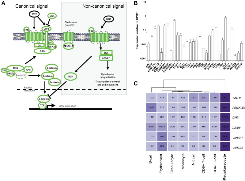
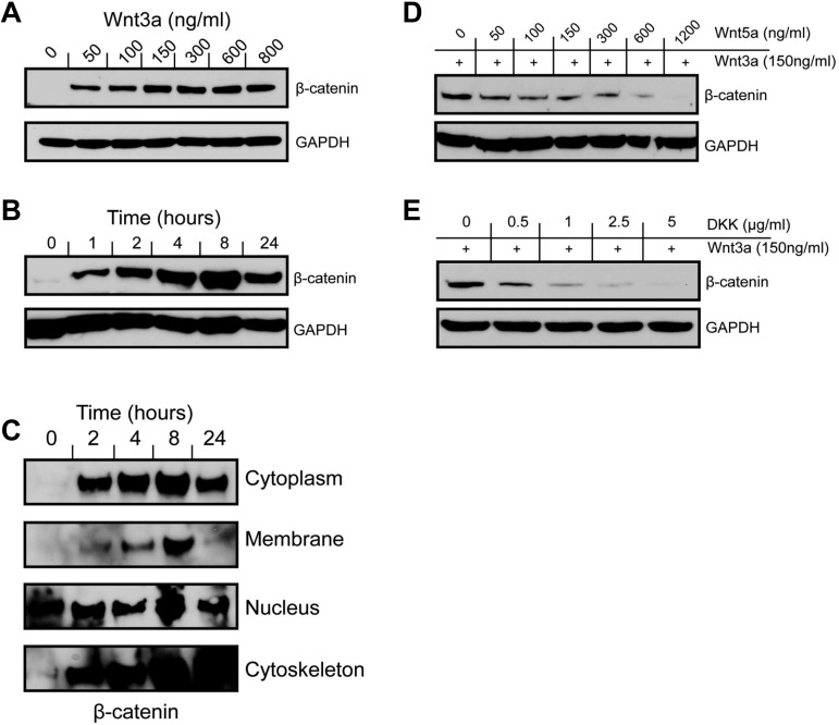
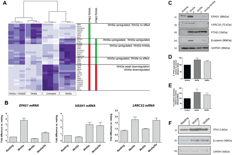
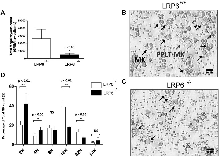
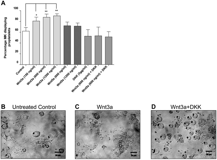
Wnt signaling is involved in numerous aspects of vertebrate development and homeostasis, including the formation and function of blood cells. Here, we show that canonical and noncanonical Wnt signaling pathways are present and functional in megakaryocytes (MKs), with several Wnt effectors displaying MK-restricted expression. Using the CHRF288-11 cell line as a model for human MKs, the canonical Wnt3a signal was found to induce a time and dose-dependent increase in β-catenin expression. β-catenin accumulation was inhibited by the canonical antagonist dickkopf-1 (DKK1) and by the noncanonical agonist Wnt5a. Whole genome expression analysis demonstrated that Wnt3a and Wnt5a regulated distinct patterns of gene expression in MKs, and revealed a further interplay between canonical and noncanonical Wnt pathways. Fetal liver cells derived from low-density-lipoprotein receptor-related protein 6-deficient mice (LRP6(-/-)), generated dramatically reduced numbers of MKs in culture of lower ploidy (2N and 4N) than wild-type controls, implicating LRP6-dependent Wnt signaling in MK proliferation and maturation. Finally, in wild-type mature murine fetal liver-derived MKs, Wnt3a potently induced proplatelet formation, an effect that could be completely abrogated by DKK1. These data identify novel extrinsic regulators of proplatelet formation, and reveal a profound role for Wnt signaling in platelet production.


Figures

Figure 1
Figure 1
Expression of Wnt signaling pathway components in the megakaryocyte. (A) Overview of the canonical and noncanonical Wnt signaling pathways. All components shown in green were detected at the transcript level in our previous microarray studies of in vitro differentiated MKs. (B) Expression of Wnt signaling pathway components in in vitro differentiated MKs. Data are presented as the mean and range of the expression relative to the HPRT gene from 2 biologic replicates. (C) Heatmap displaying MK-specific components of the Wnt signaling pathway. The intensity of the shading reflects the level of expression of each gene in each cell type. Numbers shown indicate the normalized expression value of each gene as determined in Watkins et al.
Figure 2
Figure 2
Canonical Wnt signaling is functional in MKs. Incubation of CHRF288-11 cells with Wnt3a results in a dose (A) and time (B; dose 150 ng/mL) dependent increase in cellular β-catenin levels as detected by Western blotting of whole cell lysates using an anti–β-catenin antibody. (C) Subcellular fractionation of cells treated with Wnt3a indicates that β-catenin accumulation occurs in subcellular fractions during a 24 hour period. (D) β-catenin accumulation in response to Wnt3a treatment is inhibited in a dose-dependent manner by the noncanonical agonist Wnt5a. (E) The canonical antagonist DKK1 inhibits β-catenin accumulation in a dose-dependent manner at an 8 hour time point. All blots are representative of at least 3 or more independent experiments.
Figure 3
Figure 3
Transcriptional response of MKs to signaling via Wnt3a and Wnt5a. (A) Heatmap representation of genes differentially expressed in CHRF288-11 cells in response to Wnt3a, Wnt5a, or both in combination. The data from 3 replicates for each experimental group are shown. Clustering of these data identifies subsets of genes that are regulated by either one or both of the ligands. Genes shown in bold are known targets of Wnt3a signaling. (B) Confirmation of observed patterns of expression by RT-PCR for transcripts encoding EPAS1, VASH1, and LRRC32 in response to treatment with Wnt ligands. Data are presented as mean ± SD (n = 3) of fold difference versus expression in untreated cells. (C) Confirmation of changes in protein expression levels of EPAS1, LRRC32, and PTH2 in response to treatment with Wnt ligands. (D-F) PTH2 and β-catenin expression in fetal liver derived murine MKs in response to Wnt3a or Wnt5a. Data shown in panels D and E represent mean detection level (± SD) as determined by Western blotting followed by densitometry. (F) Representative blots showing PTH2 (i), β-catenin (ii), and GAPDH (iii) expression in fetal liver MKs after Wnt treatment.
Figure 4
Figure 4
Ex vivo analysis of fetal liver megakaryopoiesis in LRP6−/− mice. (A) Absolute numbers of MK cells (GPIIb/IIIa+) cells in MK cultures from fetal liver cells. Data shown are the mean ± SD from replicate cultures of LRP6+/+ (n = 5) and LRP6−/− (n = 4) cells. (B-C) Representative images from LRP6+/+ and LRP6−/− cultures, respectively. (D) Ploidy analysis of MKs derived from LRP6+/+ and LRP6−/− cultures. Data represent mean ± SD from replicate cultures of LRP6+/+ (n = 5) and LRP6−/− (n = 3) cells.
Figure 5
Figure 5
Wnt signaling stimulates proplatelet production in fetal liver MKs ex vivo. (A) Percentage of cultured MKs extending proplatelets in cells treated with Wnt3a, Wnt5a, and DKK1. Data are presented as mean ± SD from 3 individual experiments. P values and significance have been determined by 1-way ANOVA followed by Dunnett posttest to compare all groups with untreated cells. (B-D) Representative images of proplatelet-producing MKs in untreated, Wnt3a, and Wnt5a treated samples, respectively.

References

    1. Gordon MD, Nusse R. Wnt signaling: multiple pathways, multiple receptors, and multiple transcription factors. J Biol Chem. 2006;281(32):22429–22433. - PubMed
    1. Semenov MV, Habas R, Macdonald BT, He X. SnapShot: Noncanonical Wnt Signaling Pathways. Cell. 2007;131(7):1378. - PubMed
    1. Kokolus K, Nemeth MJ. Non-canonical Wnt signaling pathways in hematopoiesis. Immunol Res. 2010;46(1-3):155–164. - PubMed
    1. Malhotra S, Baba Y, Garrett KP, Staal FJ, Gerstein R, Kincade PW. Contrasting responses of lymphoid progenitors to canonical and noncanonical Wnt signals. J Immunol. 2008;181(6):3955–3964. - PMC - PubMed
    1. Staal FJ, Sen JM. The canonical Wnt signaling pathway plays an important role in lymphopoiesis and hematopoiesis. Eur J Immunol. 2008;38(7):1788–1794. - PMC - PubMed

Publication types

MeSH terms