. 2012 Dec 20;492(7429):428-32.
doi: 10.1038/nature11617.

A prefrontal cortex-brainstem neuronal projection that controls response to behavioural challenge

Affiliations

A prefrontal cortex-brainstem neuronal projection that controls response to behavioural challenge

Melissa R Warden et al. Nature. .

Abstract

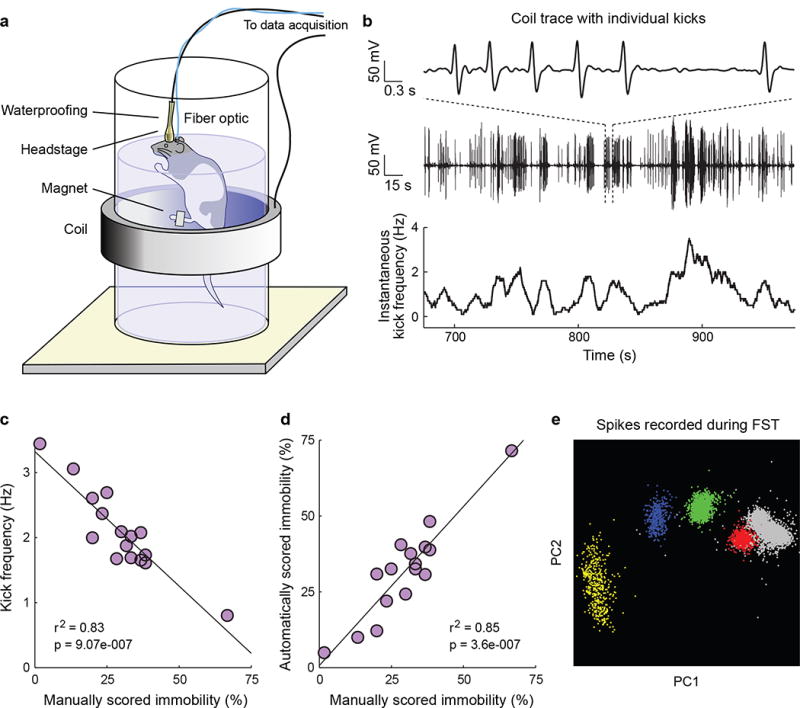
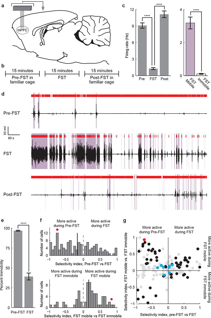
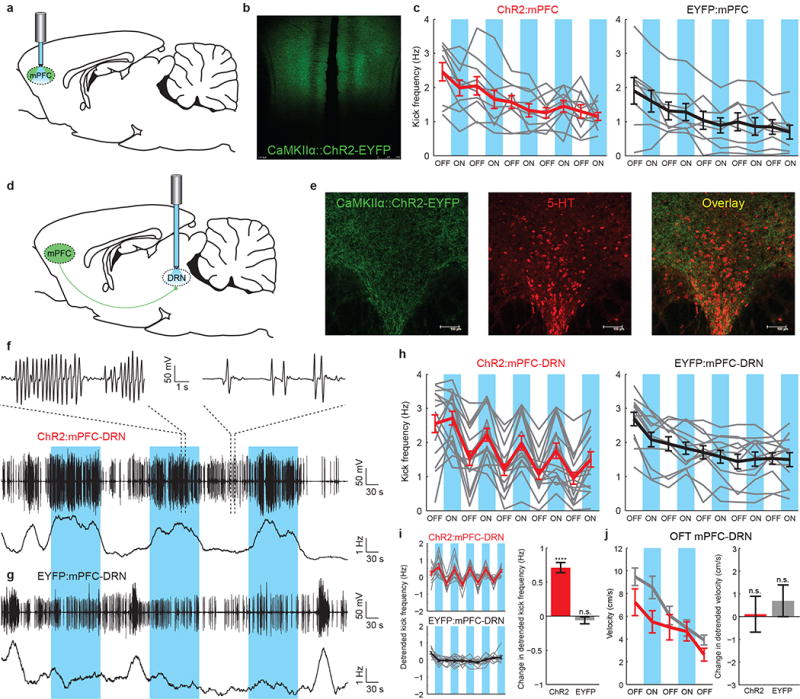
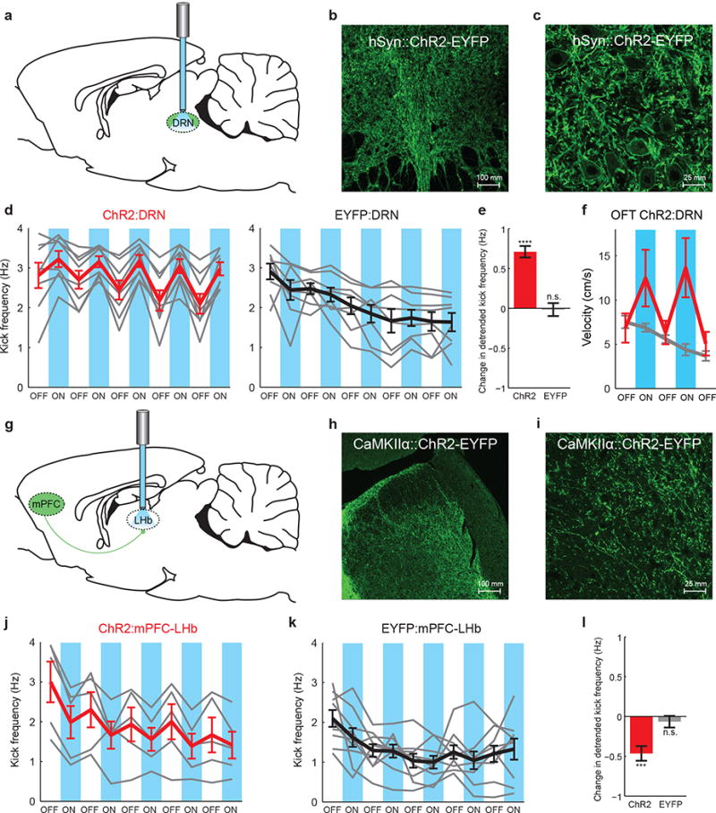
The prefrontal cortex (PFC) is thought to participate in high-level control of the generation of behaviours (including the decision to execute actions); indeed, imaging and lesion studies in human beings have revealed that PFC dysfunction can lead to either impulsive states with increased tendency to initiate action, or to amotivational states characterized by symptoms such as reduced activity, hopelessness and depressed mood. Considering the opposite valence of these two phenotypes as well as the broad complexity of other tasks attributed to PFC, we sought to elucidate the PFC circuitry that favours effortful behavioural responses to challenging situations. Here we develop and use a quantitative method for the continuous assessment and control of active response to a behavioural challenge, synchronized with single-unit electrophysiology and optogenetics in freely moving rats. In recording from the medial PFC (mPFC), we observed that many neurons were not simply movement-related in their spike-firing patterns but instead were selectively modulated from moment to moment, according to the animal's decision to act in a challenging situation. Surprisingly, we next found that direct activation of principal neurons in the mPFC had no detectable causal effect on this behaviour. We tested whether this behaviour could be causally mediated by only a subclass of mPFC cells defined by specific downstream wiring. Indeed, by leveraging optogenetic projection-targeting to control cells with specific efferent wiring patterns, we found that selective activation of those mPFC cells projecting to the brainstem dorsal raphe nucleus (DRN), a serotonergic nucleus implicated in major depressive disorder, induced a profound, rapid and reversible effect on selection of the active behavioural state. These results may be of importance in understanding the neural circuitry underlying normal and pathological patterns of action selection and motivation in behaviour.


Figures

Figure 1
Figure 1. The automated FST provides a high temporal resolution behavioral readout that can be synchronized with simultaneously recorded neural data
a) A schematic of the automated FST. A coil of wire surrounds the tank of water and a magnet is attached to the rat’s back paw. Movement of the magnet within the coil during swimming induces a current that can be recorded. To permit concurrent neural recordings the headstage is waterproofed. An optical fiber can be included for simultaneous optical stimulation. b) Example FST coil voltage traces. Top: a 6-second coil trace showing individual kicks. Middle: a 5-minute coil trace. Bottom: Instantaneous kick frequency estimated from the 5-minute coil trace. c) Average kick frequency corresponds well to manually scored immobility estimates. d) Estimates of FST immobility derived from the induction coil correspond tightly to manually scored immobility estimates. e) 4 well-isolated single mPFC units recorded during the FST.
Figure 2
Figure 2. Prefrontal neuronal activity encodes FST behavioral state
a) A tetrode microdrive or fixed wire array was implanted over the mPFC. b) 15 minutes of data were recorded pre-FST, 15 minutes during the FST, and 15 minutes post-FST. c) Bar plot of an example neuron that is inhibited during immobile states in the FST. (Mann-Whitney U test, * p<0.05; ** p<0.01; *** p<0.001; **** p<0.0001). d) Raster plot of the same neuron. Coil voltage in black, mobile states in purple, spikes in red. Top: pre-FST activity. Middle: activity during the FST. Bottom: post-FST activity. e) Immobility during the pre-FST and FST test epochs (11 rats). f) Distribution of population selectivity indices (Supplementary Methods). Top: pre-FST vs. FST epochs. All neurons significantly selective for pre-FST vs. FST are shown. Bottom: mobile vs. immobile FST states. All neurons significantly selective for mobile vs. immobile FST state are shown. g) Joint distribution of selectivity indices. Black circles: neurons selective for both task epoch and mobility. Red circle: example neuron. Blue circles: putative inhibitory fast-spiking neurons. Gray circles: non-significantly selective neurons. All recorded neurons are shown. Error bars indicate s.e.m.
Figure 3
Figure 3. Optogenetic stimulation of mPFC axons in the DRN, but not excitatory mPFC cell bodies, induces behavioral activation
a) ChR2-EYFP or EYFP-expressing mPFC principal neurons were directly illuminated. b) ChR2-EYFP fluorescence in the mPFC. c) FST kick frequency for ChR2:mPFC (left, n=10) and EYFP:mPFC (right, n=8) rats. Gray lines: individual rats. Thick lines: average for ChR2:mPFC (red) or EYFP:mPFC (black) rats. Blue bars: light on. d) A fiber optic was implanted over the DRN after mPFC injection. e) ChR2-EYFP fluorescence in mPFC axons in the DRN (immunostained for 5-HT). f) FST behavioral data from one ChR2:mPFC-DRN rat. Top, middle: coil voltage. Bottom: kick frequency. g) FST data from one EYFP:mPFC-DRN rat. h) FST kick frequency for all rats. Left: ChR2:mPFC-DRN rats (n=16). Right: EYFP:mPFC-DRN rats (n=12). i) Left: exponentially detrended data from h. Right: change in detrended kick frequency from light-off to light-on epochs, ChR2:mPFC-DRN (red) and EYFP:mPFC-DRN (gray) rats. j) Left: velocity during stimulation in the open field test. Red: ChR2:mPFC-DRN rats (n=12). Gray: EYFP:mPFC-DRN rats (n=12). Right: change in detrended velocity from light-off to light-on epochs. Error bars indicate s.e.m.
Figure 4
Figure 4. Behavioral activation resulting from stimulation of DRN-projecting mPFC axons is specific to the mPFC-DRN synapse
a) A fiber optic was implanted over ChR2- or EYFP-expressing neurons in the DRN. b) 20× image of ChR2-EYFP-expressing DRN neuronal cell bodies. c) 40×2× DRN image. d) FST kick frequency for all rats. Left: ChR2:DRN rats (n=8). Right: EYFP:DRN rats (n=8). Gray lines: individual rats. Thick lines: average for ChR2:DRN (red) or EYFP:DRN (black) rats. Blue bars: light on. e) Detrended change in kick frequency from light-off to light-on epochs, ChR2:DRN (red) and EYFP:DRN (gray) rats. f) Velocity during stimulation in the open field test. Red: ChR2:DRN (n=12), Gray:EYFP:DRN (n=12) rats. g) Fiber optics were implanted bilaterally over the LHb to activate ChR2-expressing LHb-projecting mPFC axons. h) 20× image of ChR2-EYFP-expressing mPFC axons in the LHb. i) 40×2× LHb image. j) FST kick frequency for all rats. Left: ChR2:mPFC-LHb rats (n=5). Right:EYFP:mPFC-LHb rats (n=9). k) Detrended change in kick frequency from light-off to light-on epochs in the FST. Error bars indicate s.e.m.

Comment in

References

    1. McGuire JT, Botvinick MM. Prefrontal cortex, cognitive control, and the registration of decision costs. Proceedings of the National Academy of Sciences of the United States of America. 2010;107:7922–6. - PMC - PubMed
    1. Ridderinkhof KR, van den Wildenberg WPM, Segalowitz SJ, Carter CS. Neurocognitive mechanisms of cognitive control: the role of prefrontal cortex in action selection, response inhibition, performance monitoring, and reward-based learning. Brain and cognition. 2004;56:129–40. - PubMed
    1. Mayberg HS, et al. Reciprocal limbic-cortical function and negative mood: converging PET findings in depression and normal sadness. The American journal of psychiatry. 1999;156:675–82. - PubMed
    1. Maes M, Meltzer H. The serotonin hypothesis of major depression. Psychopharmacology: the fourth generation of progress. 1995:933–44.
    1. Kessler RC, et al. Lifetime prevalence and age-of-onset distributions of mental disorders in the World Health Organization’s World Mental Health Survey Initiative. World psychiatry: official journal of the World Psychiatric Association (WPA) 2007;6:168–76. - PMC - PubMed

Publication types